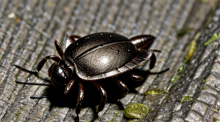
Tick Anatomy and Physiology
The Tick's Nervous System
Ganglia and Nerve Clusters
Ticks possess a decentralized nervous system composed of paired ganglia and peripheral nerve clusters. Each ganglion contains neuronal cell bodies that coordinate motor activity, sensory processing, and reflexes for the body segment it serves. The ventral nerve cord links the ganglia, allowing signal transmission along the abdomen.
When the head is removed, the anterior ganglia are lost, but the remaining posterior ganglia retain functional autonomy. They continue to regulate muscle contraction, blood‑feeding behavior, and basic metabolic processes. Evidence shows that detached bodies can complete a blood meal, excrete waste, and maintain movement for several days despite the absence of the brain.
Key observations:
- Posterior ganglia sustain rhythmic locomotion and attachment mechanisms.
- Peripheral nerve clusters transmit sensory input from the cuticle to the remaining ganglia.
- Metabolic control persists through hormonal signaling independent of the cranial region.
The capacity of a tick to survive without its head therefore depends on the residual ganglia’s ability to maintain essential physiological functions. This autonomy explains why decapitated specimens can remain alive for a limited period, although long‑term viability declines without the central brain structures.
Decentralized Control
Ticks possess a series of ventral nerve cords and paired ganglia that regulate locomotion, feeding and sensory responses. The absence of a cephalic segment does not eliminate these ganglia, allowing basic motor patterns to persist for a limited period. This distributed neural architecture exemplifies decentralized control, where command functions are shared among multiple nodes rather than concentrated in a single brain.
Key characteristics of decentralized control in ticks include:
- Redundant ganglionic clusters along the body axis
- Localized processing of sensory inputs at each segment
- Autonomous generation of rhythmic motor outputs without central oversight
The same principles guide engineered systems that emulate arthropod resilience. Robots equipped with multiple micro‑controllers can maintain locomotion after loss of a primary processor, mirroring the tick’s ability to function despite head removal. Decentralized control thus provides fault tolerance, scalability and adaptability in both biological and artificial contexts.
In summary, the tick’s survival after decapitation illustrates how distributed neural networks sustain essential behaviors, reinforcing the value of decentralized control strategies for robust system design.
Survival Mechanisms of Ticks
Respiration and Metabolism
Spiracles and Tracheal System
Ticks breathe through a pair of posterior openings called «spiracles», which connect to a network of thin tubes termed the «tracheal system». These structures penetrate the cuticle and terminate in minute air sacs that deliver oxygen directly to internal tissues. The spiracular valves regulate airflow, preventing water loss while allowing gas exchange.
The head of a tick houses sensory organs and mouthparts but does not contain primary respiratory openings. Consequently, removal of the cephalothorax does not immediately disrupt the spiracular pathway. Oxygen can continue to reach the body’s cells as long as the posterior cuticle remains intact.
Survival without the head is limited by several factors:
- Loss of feeding capability halts nutrient intake.
- Disruption of neural control impairs coordination of spiracular opening and closing.
- Degradation of internal organs proceeds unchecked, leading to systemic failure.
In practice, decapitated ticks may exhibit brief activity, sustained by residual oxygen supplied through the functional spiracles, but eventual death occurs as metabolic demands exceed the limited respiratory capacity.
Low Metabolic Rate
Ticks possess an exceptionally low metabolic rate compared with most arthropods. Energy consumption remains minimal during periods of inactivity, allowing the organism to endure prolonged intervals without a blood meal.
The low metabolic demand reduces the requirement for rapid circulatory and respiratory function. Heartbeats occur at a slow pace, and oxygen uptake is limited, which together diminish the risk of tissue necrosis when the central nervous system is compromised.
If the cephalic region is removed, the remaining body retains the capacity to sustain basic cellular processes for a limited time. The reduced energy requirement slows degradation of internal organs, permitting survival for days to weeks despite the loss of feeding apparatus and sensory input.
Key effects of the low metabolic rate in this context:
- Extended tolerance to fasting conditions.
- Preservation of vital organ function in the absence of head‑derived hormonal regulation.
- Delayed onset of systemic failure, extending the window for potential regeneration or external intervention.
Consequently, the tick’s inherently sluggish metabolism constitutes the primary factor that enables temporary survival after decapitation, though long‑term viability remains unattainable without the head’s essential functions.
Sensory Functions
Palps and Leg Receptors
Palps, located on the anterior gnathosoma, contain chemosensory and mechanosensory sensilla that detect host odors, carbon‑dioxide gradients, and tactile cues. These structures guide the tick toward a suitable attachment site and initiate the probing phase of blood‑feeding. Damage or removal of the cephalic capsule eliminates palpal input, depriving the organism of essential host‑locating signals.
Leg receptors, distributed along the eight ambulacral appendages, comprise Haller’s organ on the first pair and numerous cuticular sensilla on the remaining legs. Haller’s organ integrates thermal, olfactory, and humidity information, while the distal sensilla monitor substrate texture and movement. Together they coordinate locomotion, questing behavior, and attachment stability.
Consequences of head loss include:
- Absence of palpal chemosensory feedback, preventing host detection.
- Disruption of Haller’s organ function, eliminating thermal and olfactory guidance.
- Loss of mechanoreceptive input required for coordinated movement and questing posture.
Without these sensory systems, a tick cannot locate a host, secure attachment, or regulate feeding, rendering long‑term survival implausible.
Chemical and Thermal Sensing
Ticks rely on two principal sensory systems to locate hosts: chemoreception and thermoreception. Both systems are concentrated in the anterior appendages, especially the Haller’s organ on the forelegs and the mouthparts. Removal of the head eliminates these structures, directly impairing signal acquisition from the environment.
Chemoreception operates through a suite of receptor proteins that bind volatile compounds such as carbon‑dioxide, ammonia and host skin odors. Binding triggers neuronal activation in the Haller’s organ, guiding the tick toward a potential host. Without the gnathosoma, the receptor array is absent, preventing detection of chemical cues that initiate questing behavior.
Thermoreception depends on temperature‑sensitive neurons embedded in the palpal sensilla. These neurons respond to minute temperature gradients generated by a warm‑blooded host, providing directional information. Loss of the head removes the palpal sensilla, abolishing thermal detection.
Consequences of head loss for a tick:
- Absence of chemosensory receptors → inability to sense host odorants or carbon‑dioxide.
- Absence of thermosensory neurons → inability to detect host heat.
- Disruption of questing behavior → reduced probability of locating a blood meal.
- Increased mortality risk due to failure to feed.
Headlessness in Invertebrates
Regenerative Capabilities
Ticks possess a limited capacity for tissue repair after traumatic injury. The nervous system of acariform arachnids lacks the extensive regenerative pathways found in some annelids and planarians, restricting recovery of lost structures. When the cephalic capsule is removed, the following outcomes are typical:
- Hemolymph loss leads to rapid desiccation; the cuticle cannot reseal without the dorsal shield provided by the head.
- Salivary glands, located primarily in the anterior region, cease secretion, depriving the organism of essential enzymes for blood‑feeding.
- Central ganglia, essential for motor coordination, are absent, preventing locomotion and host‑attachment.
Regeneration of the mouthparts and sensory organs has not been documented in any ixodid species. Experimental observations indicate that decapitated nymphs and adults exhibit immediate cessation of feeding behavior and die within hours. No evidence supports the formation of a functional replacement head or the restoration of neural circuits.
Consequently, while ticks can repair minor cuticular wounds, they lack the biological mechanisms required to reconstruct a head and resume life processes. Survival without the cephalic region is therefore untenable.
Short-Term Bodily Functions
The removal of a tick’s cephalic region does not instantly terminate all physiological activity. Blood circulation persists through the dorsal vessel for several minutes, delivering oxygen and nutrients to peripheral tissues. Muscular contraction in the legs continues under residual neuronal input, allowing limited movement and attachment to the host. Respiratory gas exchange via the spiracles remains functional until tracheal pressure equalizes with the external environment.
Short‑term bodily functions that operate after decapitation include:
- Hemolymph flow driven by the contractile heart tube
- Neuromuscular signaling in the thoracic ganglia
- Cuticular respiration through open spiracles
- Metabolic enzyme activity within gut cells
- Cellular ATP production sustained by stored glycogen
These processes sustain minimal viability, enabling the tick to survive briefly without its head before irreversible failure of central regulation occurs.
Implications for Tick Removal and Control
Proper Tick Removal Techniques
Proper removal of attached ticks prevents the risk of the parasite surviving after the head is detached, a scenario that can lead to continued feeding and disease transmission.
- Grasp the tick as close to the skin as possible using fine‑point tweezers or a specialized tick‑removal tool.
- Apply steady, downward pressure; avoid twisting, jerking, or crushing the body.
- Maintain traction until the mouthparts separate cleanly from the skin.
- Disinfect the bite site with an antiseptic and wash hands thoroughly.
After extraction, place the tick in a sealed container with a small amount of alcohol for identification if needed. Monitor the bite area for signs of inflammation, rash, or fever over the next two weeks. Seek medical advice promptly if any symptoms develop, as residual head fragments can still release pathogens.
Pest Control Strategies
Ticks cannot maintain vital functions after decapitation; loss of the capitulum halts feeding and leads to rapid mortality. This biological constraint underpins several pest‑control measures aimed at reducing tick populations and preventing disease transmission.
- Mechanical disruption: manual removal of engorged ticks, application of heat or freezing devices that damage the head capsule.
- Chemical acaricides: topical or systemic agents that target neural receptors located in the capitulum, ensuring rapid incapacitation.
- Biological agents: entomopathogenic fungi and nematodes that infiltrate the tick’s cuticle and impair cephalic structures.
- Environmental sanitation: regular mowing, leaf‑litter removal, and controlled burns that expose ticks to desiccation and increase head loss due to environmental stress.
- Physical traps: CO₂‑baited devices that attract questing ticks, followed by rapid decapitation mechanisms.
Effective management integrates these tactics, monitors tick density, and adjusts interventions according to seasonal activity patterns. Emphasis on strategies that directly compromise the head region maximizes mortality rates while minimizing non‑target impacts.
