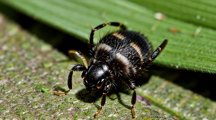
Understanding Tick Behavior
The Tick's Feeding Process
Attachment and Saliva
Ticks secure themselves by inserting their hypostome, a barbed feeding organ, into the host’s skin. The hypostome’s backward‑pointing hooks prevent backward movement, while the tick’s mouthparts lock into the tissue. Saliva is released simultaneously; it contains anticoagulants, anti‑inflammatory agents, and immunomodulators that keep blood flowing and reduce host detection. These compounds also facilitate the formation of a cement‑like matrix that reinforces the attachment.
The same saliva that maintains attachment also enables rapid detachment when the tick is disturbed. Salivary enzymes can dissolve the cement, and the tick can contract its forelegs to pull free. After a brief feeding period, the tick may disengage and drop off the host, leaving the bite site without the feeding apparatus.
Key points:
- Hypostome insertion creates a mechanical anchor.
- Saliva provides anticoagulation, immune suppression, and cement formation.
- Enzymatic activity in saliva can reverse cement formation, allowing the tick to release itself.
Duration of Feeding
Ticks attach to a host and remain attached for a defined feeding period that varies by species and life stage. The feeding cycle proceeds through three phases: attachment, slow feeding, and rapid engorgement. During the slow phase, the tick inserts its hypostome and secretes anticoagulants, maintaining a stable attachment while ingesting blood at a low rate. This stage typically lasts 2–5 days for larvae, 3–7 days for nymphs, and 5–10 days for adult females. The rapid engorgement phase follows, during which blood intake accelerates dramatically and the tick swells to its maximum size; this phase can last from a few hours up to 48 hours depending on the stage.
Several factors can cause a tick to detach before completing the feeding cycle:
- Host grooming – mechanical removal or vigorous scratching disrupts the hypostome.
- Environmental stress – temperature extremes or low humidity impair the tick’s ability to maintain attachment.
- Immune response – host inflammatory reactions can weaken the tick’s attachment sites.
- Pathogen load – heavy infection may reduce the tick’s stamina, prompting premature detachment.
When a tick disengages early, the feeding duration is truncated, and the amount of blood ingested is insufficient for full development. Consequently, the tick may drop off the host and die, or survive long enough to re‑attach elsewhere if conditions permit. Understanding the typical feeding timelines and the circumstances that interrupt them clarifies why a tick may bite, feed briefly, and then fall off.
When Ticks Detach Naturally
Full Engorgement
Full engorgement occurs when a tick has completed its blood meal and its body expands to several times its unfed size. The process typically lasts three to seven days for adult females, depending on species and host availability. During this period the tick’s abdomen swells, the cuticle stretches, and the mouthparts remain anchored to the host’s skin.
When engorgement reaches its peak, the attachment becomes less secure. The tick’s legs, which hold it in place, may loosen as the abdomen fills, increasing the likelihood that the parasite detaches spontaneously. This detachment often happens shortly after the tick has finished feeding, before it drops to the ground to lay eggs.
Key points about full engorgement and subsequent loss of attachment:
- Engorged females can weigh up to 100 mg, a hundredfold increase over their unfed mass.
- The tick’s salivary glands cease secretion once the blood meal is complete, reducing the stimulus that maintains attachment.
- Mechanical disturbance or host grooming can dislodge an engorged tick more easily than an unfed one.
- After detachment, the tick seeks a protected microhabitat to molt or, in the case of females, to deposit eggs.
Recognizing an engorged tick on a host is essential for timely removal. The abdomen appears distended, the body is pale to grayish, and the legs may be splayed. Prompt extraction with fine‑point tweezers, grasping the mouthparts close to the skin, minimizes the chance of leaving mouthparts embedded and reduces pathogen transmission risk.
In summary, full engorgement marks the final stage of a tick’s feeding cycle, during which the parasite’s grip weakens and spontaneous fall‑off becomes common. Understanding this behavior informs effective tick management and reduces the probability of disease exposure.
Environmental Factors
Ticks attach to a host when conditions favor feeding, but environmental variables determine the likelihood of attachment and subsequent detachment.
Temperature directly influences tick activity. Warm periods (20‑30 °C) increase questing behavior, while temperatures below 5 °C suppress movement and reduce bite incidence. Rapid temperature drops after attachment can cause the tick to disengage prematurely.
Humidity governs desiccation risk. Relative humidity above 80 % maintains tick hydration, supporting prolonged feeding. Low humidity accelerates water loss, prompting the tick to abandon the host to seek a more favorable microclimate.
Vegetation density shapes host encounter rates. Dense understory provides shelter and enhances questing success; open habitats expose ticks to environmental stress, increasing the chance of fall‑off.
Seasonal cycles dictate life‑stage prevalence. Larvae and nymphs are most active in spring and early summer, aligning with peak host activity. Adult ticks peak in late summer and autumn; cooler, drier conditions during these periods raise detachment probability.
Host behavior affects attachment duration. Hosts moving through moist, shaded areas remain longer in favorable zones, reducing tick drop‑off. Conversely, hosts traversing dry, exposed terrain experience higher tick loss.
Key environmental factors can be summarized:
- Temperature: optimal range for feeding, extreme fluctuations trigger detachment.
- Humidity: high levels sustain feeding, low levels induce disengagement.
- Vegetation cover: dense cover promotes attachment, sparse cover encourages loss.
- Seasonality: life‑stage activity peaks correspond with environmental suitability.
- Host movement patterns: moist, shaded routes favor prolonged attachment; dry, open routes increase drop‑off.
Understanding these variables clarifies why ticks may bite and later fall off under specific environmental conditions.
Risks and Concerns
Incomplete Detachment
Remaining Mouthparts
When a tick attaches to a host, it inserts its feeding apparatus— the capitulum— into the skin. The capitulum consists of the chelicerae, palps, and a hypostome covered with barbed teeth. After a period of feeding, the tick may detach spontaneously or be removed. In either case, the feeding apparatus can remain embedded in the tissue.
The retained mouthparts are typically the hypostome, which anchors the tick by the barbs. The chelicerae and palps may also stay lodged if they are not fully withdrawn. These fragments are usually small, measuring 0.2–0.5 mm, and may appear as a tiny, dark puncture or a raised spot.
Potential consequences of retained mouthparts include:
- Local inflammation caused by mechanical irritation.
- Possible transmission of pathogens from the remaining tissue.
- Secondary infection if the site is not kept clean.
Management steps:
- Inspect the bite site carefully after tick removal; look for a visible core or a tiny black dot.
- If a fragment is observed, attempt gentle extraction with sterilized tweezers, pulling parallel to the skin surface.
- Disinfect the area with an antiseptic solution.
- Monitor for signs of redness, swelling, or fever; seek medical evaluation if symptoms develop.
Preventive measures focus on thorough removal techniques that minimize the chance of mouthpart retention. Using fine-tipped forceps to grasp the tick near the mouthparts and applying steady, upward traction reduces the risk of breaking the capitulum.
Disease Transmission
Incubation Period
Ticks may attach, feed for several hours to days, and then detach without delivering pathogens. The time between a tick’s removal and the appearance of clinical signs—the incubation period—varies by pathogen, not by the act of detachment itself.
Typical incubation periods for the most common tick‑borne illnesses are:
- Lyme disease (Borrelia burgdorferi): 3 – 30 days; skin rash often precedes systemic symptoms.
- Rocky Mountain spotted fever (Rickettsia rickettsii): 2 – 14 days; fever and rash develop rapidly.
- Anaplasmosis (Anaplasma phagocytophilum): 5 – 21 days; flu‑like symptoms appear after a short delay.
- Babesiosis (Babesia microti): 1 – 4 weeks; hemolytic anemia may be delayed.
- Ehrlichiosis (Ehrlichia chaffeensis): 5 – 10 days; fever and headache emerge early.
The incubation period begins once the pathogen is transmitted, which can occur within minutes of attachment for some agents, but often requires at least 24 hours of feeding. Consequently, a tick that falls off after a brief attachment may not have transmitted disease, resulting in no incubation period. Conversely, a tick that remains attached for the full feeding duration can introduce pathogens, and the subsequent incubation timeline follows the ranges listed above. Monitoring for symptoms during these windows enables prompt diagnosis and treatment.
Common Tick-Borne Illnesses
Ticks often attach to a host, feed, and then detach on their own or after removal. Pathogen transmission typically requires a minimum feeding period; the longer the tick remains attached, the greater the likelihood of infection. Consequently, even a tick that drops off after a brief bite can pose a health risk if the attachment lasted long enough for pathogens to enter the bloodstream.
Common tick‑borne illnesses include:
- Lyme disease – caused by Borrelia burgdorferi; early signs are erythema migrans rash, fever, headache, and fatigue; untreated infection may progress to joint, cardiac, or neurological complications.
- Anaplasmosis – caused by Anaplasma phagocytophilum; symptoms appear within 1‑2 weeks and include fever, chills, muscle aches, and leukopenia; early antibiotic therapy prevents severe disease.
- Ehrlichiosis – caused by Ehrlichia chaffeensis; presents with fever, headache, malaise, and thrombocytopenia; prompt doxycycline treatment reduces mortality.
- Babesiosis – caused by Babesia microti; manifests as hemolytic anemia, fever, and chills; severe cases may require exchange transfusion, especially in immunocompromised patients.
- Rocky Mountain spotted fever – caused by Rickettsia rickettsii; characterized by fever, rash, and endothelial damage; early treatment with doxycycline is critical to avoid fatal outcomes.
- Tularemia – caused by Francisella tularensis; symptoms range from ulceroglandular lesions to pneumonic disease; antibiotic therapy is essential for recovery.
Risk assessment should consider tick species, geographic prevalence, and duration of attachment. Immediate removal of a feeding tick, followed by monitoring for symptoms within the typical incubation window of each disease, constitutes the primary preventive strategy.
What to Do After a Tick Bite
Safe Removal Techniques
Tools and Methods
Ticks may attach, feed, and later detach without external signs. Accurate assessment relies on specific instruments and systematic procedures.
Essential instruments include:
- Fine‑point tweezers or tick‑specific forceps for secure grasp.
- Tick removal devices with a notch to slide under the mouthparts.
- Magnifying lens or portable microscope to inspect attachment depth.
- Disposable gloves to prevent contamination.
- Transparent adhesive patches or sticky traps for collecting detached specimens.
- PCR kits or rapid antigen tests for pathogen detection in the removed tick.
Effective procedures consist of:
- Conducting a thorough skin examination within 24 hours of exposure, focusing on hidden areas such as scalp, groin, and armpits.
- Grasping the tick as close to the skin as possible, applying steady upward pressure to extract without crushing the body.
- Placing the extracted tick in a sealed container for laboratory analysis or visual confirmation of engorgement, which indicates recent feeding.
- Applying a sterile antiseptic to the bite site and monitoring for erythema, swelling, or ulceration over several days.
- Deploying adhesive patches around the bite area for 48 hours to capture any detached tick that may fall off after feeding.
- Sending the collected specimen to a diagnostic lab for molecular testing to identify transmitted pathogens and to verify that the tick has indeed separated from the host.
Combining precise tools with a step‑by‑step protocol ensures reliable detection of tick attachment, safe removal, and confirmation of subsequent detachment.
Post-Removal Care
A tick may attach, feed, and detach on its own, leaving the bite site exposed to potential infection. After removal, immediate actions reduce the risk of disease transmission and promote healing.
- Clean the area with soap and water, then apply an antiseptic such as iodine or alcohol.
- Inspect the wound for remaining mouthparts; if any fragment remains, remove it with sterile tweezers, avoiding squeezing the surrounding skin.
- Apply a sterile adhesive bandage if the bite is bleeding; otherwise, keep the site uncovered to allow air exposure.
- Observe the site for 24‑48 hours. Note redness, swelling, increasing warmth, or a rash extending beyond the bite.
- Record the date of removal and the estimated duration of attachment; this information assists healthcare providers if symptoms develop.
- Seek medical evaluation promptly if fever, headache, fatigue, muscle aches, or a bullseye‑shaped rash appear, as these may indicate tick‑borne illness.
- For individuals with known allergies to tick saliva or previous severe reactions, consider prophylactic antibiotics after consulting a clinician.
Maintaining a clean environment, limiting skin irritation, and monitoring for systemic signs constitute the core of effective post‑removal care.
When to Seek Medical Attention
Symptoms to Watch For
A tick may attach, feed, and then detach on its own or be removed. After the bite, monitor the bite site and overall health for early signs of infection or disease transmission.
- Redness spreading beyond the immediate bite area
- Swelling or warmth around the attachment point
- A rash resembling a target or bullseye pattern
- Fever, chills, or unexplained temperature elevation
- Headache, fatigue, or muscle aches
- Joint pain, especially in knees or elbows
- Nausea, vomiting, or abdominal discomfort
- Neurological symptoms such as tingling, numbness, or facial weakness
If any of these symptoms appear within days to weeks after the tick detaches, seek medical evaluation promptly. Early detection and treatment reduce the risk of severe complications.
Prevention Strategies
Ticks attach to skin, feed, and may detach after engorgement. Preventing both attachment and subsequent detachment reduces disease risk.
Wear light-colored, tightly woven clothing when entering wooded or grassy areas. Tuck shirts into pants, and secure pant legs with gaiters. Apply EPA‑registered repellents containing DEET, picaridin, or IR3535 to exposed skin and clothing. Reapply according to product instructions, especially after sweating or water exposure.
Maintain yard by trimming vegetation, removing leaf litter, and creating a barrier of wood chips or gravel between lawn and forested edges. Treat perimeters with acaricide products where appropriate, following label directions.
Inspect humans and animals immediately after outdoor activity. Conduct systematic searches: run fingers over the body, use a mirror for hard‑to‑see areas, and examine pets’ fur and ears. Prompt removal of attached ticks—grasping close to the skin with fine‑pointed tweezers and pulling steadily—prevents prolonged feeding.
Limit wildlife access to residential spaces. Install fencing, secure garbage, and discourage deer feeding. Reduce rodent habitats by storing feed in sealed containers.
Educate household members about tick habitats, seasonal activity peaks, and proper removal techniques. Provide written guidelines and visual aids in common areas.
By integrating personal protection, environmental management, regular inspections, and informed removal, the likelihood of a tick attaching and later falling off is substantially lowered.
