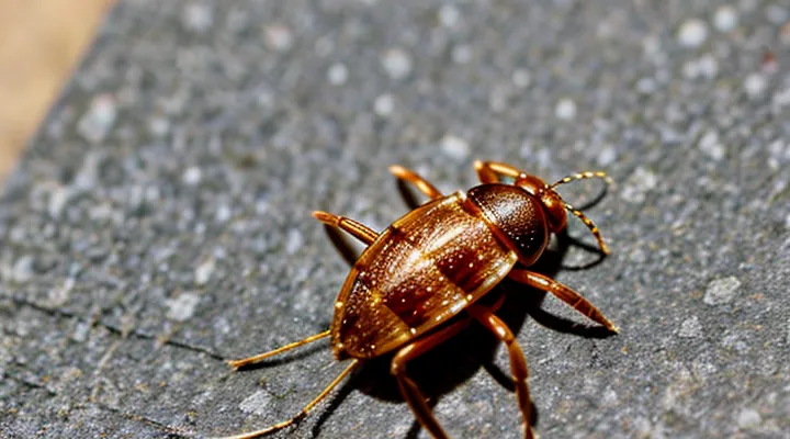
Why Improper Tick Removal Is Dangerous
Risks of Incomplete Removal
Retained Mouthparts
When a tick is detached by pulling, the mandibles and hypostome often remain embedded in the skin. These retained mouthparts act as a conduit for pathogens, allowing bacteria such as Borrelia burgdorferi or Anaplasma to enter the bloodstream even after the tick’s body is gone. The foreign material also triggers a localized inflammatory response, which can develop into an ulcer or granuloma if not removed promptly.
Key consequences of retained mouthparts:
- Increased risk of infection transmission
- Prolonged skin irritation and possible secondary bacterial infection
- Potential for chronic lesions that may require surgical excision
The most reliable removal technique involves using fine‑pointed tweezers to grasp the tick as close to the skin as possible and applying steady, upward pressure. This method minimizes the chance of mouthpart separation. If the mandibles are observed in the wound after removal, a sterile needle or scalpel should be used to extract them under clean conditions. Following extraction, disinfect the site with an antiseptic and monitor for signs of infection, such as redness, swelling, or fever. Early medical evaluation is advised if symptoms develop.
Increased Infection Risk
Removing a tick by jerking it off the skin raises the probability of infection. When the tick’s body is crushed, saliva and gut contents can be forced into the bite wound, delivering bacteria such as Borrelia burgdorferi (Lyme disease) or Anaplasma species directly into the host. If the mouthparts break off and remain embedded, they become a foreign body that can harbor bacteria and provoke local inflammation, creating a portal for secondary bacterial infection.
Key mechanisms that increase infection risk include:
- Mechanical rupture of the tick – rapid traction tears the exoskeleton, releasing infectious fluids.
- Retention of mouthparts – fragments left in the skin act as nidus for bacterial colonization.
- Tissue trauma – excessive force damages surrounding skin, compromising the barrier function and facilitating pathogen entry.
- Delayed proper treatment – improper removal often leads to incomplete extraction, requiring additional medical intervention that carries its own infection hazards.
The recommended procedure eliminates these risks. Using fine‑pointed tweezers, grasp the tick as close to the skin as possible, apply steady, gentle pressure, and pull upward without twisting. After removal, cleanse the site with antiseptic and monitor for signs of infection. This method preserves the tick’s integrity, minimizes tissue injury, and reduces the likelihood that disease‑causing organisms will be introduced.
Potential for Disease Transmission
Crushing the Tick
Crushing a tick after it has attached to skin may appear to destroy the parasite, but the method carries significant hazards. When the body of the tick is ruptured, saliva and internal fluids containing pathogens are released directly onto the wound surface. This increases the probability that bacteria, viruses, or protozoa such as Borrelia burgdorferi will penetrate the host’s tissue, potentially initiating infection.
The mechanical pressure applied during crushing often fails to detach the mouthparts, which can remain embedded in the skin. Retained hypostome fragments act as a foreign body, provoking localized inflammation and creating a conduit for microbial entry. Incomplete removal of these parts may also complicate subsequent medical assessment.
A controlled approach reduces these risks:
- Use fine‑tipped tweezers to grasp the tick as close to the skin as possible.
- Apply steady, downward pressure to extract the entire organism without twisting.
- Disinfect the bite area with an antiseptic solution after removal.
- Dispose of the tick in a sealed container for identification if needed.
If crushing is unavoidable—such as when a tick is accidentally stepped on—immediate cleansing of the area with soap and an antiseptic is essential. Monitoring the site for erythema, swelling, or flu‑like symptoms should continue for at least four weeks, with medical consultation if any signs emerge.
Overall, the release of pathogen‑laden fluids and the potential for retained mouthparts make crushing an unreliable and risky alternative to proper extraction.
Releasing Pathogens
Ticks anchor to the skin with a barbed feeding tube that penetrates the epidermis. When the insect is squeezed, twisted, or pulled by the body, the tube can be compressed, forcing saliva and gut contents into the bite wound.
Pathogens that ticks transmit—such as Borrelia burgdorferi, Anaplasma phagocytophilum, Rickettsia spp., and various viruses—reside in the salivary glands and midgut. Disruption of these structures releases infectious material directly into the host’s tissue, increasing the probability of immediate infection and raising the inoculum dose.
Typical outcomes of improper removal include:
- Direct deposition of bacteria, viruses, or protozoa into the bite site.
- Elevated risk of secondary skin irritation that facilitates pathogen entry.
- Potential for systemic spread if a large pathogen load enters the bloodstream.
Proper extraction requires:
- Fine‑tipped tweezers or a specialized tick‑removal tool.
- Grasping the tick as close to the skin as possible.
- Applying steady, upward pressure without squeezing the body.
- Disinfecting the bite area after removal.
Following these steps minimizes tissue damage, prevents the expulsion of pathogen‑laden fluids, and reduces the likelihood of disease transmission.
How to Properly Remove a Tick
Preparation Before Removal
Gather Necessary Tools
When a tick is attached, using fingers to detach it can leave mouthparts embedded, increasing the chance of infection. Proper extraction depends on having the right equipment at hand.
- Fine‑pointed tweezers (or a calibrated tick‑removal tool)
- Disposable nitrile gloves
- Antiseptic solution or wipes
- Small sealable container or zip‑lock bag for the tick
- Alcohol swab for post‑removal skin care
- Optional: magnifying glass for close inspection
Before attempting removal, wash hands, put on gloves, and disinfect the bite area. Place the container nearby so the tick can be sealed immediately after extraction. Having these items prepared eliminates the need for improvised methods and ensures the tick is removed cleanly, minimizing tissue damage and pathogen transmission.
Clean the Area
Removing a tick by simply pulling it can leave parts of the mouth embedded in the skin, creating a portal for bacteria and viruses. After the tick is detached, the surrounding tissue must be treated to close that portal and reduce the chance of secondary infection.
- Wash the site with soap and running water for at least 30 seconds.
- Apply an antiseptic such as povidone‑iodine or chlorhexidine; let it dry.
- Cover the area with a clean, non‑adhesive dressing if irritation is expected.
- Observe the spot for redness, swelling, or discharge for the next 48 hours; seek medical advice if symptoms develop.
Cleaning the area also removes residual tick saliva, which can contain pathogens that survive briefly on the skin surface. Proper decontamination therefore complements careful removal and minimizes health risks.
The Correct Removal Technique
Grasping the Tick
Grasping a tick requires a method that prevents the mouthparts from breaking off inside the skin. Using only fingers or a rough tug can compress the abdomen, causing the tick to inject additional saliva and increase the risk of pathogen transmission. A fine‑pointed, non‑toothed instrument—such as tweezers, a tick‑removal device, or a small hook—provides the control needed to secure the tick close to the skin surface.
The correct technique involves the following steps:
- Position the tool so that the tips are as close to the skin as possible, encircling the tick’s head.
- Apply steady, even pressure while pulling upward in a straight line.
- Avoid twisting, jerking, or squeezing the body.
- After removal, disinfect the bite area and the tool.
- Preserve the tick in a sealed container if identification or testing is required.
Improper removal often leaves fragments of the hypostome embedded, which can lead to localized inflammation, secondary infection, or prolonged exposure to tick‑borne agents. Using a proper gripping tool and a controlled motion eliminates these hazards and ensures complete extraction.
Gentle, Steady Pull
A tick must be detached with a controlled, continuous traction rather than a sudden jerk. This method keeps the parasite’s mouthparts intact, limits the amount of saliva or infected fluid that can enter the wound, and minimizes damage to surrounding skin.
- Mouthparts remain whole, preventing embedded fragments that may cause local irritation.
- Salivary exchange is reduced, lowering the risk of transmitting bacteria, viruses, or protozoa.
- Tissue disruption is minimized, decreasing the chance of secondary infection.
The procedure requires fine‑point tweezers positioned as close to the skin surface as possible. Apply a gentle, steady pressure directly outward until the tick releases. Avoid twisting, squeezing, or crushing the body, as these actions increase the likelihood of mouthpart separation and pathogen exposure.
Aftercare and Monitoring
Disinfecting the Bite Site
Disinfecting the bite area is a critical step after a tick attachment because residual mouthparts can serve as a portal for bacteria and viruses. Proper antiseptic treatment reduces the likelihood that pathogens introduced during feeding will establish an infection.
Effective agents include 70 % isopropyl alcohol, povidone‑iodine solution (10 % concentration), and chlorhexidine gluconate (0.5 % concentration). Each rapidly destroys a broad spectrum of microorganisms on the skin surface, limiting colonization at the wound site.
Recommended procedure:
- Rinse the area with clean running water and mild soap; remove visible debris.
- Apply a generous amount of the chosen antiseptic, ensuring full coverage of the puncture.
- Allow the solution to remain in contact for at least 30 seconds before gently blotting excess liquid.
- If the bite will be exposed to further irritation, place a sterile adhesive bandage over the treated skin.
Combining thorough disinfection with correct removal technique—grasping the tick as close to the skin as possible with fine‑tipped tweezers and applying steady, even pressure—prevents mouthpart retention and minimizes the risk of disease transmission.
Observing for Symptoms
Observing for symptoms after a tick encounter is essential for early detection of tick‑borne diseases. Immediate removal with improper technique can leave mouthparts embedded, increasing pathogen transmission risk. The correct approach involves a careful extraction followed by systematic monitoring.
Key signs to watch for during the observation period:
- Expanding redness or a target‑shaped rash at the bite site
- Fever, chills, or unexplained fatigue
- Headache, neck stiffness, or muscle aches
- Joint pain, especially in knees or elbows
- Nausea, vomiting, or abdominal pain
If any of these manifestations appear within 2–4 weeks after the bite, seek medical evaluation promptly. Documentation of the bite date, tick size, and attachment duration aids clinicians in selecting appropriate diagnostic tests and treatment regimens. Continuous observation, combined with proper removal, reduces the likelihood of severe complications.
When to Seek Medical Attention
Signs of Infection
Redness and Swelling
Redness and swelling often appear after a tick is forcibly extracted. The bite site becomes inflamed because the tick’s mouthparts can remain embedded, releasing saliva that contains anticoagulants and inflammatory proteins. These substances trigger a localized immune response, producing heat, erythema, and edema.
If the tick’s head or hypostome stays in the skin, the body may treat the retained fragment as a foreign object. The ongoing release of saliva can prolong inflammation, increase swelling, and raise the risk of secondary bacterial infection. Persistent redness may indicate that the tick was not removed cleanly.
Proper removal minimizes tissue trauma and reduces the likelihood of these symptoms. The recommended technique involves:
- Grasping the tick as close to the skin as possible with fine‑point tweezers.
- Applying steady, downward pressure to pull straight out without twisting.
- Disinfecting the bite area after extraction.
- Monitoring the site for several days; seeking medical attention if redness expands, swelling worsens, or a rash develops.
When redness and swelling are observed after an attempt to pull a tick, they signal that the bite may have been mishandled, and prompt corrective measures are necessary to prevent complications.
Rash or Fever
Removing a tick by pulling it out can trigger a rash or fever. The tick’s mouthparts often remain embedded when the insect is yanked, creating a breach that allows bacteria and viruses to enter the bloodstream. Local skin irritation appears as a red, sometimes itchy, area around the bite; systemic infection may develop as a fever within days.
Common pathogens transmitted through incomplete removal include:
- Borrelia burgdorferi – causes Lyme disease, characterized by a spreading rash and elevated temperature.
- Rickettsia species – produce spotted fevers with rash and chills.
- Anaplasma phagocytophilum – leads to fever, muscle aches, and sometimes a mild rash.
Symptoms usually emerge 3–10 days after the bite. Early signs are:
- Redness that expands beyond the bite site.
- Warmth and swelling at the attachment point.
- Fever exceeding 38 °C (100.4 °F).
- Headache, fatigue, or joint pain.
Prompt, proper removal—grasping the tick close to the skin with fine‑point tweezers and applying steady pressure—reduces the likelihood of these reactions. If a rash or fever develops after a bite, seek medical evaluation; early antibiotic therapy can prevent complications.
Persistent Symptoms
Concerns After Removal
Removing a tick does not end the health risk; the period following extraction demands careful observation. Incomplete mouthpart removal can leave a foreign body that irritates tissue and serves as a gateway for pathogens. Even when the whole tick is extracted, bacteria or viruses transmitted during attachment may remain dormant before causing symptoms.
Key concerns after removal include:
- Residual mouthparts embedded in skin
- Development of erythema migrans or localized rash within days to weeks
- Fever, headache, fatigue, or joint pain emerging weeks after the bite
- Secondary bacterial infection at the excision site
Each symptom warrants prompt medical evaluation, especially if it appears within the typical incubation window for Lyme disease, anaplasmosis, or babesiosis. Documentation of the bite date, tick appearance, and any evolving signs improves diagnostic accuracy.
If the bite site shows persistent redness, swelling, or discharge, seek professional care. A physician may prescribe prophylactic antibiotics, order serologic testing, or recommend follow‑up visits to track disease progression. Maintaining a log of temperature readings and symptom onset supports timely intervention.
Unusual Reactions
Removing a tick by forceful pulling can trigger atypical physiological responses that complicate diagnosis and treatment. When the mouthparts detach from the host’s skin, they may embed deeper, releasing additional saliva and pathogens that provoke unexpected immune reactions.
Unusual reactions observed after improper extraction include:
- Rapid onset of localized swelling that exceeds typical erythema, indicating an allergic response to tick saliva proteins.
- Development of a necrotic lesion at the bite site, suggesting tissue damage from retained mouthparts.
- Systemic symptoms such as fever, headache, and joint pain appearing within days, reflecting early dissemination of tick‑borne microbes.
- Appearance of a rash with vesicular or bullous formations, a sign of hypersensitivity uncommon in standard bite presentations.
These outcomes arise because violent traction can compress the tick’s salivary glands, forcing a larger volume of biologically active substances into the host. The resulting exposure elevates the risk of severe allergic manifestations and accelerates pathogen transmission, underscoring the need for controlled removal techniques.
