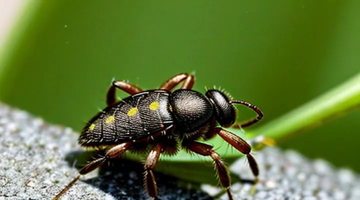
The Anatomy of a Tick Bite
Tick Saliva: A Complex Cocktail
Anesthetic Properties
Ticks inject saliva that contains several anesthetic agents, such as salivary gland-derived proteins and neuroactive peptides. These substances bind to voltage‑gated sodium channels on peripheral nerves, temporarily blocking action potentials and preventing the host from feeling the initial puncture. The anesthetic effect reduces immediate pain, allowing the tick to remain attached for days.
After the tick detaches, the anesthetic compounds degrade and the immune system encounters residual salivary proteins. This triggers a localized inflammatory response characterized by mast‑cell degranulation and release of histamine, prostaglandins, and cytokines. The resulting irritation of cutaneous nerve endings produces the characteristic itch at the bite site.
Key anesthetic components in tick saliva:
- Ixolaris‑like peptides: inhibit sodium channel activation.
- Salp15: interferes with neuronal signaling and modulates immune detection.
- Evasins: bind chemokines, dampening early inflammatory signals.
The combination of initial numbness and subsequent inflammatory mediators explains why the bite area becomes itchy after the anesthetic effect subsides.
Anticoagulant Factors
Ticks inject saliva containing a suite of anticoagulant proteins that disrupt normal clotting cascades. By inhibiting thrombin, factor Xa, and platelet aggregation, these molecules keep blood fluid long enough for the arthropod to feed. The same biochemical interference alerts the host’s immune system, producing the characteristic pruritic reaction at the bite site.
Salivary anticoagulant factors include:
- Ixolaris – binds factor Xa‑Va complex, preventing conversion of prothrombin to thrombin.
- Salp14 – inhibits factor Xa directly, reducing fibrin formation.
- Antithrombin‑like protein – neutralizes thrombin activity, prolonging bleeding.
- Apyrase – degrades ADP, limiting platelet activation.
- Histamine‑binding protein – sequesters host histamine, modulating inflammation but also altering mast‑cell signaling.
These agents provoke itch through several mechanisms. Inhibition of clotting generates fibrin‑degradation products that act as chemotactic signals for neutrophils and macrophages. Recruited cells release cytokines (IL‑1β, TNF‑α) that stimulate mast cells to degranulate, liberating histamine and other pruritogenic mediators. Additionally, some anticoagulants bind directly to host receptors, triggering intracellular pathways that sensitize peripheral nerve endings.
The combined effect of prolonged bleeding, inflammatory cell influx, and mediator release creates a localized hypersensitivity response. The result is the persistent itching commonly observed after a tick attachment.
Immunomodulatory Substances
Ticks inject saliva that contains a complex mixture of immunomodulatory compounds. These agents interfere with the host’s immediate defense mechanisms, creating a local environment that favors prolonged feeding. The same alterations provoke sensory nerve activation, producing the characteristic itch at the bite site.
Key substances in tick saliva include:
- Salivary cystatins: inhibit cysteine proteases, dampen antigen processing, prolong inflammation.
- Prostacyclin and prostaglandin E₂: dilate blood vessels, sensitize peripheral nociceptors, enhance pruritic signaling.
- Histamine‑binding proteins: sequester host histamine, yet trigger alternative pathways that release other pruritogenic mediators.
- Salp15 and other immunosuppressive proteins: suppress T‑cell activation, shift cytokine balance toward Th2 responses, which are associated with itch.
- Anticoagulant peptides (e.g., ixolaris): maintain blood flow, indirectly sustain inflammatory cell recruitment.
The combined effect of these molecules suppresses early immune clearance while promoting a Th2‑dominant milieu. Elevated interleukin‑4, interleukin‑13, and chemokine release sensitize cutaneous C‑fibers, translating the biochemical disturbance into a persistent pruritic sensation.
The Immune System's Response
Histamine Release
Ticks inject saliva containing anticoagulants and proteins that trigger a rapid immune response. The immediate reaction involves activation of resident mast cells and basophils at the bite site. These cells degranulate, releasing histamine into the surrounding tissue.
Histamine binds to H1 receptors on peripheral sensory nerves, causing depolarization and the perception of itch. Simultaneously, histamine induces:
- Vasodilation, increasing blood flow and edema that stretch skin fibers;
- Increased vascular permeability, allowing plasma proteins to infiltrate the tissue and amplify irritation;
- Recruitment of additional immune cells, sustaining the inflammatory milieu.
The combined effect of nerve sensitization and tissue swelling produces the characteristic pruritus observed after a tick attachment.
Inflammatory Mediators
A tick bite introduces saliva containing proteins that trigger a rapid immune response. The immediate reaction involves activation of resident mast cells and keratinocytes, leading to the release of soluble factors that sensitize peripheral nerve endings. This sensitization generates the characteristic pruritus at the bite site.
Key inflammatory mediators responsible for the itch include:
- Histamine released from mast cell degranulation, binds H1 receptors on sensory fibers.
- Prostaglandin E₂ and prostaglandin D₂, produced by cyclooxygenase activity, lower the activation threshold of itch neurons.
- Leukotriene C₄, generated via the 5‑lipoxygenase pathway, amplifies neurogenic inflammation.
- Cytokines such as interleukin‑31, interleukin‑4, and interleukin‑13, secreted by Th2 lymphocytes, directly excite pruriceptors.
- Substance P, released from sensory nerves, promotes vasodilation and further mast cell activation.
- Tryptase, a protease from mast cells, cleaves protease‑activated receptors on nerve terminals.
These mediators act synergistically, producing vasodilation, edema, and heightened neuronal excitability that manifest as itching. The combined effect sustains the pruritic signal until the inflammatory cascade resolves.
Allergic Reactions
The itch that follows a tick bite frequently results from an allergic reaction to substances introduced by the arthropod. Salivary proteins, anticoagulants, and other bioactive molecules act as allergens that trigger the host’s immune system.
Immediate hypersensitivity (type I) involves IgE antibodies bound to mast cells. Upon re‑exposure to the tick’s antigens, cross‑linking of IgE induces rapid degranulation, releasing histamine, prostaglandins, and leukotrienes. These mediators increase vascular permeability and stimulate peripheral nerve endings, producing the characteristic burning or itching sensation within minutes to hours.
Delayed hypersensitivity (type IV) develops over 24–72 hours. Antigen‑presenting cells process tick proteins and present them to sensitized T lymphocytes. Activated T cells release interferon‑γ and other cytokines that attract macrophages and neutrophils. The ensuing inflammation damages epidermal structures, prolonging pruritus and often producing a raised, erythematous papule.
Additional allergic mechanisms include:
- Cross‑reactivity with other arthropod allergens, heightening sensitivity after prior exposures.
- Production of IgG antibodies that form immune complexes, contributing to local complement activation.
- Individual variations in skin barrier integrity, which amplify allergen penetration.
Clinically, the allergic component presents as:
- Localized erythema and swelling.
- Pruritic papule or wheal at the bite site.
- Possible secondary excoriation from scratching.
Management focuses on interrupting the allergic cascade:
- Oral antihistamines (e.g., cetirizine, diphenhydramine) to block histamine receptors.
- Topical corticosteroids to reduce T‑cell mediated inflammation.
- Cold compresses to diminish nerve activation and vasodilation.
- Avoidance of scratching to prevent secondary infection.
Recognizing the allergic basis of tick‑bite pruritus guides appropriate pharmacologic intervention and reduces the risk of complications.
Factors Influencing Itch Intensity
Tick Species and Saliva Composition
Ticks that bite humans belong mainly to three genera. Ixodes scapularis (black‑legged tick), Dermacentor variabilis (American dog tick) and Amblyomma americanum (lone‑star tick) are responsible for the majority of bites in temperate regions. Each species secretes a characteristic blend of salivary compounds that interacts with the host’s skin and immune system.
Saliva of these ectoparasites contains several biologically active molecules:
- Anticoagulants (e.g., salp14, ixolaris) prevent clot formation, allowing prolonged feeding.
- Immunomodulators (e.g., prostaglandin E₂, evasins) suppress local immune responses.
- Histamine‑binding proteins (e.g., histamine‑binding protein‑1) sequester host histamine, delaying detection.
- Proteases and protease inhibitors (e.g., cystatins, metalloproteases) degrade skin proteins and modulate inflammation.
- Vasodilators (e.g., apyrase) increase blood flow to the feeding site.
The interaction of these components with cutaneous nerve endings triggers a cascade of events. Anticoagulants and vasodilators create a moist, inflamed microenvironment that sensitizes peripheral sensory neurons. Immunomodulators alter cytokine profiles, leading to recruitment of mast cells and eosinophils. When mast cells degranulate, they release histamine and other pruritogenic mediators. Although some tick proteins bind histamine, the overall imbalance favors a net increase in free histamine, producing the characteristic itch at the bite site. Differences in salivary composition among species explain variations in itch intensity and duration observed after bites.
Individual Sensitivities
Individual reactions to a tick bite differ because each person’s immune system responds uniquely to the saliva proteins introduced during feeding. Some people mount a robust inflammatory response that quickly produces histamine, prostaglandins, and other mediators, leading to pronounced pruritus at the bite site. Others exhibit minimal mediator release, resulting in little or no itching.
Key factors that shape personal sensitivity include:
- Genetic variations affecting cytokine production and IgE synthesis.
- Prior sensitization to tick salivary antigens, which can amplify allergic pathways on subsequent exposures.
- Skin barrier integrity; compromised epidermis permits deeper antigen penetration and stronger irritation.
- Age‑related immune shifts; children and elderly individuals often display altered inflammatory profiles.
- Co‑existing conditions such as atopic dermatitis or allergic rhinitis, which predispose to heightened itch responses.
When a sensitized individual encounters tick saliva, antigen‑presenting cells trigger Th2‑dominant pathways, prompting IgE‑mediated mast cell degranulation. The resulting surge of histamine, leukotrienes, and proteases activates peripheral nerve fibers, producing the characteristic itch. In contrast, a Th1‑biased response may generate a milder, non‑pruritic inflammation.
Recognizing these personal determinants guides clinical management. Assessment should consider patient history of allergies, skin health, and previous tick exposures. Antihistamines, topical corticosteroids, or immune‑modulating agents can be selected based on the identified sensitivity profile, improving symptom control and reducing the risk of secondary complications.
Duration of Attachment
Ticks that remain attached for several days introduce larger volumes of saliva into the host’s skin. Saliva contains anticoagulants, anesthetics, and proteins that suppress immediate immune detection. The longer the attachment, the greater the cumulative exposure to these agents, which provokes a delayed hypersensitivity reaction manifested as itching.
Extended feeding also allows the tick’s mouthparts to embed deeper into the epidermis and dermis. Mechanical disruption of nerve endings during prolonged insertion generates local irritation. As the tick expands, tissue stretching intensifies, contributing further to the pruritic sensation.
The host’s immune system responds to tick antigens over time. Repeated exposure during a multi‑day attachment leads to:
- Increased production of IgE antibodies specific to tick salivary proteins.
- Activation of mast cells and release of histamine and other pruritogenic mediators.
- Recruitment of eosinophils that sustain inflammation around the bite site.
When the tick detaches, residual salivary components persist in the skin. Their gradual degradation sustains the itch for days to weeks, especially after prolonged attachment periods. Prompt removal within 24 hours limits saliva volume, reduces tissue trauma, and shortens the duration of post‑bite pruritus.
Potential Complications and When to Seek Medical Attention
Secondary Infections
A tick bite creates a small breach in the skin that can become colonized by microorganisms present on the tick’s mouthparts or on the host’s surface. When bacteria such as Staphylococcus aureus or Streptococcus pyogenes infiltrate the wound, they multiply and trigger a local inflammatory response. Cytokines and histamine released by immune cells irritate nerve endings, producing the characteristic itch.
Fungal organisms, particularly dermatophytes, may also settle in the moist environment of the bite site. Their enzymes degrade keratin, provoking an allergic-type reaction that adds to the pruritus. Viral agents are less common but can contribute to dermatitis when secondary infection follows the primary bite.
Typical signs accompanying infection‑induced itching include:
- Redness that expands beyond the original puncture
- Swelling or palpable warmth
- Purulent discharge or crust formation
- Tenderness that worsens with scratching
Prompt antimicrobial therapy reduces bacterial load, diminishes inflammation, and alleviates itch. Topical antiseptics (e.g., chlorhexidine) applied after removal of the tick can interrupt colonization. If infection is established, oral antibiotics targeting gram‑positive cocci (such as cephalexin or dicloxacillin) are standard. Antifungal creams containing terbinafine or clotrimazole address fungal secondary infection. Adjunctive antihistamines or topical corticosteroids may control itch while the antimicrobial regimen resolves the underlying infection.
Preventive measures focus on early tick removal, thorough cleansing of the bite area with soap and water, and monitoring for signs of infection over the subsequent 24–48 hours. Recognizing secondary infection as a source of itch enables timely treatment and prevents complications such as cellulitis or systemic spread.
Allergic Hypersensitivity
Allergic hypersensitivity is a primary factor that makes a tick‑bite area itch. When a tick inserts its mouthparts, it releases saliva containing proteins that the immune system recognises as foreign. In sensitised individuals, these proteins trigger an IgE‑mediated response. IgE bound to mast cells cross‑links upon exposure to the salivary antigens, causing rapid degranulation and release of histamine, prostaglandins, and leukotrienes. These mediators increase vascular permeability, stimulate peripheral nerves, and produce the characteristic pruritus.
In addition to the immediate reaction, a delayed‑type (type IV) hypersensitivity can develop. Antigen‑presenting cells process tick salivary proteins and present them to T lymphocytes, which secrete cytokines that attract macrophages and eosinophils. The ensuing inflammation sustains itch for days after the bite.
Key elements of the allergic pathway:
- Saliva‑borne antigens introduced during feeding
- IgE binding on mast cells (immediate phase)
- Histamine and other vasoactive substances released within minutes
- T‑cell activation and cytokine release (delayed phase)
- Recruitment of eosinophils and macrophages, prolonging inflammation
The combined effect of these mechanisms explains the persistent itching at the site of a tick bite.
Lyme Disease and Other Tick-Borne Illnesses
A tick bite often becomes itchy because the skin reacts to saliva proteins, to inflammation caused by pathogen invasion, and to the body’s immune response. When Borrelia burgdorferi, the bacterium that causes Lyme disease, is transmitted, the local reaction can intensify. The spirochete triggers cytokine release, increasing histamine levels and producing a persistent pruritus that may last days to weeks. Other tick‑borne agents—such as Anaplasma phagocytophilum, Ehrlichia chaffeensis, Babesia microti, and Rickettsia rickettsii—also provoke inflammatory cascades that manifest as itching at the attachment site.
Key points linking itch to specific illnesses:
- Lyme disease: erythema migrans often begins as a small, itchy papule that expands into a ring‑shaped rash; itching may precede the classic bull’s‑eye appearance.
- Anaplasmosis: early skin irritation is less common but can present as a mild, itchy erythema near the bite.
- Ehrlichiosis: localized itching may accompany a maculopapular rash that appears later in the disease course.
- Babesiosis: skin symptoms are rare; itch usually results from co‑infection with another pathogen.
- Rocky‑mountain spotted fever: a petechial rash may be preceded by an itchy bite area, reflecting vascular inflammation.
The itch arises from several mechanisms:
- Histamine release from mast cells activated by tick saliva proteins.
- Cytokine production (IL‑1, IL‑6, TNF‑α) triggered by bacterial or protozoan invasion.
- Neurogenic inflammation where sensory nerves become sensitized, prolonging pruritic signaling.
- Secondary bacterial infection that can develop if the bite is scratched, adding another source of irritation.
Recognition of these patterns assists clinicians in distinguishing benign bite reactions from early signs of tick‑borne disease. Prompt evaluation, serologic testing, and, when indicated, antibiotic therapy reduce the risk of systemic complications while alleviating the localized itch.
