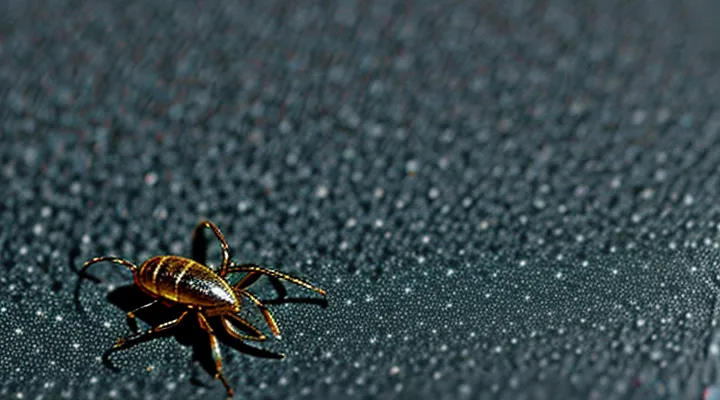
Why Submit a Tick for Analysis?
Identifying Disease Risk
When a tick is removed, the site of attachment influences the assessment of infection risk. Ticks attached to areas with thin skin—such as the scalp, behind the ears, neck, armpits, groin, and behind the knees—are more likely to remain attached longer, increasing the chance of pathogen transmission. Conversely, ticks found on the torso or limbs usually detach sooner, reducing exposure time.
Submitting the specimen for laboratory analysis requires accurate documentation of the attachment location. This information assists diagnosticians in estimating the probability of diseases such as Lyme borreliosis, anaplasmosis, or Rocky Mountain spotted fever. The following points summarize the critical considerations for risk identification:
- Record the exact anatomical region (e.g., scalp, axilla, inguinal fold).
- Note the duration of attachment, if known; longer than 24 hours markedly raises transmission risk.
- Preserve the tick in a sealed container with a damp cotton ball to maintain viability.
- Include a brief description of the tick’s developmental stage (larva, nymph, adult) and species, if identifiable.
Laboratories use the location data alongside species identification to prioritize testing for specific pathogens. For example, nymphal Ixodes scapularis removed from the scalp often triggers comprehensive PCR panels for Borrelia burgdorferi, whereas adult Dermacentor variabilis from the lower leg may prompt testing for Rickettsia rickettsii. Accurate site reporting therefore enhances diagnostic precision and guides appropriate clinical management.
Peace of Mind
Peace of mind stems from confidence that a tick removed from a person is handled correctly for laboratory examination. Confidence begins with accurate identification of the attachment site, because the location influences removal technique and documentation.
Typical attachment sites include:
- Scalp and hairline
- Neck and behind the ears
- Underarms
- Groin and genital region
- Behind the knees
- Waistline and belt area
- Between toes or on the feet
To maintain assurance, follow these steps:
- Use fine‑tipped tweezers to grasp the tick as close to the skin as possible.
- Pull upward with steady pressure, avoiding squeezing the body.
- Place the intact specimen in a sealed, labeled container (e.g., a small vial with a damp cotton ball).
- Record the date, exact body site, and any observed symptoms.
Submit the sealed container to a qualified public‑health laboratory or veterinary clinic. Provide the accompanying label information; most labs request a brief questionnaire about exposure history. Turnaround time for species identification and pathogen testing ranges from 3 to 7 days.
Lab results confirm whether the tick carries disease agents. A definitive report eliminates uncertainty, allowing timely medical decisions and reducing anxiety about potential infection.
Preparing a Tick for Submission
Safe Tick Removal Techniques
Ticks attached to the scalp, armpits, groin, and behind the knees are the most common locations for submission to laboratory analysis. Prompt removal reduces the risk of pathogen transmission and preserves the specimen for accurate identification.
Use fine‑point tweezers or a specialized tick‑removal tool. Grasp the tick as close to the skin as possible, applying steady pressure to pull straight upward. Avoid twisting or squeezing the body, which can cause the mouthparts to remain embedded and increase contamination. After extraction, place the tick in a sealed container with a damp cotton ball to maintain humidity, then label with the removal site, date, and patient information before sending to the diagnostic laboratory.
Key steps for safe removal:
- Disinfect hands and tools with alcohol.
- Secure the tick at the head, not the abdomen.
- Pull upward with even force until the entire organism detaches.
- Inspect the bite site for retained parts; if present, remove with sterilized tweezers.
- Store the tick in a breathable container, keep cool, and ship promptly.
Proper handling ensures reliable laboratory results and minimizes health risks.
Proper Storage of the Tick
When a tick is removed for laboratory examination, its condition determines the reliability of species identification and pathogen testing. Preserve the specimen immediately to prevent degradation.
Place the tick in a small, sealable container such as a screw‑cap microtube or a zip‑lock bag. Add a damp cotton ball or a few drops of sterile saline to keep the arthropod moist, but avoid excess liquid that could cause drowning. If a preservative is required, submerge the tick in 70 % ethanol; this method is suitable for DNA analysis but may impair morphological assessment, so note the intended testing.
Store the container in a refrigerator at 4 °C if analysis will occur within 24–48 hours. For longer intervals, maintain the specimen at −20 °C or in a freezer. Label the package with the date of removal, the exact body location (e.g., scalp, forearm, groin), and any relevant patient information.
Before sending, ensure the container is sealed, protected from crushing, and accompanied by a brief request form specifying the desired diagnostic tests. Proper handling from removal to submission maximizes the likelihood of accurate results.
Information to Include with the Tick
When a tick is removed for laboratory evaluation, the accompanying data must be precise and complete. The information enables accurate identification of the species, assessment of disease risk, and appropriate medical advice.
The submission packet should contain:
- Date of removal, recorded in day‑month‑year format.
- Exact anatomical site of attachment (e.g., scalp, right axilla, lower back).
- Approximate duration of attachment, expressed in hours or days if known.
- Patient demographics: age, sex, and any relevant medical conditions (immunosuppression, pregnancy).
- Recent travel history, especially visits to endemic regions within the past six months.
- Current medications, with emphasis on antibiotics or antiparasitic agents.
- Presence of symptoms such as fever, rash, or joint pain at the time of removal.
- Photographs of the bite site, if available, to document erythema or inflammation.
Include a brief label on the specimen vial that repeats the above details, ensuring legibility. Attach a signed consent form authorizing the analysis and any subsequent reporting of findings. Providing this comprehensive dataset eliminates ambiguity and accelerates diagnostic turnaround.
Where to Submit a Tick
Local Health Departments
Local health departments operate laboratories that receive ticks removed from humans for species identification, pathogen testing, and public‑health reporting. When a tick is found attached, the removal site on the body must be recorded because infection risk varies with anatomical location.
Typical submission sites include:
- Scalp or hairline
- Behind the ears
- Neck or collarbone area
- Axilla (underarm)
- Groin or genital region
- Inner thigh or knee crease
- Behind the knee
- Ankle or foot
- Any other area where the tick is firmly attached
The specimen should be placed in a sealed, labeled container. Labels must contain the exact body location, date of removal, and, if known, the duration of attachment. Health department staff use this information to prioritize testing and to contribute data to regional tick‑borne disease surveillance.
University Extension Offices
University Extension Offices function as community resources for vector‑borne disease monitoring. They accept tick specimens collected from individuals who suspect exposure, providing clear instructions on the appropriate anatomical sites for submission.
Ticks removed from the following locations are most suitable for laboratory analysis:
- Scalp or hairline
- Neck or back of the ears
- Axillary (underarm) region
- Groin or inner thigh
- Abdomen, particularly near the waistline
- Legs, especially the lower calf or ankle
- Hands and fingers
Extension staff advise that the tick should be placed in a sealed container with a moist cotton ball, labeled with the exact body site, date of removal, and any symptoms observed. Samples are then forwarded to the university’s entomology or public health laboratory for species identification and pathogen testing. The offices also disseminate educational materials on proper removal techniques and preventive measures, reinforcing community awareness of tick‑related health risks.
Private Laboratories
Private laboratories that specialize in arthropod diagnostics accept tick specimens collected from any part of the human body where the parasite is attached. Laboratories typically request that the tick be removed with fine tweezers, placed in a secure container, and accompanied by a brief description of the attachment site.
Common attachment locations include:
- Scalp and hairline
- Behind the ears
- Neck and collarbone region
- Axillary (underarm) area
- Groin and genital region
- Waistline and abdominal folds
- Behind the knees and popliteal fossa
- Feet, especially between toes
When submitting a sample, follow these steps:
- Preserve the tick in a ventilated, sealable tube or a small vial with a moist cotton ball to maintain viability.
- Label the container with the exact body site, date of removal, and any observed symptoms.
- Include a completed request form that specifies the purpose of analysis (e.g., species identification, pathogen testing).
- Ship the specimen promptly, using a courier service that guarantees delivery within 24–48 hours.
Private facilities often provide quicker results than public health labs, employ entomologists with focused expertise, and maintain strict confidentiality for client records. Their streamlined processes enable timely identification of tick species and detection of associated pathogens, facilitating appropriate medical response.
Online Tick Testing Services
Online tick testing services require specimens that retain diagnostic value. Proper collection from the host’s skin ensures accurate pathogen detection.
Acceptable anatomical locations include:
- Scalp or hairline where the tick is attached near the hair root
- Neck, especially the posterior region where ticks often latch
- Arms, particularly the forearm or elbow crease
- Legs, focusing on the thigh, calf, or ankle area
- Torso, such as the back or chest, if the tick is visibly attached
- Groin or genital region, provided the area can be safely accessed
When removing a tick, grasp the mouthparts with fine‑point tweezers as close to the skin as possible, pull straight upward, and avoid crushing the body. Place the intact specimen in a sealed, sterile container with a damp paper towel to prevent desiccation. Include a brief note describing the attachment site, date of removal, and any symptoms experienced.
After packaging, submit the sample through the provider’s online portal, attach the required documentation, and select the appropriate testing panel. Results are typically delivered electronically within a few days, allowing timely medical decision‑making.
Interpreting Analysis Results
Understanding Potential Pathogens
Ticks attach to skin areas where the cuticle is thin or the skin folds, providing easy access to blood. Frequently observed sites include the scalp, behind the ears, the neck, under the arms, the groin, the waistline, the back of the knees, and the inner elbows. Less common locations are the hands, feet, and genital region. Documenting the precise region of attachment is essential for accurate laboratory submission.
Understanding the microorganisms that a tick may harbor guides clinical decision‑making. The most relevant agents are:
- Borrelia burgdorferi – causative agent of Lyme disease.
- Anaplasma phagocytophilum – responsible for human granulocytic anaplasmosis.
- Ehrlichia chaffeensis – causes human monocytic ehrlichiosis.
- Rickettsia spp. – includes agents of spotted fever rickettsiosis.
- Babesia microti – protozoan that produces babesiosis.
- Powassan virus – neuroinvasive flavivirus.
Submitting the tick for analysis enables species identification and molecular detection of these pathogens. Proper preparation involves:
- Removing the tick with fine‑pointed tweezers, avoiding compression of the body.
- Placing the specimen in a sterile, airtight container (e.g., a sealed tube or zip‑lock bag).
- Labeling the container with the date of removal, the exact body region, and any relevant patient information.
- Sending the sample to a certified public health or veterinary laboratory equipped for PCR or culture assays.
Specifying the attachment site assists the laboratory in assessing the likelihood of exposure to particular pathogens, because certain tick species demonstrate regional preferences on the host. Accurate submission therefore improves diagnostic clarity and informs appropriate therapeutic measures.
Next Steps After Receiving Results
After the laboratory report arrives, verify that the identification of the specimen matches the location of the bite. Compare the tick species and any pathogen findings with the information recorded at the time of removal.
If the analysis detects an infectious agent, schedule an appointment with a medical professional promptly. Discuss the results, request a prescription if indicated, and follow the treatment plan without delay. Record the date of therapy initiation and any follow‑up instructions.
When the report is negative for disease, still inform a healthcare provider. Ask whether prophylactic measures are recommended based on the tick’s species, geographic origin, and the duration it remained attached. Retain the documentation for future reference.
Maintain a personal health log that includes:
- Date of bite and removal site
- Species identification and test outcomes
- Symptoms observed after the bite
- Medical consultations and prescribed actions
Review the log regularly. Seek immediate care if fever, rash, joint pain, or other atypical symptoms develop, even if the initial test was negative. Notify local public‑health authorities if the tick species is uncommon or if multiple cases arise in the area.
