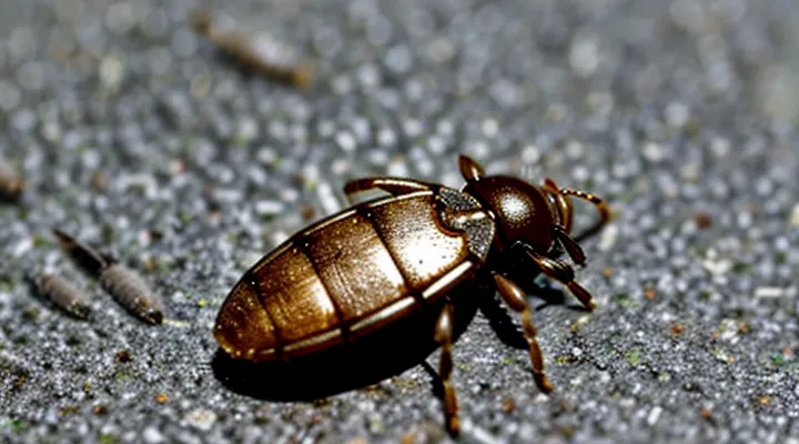
Immediate Actions After Finding a Tick
How to Safely Remove the Tick
Gathering Your Tools
When a tick embeds itself in the skin, immediate removal reduces the risk of disease transmission. Preparation begins with assembling the necessary equipment before attempting extraction.
- Fine‑point tweezers or forceps, preferably stainless steel, to grasp the tick close to the skin.
- Disposable nitrile gloves to protect hands and prevent contaminating the bite site.
- Antiseptic solution (e.g., 70 % isopropyl alcohol or povidone‑iodine) for cleaning the area before and after removal.
- Sterile gauze pads or cotton swabs for applying pressure and absorbing blood.
- Small, sealable container (plastic tube or zip‑lock bag) with a dab of alcohol for preserving the tick in case identification is required.
- Optional: a magnifying lens to improve visibility of the tick’s mouthparts.
Place all items within arm’s reach to avoid interruptions during the procedure. Verify that each tool is intact; damaged tweezers can crush the tick and increase the chance of mouthpart retention. After gathering the supplies, proceed directly to the removal steps.
The Proper Removal Technique
Finding a tick attached to your skin demands prompt, precise removal to limit pathogen transmission.
Gather the following items before beginning: fine‑pointed tweezers or a tick‑removal tool, disposable gloves, antiseptic solution (e.g., alcohol or iodine), a small sealable container, and a clean tissue.
- Put on gloves to avoid direct contact.
- Position the tweezers as close to the skin surface as possible, grasping the tick’s head or mouthparts without squeezing the body.
- Apply steady, upward pressure; pull straight out with even force, avoiding twisting or jerking motions.
- Release the tick into the container, seal it, and label with date and location if future identification is required.
- Clean the bite area with antiseptic; wash hands thoroughly after glove removal.
Observe the site for redness, swelling, or a rash over the next several weeks. If any of these signs appear, or if the tick could not be removed completely, seek medical evaluation promptly.
Do not use petroleum jelly, heat, or folk remedies to detach the tick; such methods increase the risk of incomplete extraction and pathogen release.
What Not to Do When Removing a Tick
Finding a tick embedded in your skin requires immediate, correct action. Incorrect removal techniques can increase the risk of infection, leave mouthparts behind, or facilitate pathogen transmission. The following practices must be avoided.
- Do not use bare fingers or tweezers to pinch the tick’s body. Squeezing the abdomen can force harmful fluids into the wound.
- Do not apply heat, such as a lit match, a candle flame, or a hot surface, to make the tick detach. Burning the parasite does not guarantee death and can cause it to release saliva.
- Do not smear petroleum jelly, nail polish, or other chemicals on the tick. These substances do not kill the tick quickly and may encourage it to secrete more saliva.
- Do not twist, jerk, or pull the tick sharply. Abrupt motion can break the tick, leaving parts of the mouth embedded in the skin.
- Do not crush the tick’s body against the skin. Crushing increases the chance of pathogen release into the bite site.
- Do not wait for the tick to fall off on its own. Leaving it attached for hours raises the probability of disease transmission.
After avoiding these errors, use fine‑point tweezers to grasp the tick as close to the skin as possible, pull upward with steady, even pressure, and disinfect the area afterward. Keep the removed tick in a sealed container for identification if symptoms develop.
Post-Removal Care
Cleaning the Bite Area
When a tick has been removed, the surrounding skin must be disinfected promptly to reduce the risk of infection and to remove any residual saliva that may contain pathogens. Use a clean paper towel or disposable gauze to wipe away blood, then apply an antiseptic solution such as 70% isopropyl alcohol, povidone‑iodine, or chlorhexidine. Allow the antiseptic to dry; do not rinse it off immediately, as this can dilute its effect.
After the area dries, inspect the site for signs of irritation, redness extending beyond the bite, or a small, raised bump. If any of these symptoms appear, seek medical advice without delay. Keep the wound covered with a sterile adhesive bandage for 24–48 hours, then replace it with a clean dressing if the site remains moist or if further irritation occurs.
Steps for cleaning the bite area
- Pat the skin dry with a disposable cloth.
- Apply a single‑use antiseptic swab, covering the entire bite margin.
- Let the solution air‑dry; avoid wiping it off.
- Place a sterile, non‑adhesive gauze pad over the site.
- Secure with a hypoallergenic tape or light bandage for up to two days.
- Monitor daily for swelling, redness, or discharge; document any changes.
Maintain the cleaning routine until the skin fully heals, typically within a week. Persistent inflammation or a spreading rash warrants immediate professional evaluation.
Monitoring for Symptoms
After extracting the tick, observe the bite site and overall health for several weeks. Early detection of complications relies on recognizing specific signs.
- Redness or swelling extending beyond the immediate area of the bite.
- Persistent itching, burning, or pain at the attachment point.
- Development of a circular rash, often with a clear center (“bull’s‑eye” appearance).
- Fever, chills, headache, muscle aches, or joint pain that appear within days to weeks.
- Nausea, vomiting, or unusual fatigue accompanying any of the above.
If any of these manifestations emerge, contact a healthcare professional promptly. Document the date of removal, the tick’s appearance, and the progression of symptoms to aid diagnosis. Continue monitoring for at least four weeks, as some tick‑borne illnesses have delayed onset. Absence of symptoms during this period generally indicates uncomplicated removal.
Potential Risks and When to Seek Medical Attention
Understanding Tick-Borne Diseases
Common Tick-Borne Illnesses
When a tick embeds itself in the skin, it can introduce a range of bacterial, viral, or protozoan agents. Recognizing the most frequent illnesses helps guide timely medical evaluation.
-
Lyme disease – Caused by Borrelia burgdorferi. Early signs include a expanding erythema migrans rash, fever, headache, and fatigue. Untreated infection may progress to joint, cardiac, or neurologic involvement.
-
Anaplasmosis – Result of Anaplasma phagocytophilum infection. Symptoms appear within 1‑2 weeks and often involve fever, chills, muscle aches, and mild leukopenia. Prompt doxycycline therapy prevents complications.
-
Ehrlichiosis – Triggered by Ehrlichia chaffeensis or related species. Presents with fever, headache, malaise, and thrombocytopenia. Early antimicrobial treatment reduces risk of severe organ dysfunction.
-
Babesiosis – Protozoan parasite Babesia microti infects red blood cells. Typical manifestations are hemolytic anemia, fever, chills, and fatigue. Severe cases may require combination therapy with atovaquone and azithromycin or clindamycin plus quinine.
-
Rocky Mountain spotted fever – Caused by Rickettsia rickettsii. Hallmarks include high fever, headache, and a maculopapular rash that often spreads from wrists and ankles to the trunk. Immediate doxycycline administration is essential to avoid fatal outcomes.
-
Tularemia – Francisella tularensis infection can follow tick bites. Early presentation includes ulcerative skin lesions, regional lymphadenopathy, and systemic symptoms such as fever and chills. Effective treatment relies on streptomycin or gentamicin.
-
Tick-borne relapsing fever – Multiple Borrelia species cause recurrent febrile episodes separated by afebrile periods. Diagnosis requires microscopy of blood smears; treatment with tetracycline or penicillin shortens disease duration.
Each illness has a characteristic incubation window ranging from a few days to several weeks. If a tick is discovered attached, remove it promptly with fine‑tipped tweezers, clean the site, and monitor for rash, fever, or malaise. Seek medical attention if any of the above symptoms develop, providing the date of the bite and geographic exposure to facilitate accurate diagnosis and treatment.
Symptoms to Watch For
If a tick remains attached, monitor the site and overall health for specific signs that may indicate infection.
The bite area should be examined daily. Look for a circular, expanding redness, often described as a “bull’s‑eye” pattern. The rash may appear 3–30 days after the bite and can enlarge up to several centimeters. Absence of a rash does not rule out disease; systemic symptoms often develop in parallel.
Key symptoms to watch for include:
- Fever or chills
- Severe headache, especially if accompanied by neck stiffness
- Muscle or joint aches, notably in the knees or elbows
- Unexplained fatigue or malaise
- Nausea, vomiting, or abdominal pain
- Swelling of lymph nodes near the bite
- Respiratory difficulties or chest pain (rare, but indicative of serious complications)
Some tick‑borne illnesses produce distinctive patterns. Rocky Mountain spotted fever may cause a rash that starts on wrists and ankles before spreading centrally. Anaplasmosis often presents with sudden fever, headache, and muscle pain without a rash. Babesiosis can lead to hemolytic anemia, resulting in jaundice and dark urine.
Any combination of these signs, especially when occurring within two weeks of attachment, warrants immediate medical evaluation. Early diagnosis and treatment reduce the risk of long‑term complications.
When to Consult a Doctor
Specific Warning Signs
If a tick is still attached, watch for immediate reactions around the bite site. Redness that expands rapidly, swelling that exceeds a few centimeters, or intense itching may signal an allergic response or local infection. A small blister or pus formation indicates secondary bacterial involvement and requires prompt medical attention.
Systemic warning signs appear within days to weeks after removal. Fever, chills, or a sudden rise in body temperature suggest an infection spreading beyond the skin. Headache, muscle aches, or joint pain, especially if they are severe or persistent, often precede Lyme disease or other tick‑borne illnesses. A distinctive circular rash, known as erythema migrans, typically emerges 3–30 days after the bite; it begins as a red spot and expands to a bull’s‑eye pattern, reaching up to 12 cm in diameter.
Neurological symptoms demand immediate evaluation. Numbness, tingling, facial drooping, or difficulty concentrating indicate possible neuroborreliosis or other complications. Cardiac manifestations, such as palpitations, irregular heartbeat, or chest discomfort, may reflect Lyme carditis and should be treated as an emergency.
Key warning signs can be summarized:
- Rapidly enlarging redness or swelling at the bite site
- Persistent fever, chills, or night sweats
- Severe headache, muscle or joint pain
- Bull’s‑eye rash (circular, expanding)
- Numbness, facial weakness, or cognitive changes
- Irregular heart rhythm, palpitations, or chest pain
Presence of any of these symptoms warrants consultation with a healthcare professional without delay. Early diagnosis and treatment reduce the risk of long‑term complications.
Travel History and Tick Exposure
When a tick is discovered attached to the skin, the places you have visited in recent weeks become a critical factor in assessing infection risk. Record every destination, including rural or forested areas, parks, and any outdoor activities, and note the dates of arrival and departure. This timeline establishes the window during which tick exposure could have occurred and helps clinicians match potential pathogens to the geographic distribution of tick species.
Different regions harbor distinct tick vectors and associated diseases. For example, Ixodes scapularis is prevalent in the northeastern United States and transmits Lyme disease, whereas Dermacentor spp. dominate in parts of the southeastern United States and can spread Rocky Mountain spotted fever. In Europe, Ixodes ricinus carries both Lyme disease and tick-borne encephalitis. Knowing which pathogens are endemic to the areas you visited narrows the diagnostic focus and informs the urgency of treatment.
Provide healthcare providers with the following information:
- List of countries, states, or provinces visited
- Specific locations (e.g., national parks, hiking trails)
- Dates of exposure window (typically 7–14 days before tick detection)
- Any observed tick bites or skin lesions during travel
- Symptoms such as fever, rash, joint pain, or headache that have appeared since returning
After documenting travel history, remove the tick promptly using fine‑point tweezers, grasping as close to the skin as possible, and pulling upward with steady pressure. Clean the bite site with antiseptic. Contact a medical professional within 24 hours, presenting the travel and exposure details. The clinician may order serologic tests or initiate prophylactic antibiotics based on the identified risk profile. Follow-up appointments should be scheduled to monitor for delayed symptoms, as some tick‑borne illnesses manifest weeks after the bite.
