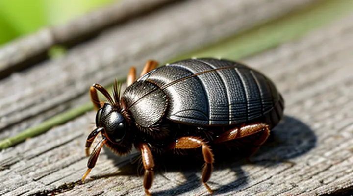
The Nature of Tick Infection
How Ticks Acquire Pathogens
Ticks become vectors for disease through three primary mechanisms. First, they acquire pathogens while feeding on an infected host. During the blood meal, the tick’s mouthparts penetrate the skin, allowing pathogens present in the host’s bloodstream to enter the tick’s gut. Second, transstadial transmission ensures that pathogens persist as the tick molts from larva to nymph to adult. The microorganism remains viable within the tick’s tissues, enabling infection of subsequent hosts. Third, transovarial transmission passes pathogens from an infected female to her offspring, seeding the next generation with the disease agent.
The probability that a tick carries a pathogen depends on several factors. Host infection prevalence determines the likelihood of acquisition during feeding; higher rates in reservoir species increase the chance of tick infection. Tick species differ in competence, reflecting variations in immune defenses and gut environment that affect pathogen survival. Environmental conditions, such as temperature and humidity, influence tick activity and host‑contact frequency, indirectly altering infection rates. Geographic distribution of both ticks and reservoir hosts further shapes regional infection probabilities.
Key variables influencing infection risk can be summarized:
- Host infection prevalence in the local ecosystem
- Tick species’ vector competence
- Frequency of blood meals across life stages
- Environmental parameters that affect tick survival and host encounters
- Presence of transovarial transmission within the tick population
Understanding these acquisition pathways clarifies why infection rates vary among tick populations and provides a framework for assessing the likelihood of encountering an infected tick in a given area.
Common Tick-borne Pathogens
Ticks transmit a limited set of microorganisms that cause most human tick‑borne illnesses. Understanding which agents are most frequently carried clarifies the probability that any given tick is infected.
- Borrelia burgdorferi – agent of Lyme disease; prevalence in Ixodes scapularis and I. pacificus ranges from 10 % to 30 % in endemic regions of the United States and up to 50 % in parts of Europe.
- Anaplasma phagocytophilum – causes human granulocytic anaplasmosis; infection rates typically 5 %–15 % in the same Ixodes species, higher in the Upper Midwest.
- Babesia microti – responsible for babesiosis; detected in 1 %–5 % of Ixodes ticks in the Northeastern United States, occasional reports of 10 % in hotspot areas.
- Rickettsia rickettsii – Rocky Mountain spotted fever vector, mainly Dermacentor variabilis and D. andersoni; prevalence usually below 1 % but can exceed 5 % during epidemic cycles.
- Ehrlichia chaffeensis – agent of human monocytic ehrlichiosis; found in Amblyomma americanum with infection rates of 2 %–8 % across the Southern United States.
- Powassan virus – flavivirus transmitted by Ixodes ticks; prevalence generally <1 % but rising in certain Canadian provinces.
Factors that modify infection likelihood include:
- Geographic region – endemic zones show higher pathogen loads.
- Tick species – each species harbors a characteristic pathogen spectrum.
- Life stage – nymphs often exhibit higher infection rates than larvae; adults may accumulate multiple agents.
- Seasonality – peak activity periods correspond with increased pathogen transmission.
- Host availability – presence of competent reservoir animals raises infection probability.
Accurate knowledge of these pathogen frequencies enables precise estimation of tick infection risk, informs public‑health advisories, and guides preventive measures such as targeted tick checks and habitat management.
Factors Influencing Tick Infection Rates
Geographic Variation and Endemic Areas
Geographic distribution strongly influences the probability that a tick carries a pathogen. Regions where competent reservoir hosts and suitable climate coexist generate higher infection rates, while areas lacking one or both factors show markedly lower prevalence.
- Northeastern United States: 30‑45 % of adult Ixodes scapularis infected with Borrelia burgdorferi; 10‑20 % harbor Anaplasma phagocytophilum.
- Upper Midwest (Wisconsin, Minnesota): 20‑35 % of adult Ixodes infected with B. burgdorferi; 5‑12 % carry Babesia microti.
- Central Europe (Germany, Czech Republic): 15‑25 % of Ixodes ricinus infected with B. burgdorferi sensu lato; 3‑8 % with Tick‑borne encephalitis virus.
- Southern England: 5‑10 % of Ixodes ricinus infected with B. burgdorferi; 1‑3 % with Anaplasma spp.
- Mediterranean basin: 2‑7 % of Rhipicephalus sanguineus infected with Rickettsia conorii; sporadic detection of Coxiella burnetii.
Climate, vegetation, and host density drive these patterns. Warm, humid environments support longer questing periods, increasing tick‑host contact. Abundant small mammals, especially rodents, serve as reservoirs for Borrelia and Anaplasma species, elevating local infection pressure. Conversely, arid zones limit tick survival and reduce pathogen transmission.
Understanding regional prevalence enables targeted public‑health interventions. Risk assessments should incorporate local infection data, seasonal tick activity, and habitat characteristics to refine preventive recommendations and diagnostic strategies.
Tick Species and Pathogen Specificity
Tick species differ markedly in their capacity to acquire and transmit pathogens, a factor that directly influences the probability of finding an infected individual. Each species exhibits a distinct ecological niche, host‑preference pattern, and competence for specific microorganisms, resulting in highly variable infection rates across regions and habitats.
Key tick–pathogen associations include:
- Ixodes scapularis – primary vector of Borrelia burgdorferi (Lyme disease), Anaplasma phagocytophilum, and Babesia microti; infection prevalence often exceeds 30 % in endemic areas.
- Dermacentor variabilis – transmits Rickettsia rickettsii (Rocky Mountain spotted fever) and Francisella tularensis; typical infection rates range from 5 % to 15 % depending on local wildlife reservoirs.
- Amblyomma americanum – associated with Ehrlichia chaffeensis, Ehrlichia ewingii, and Heartland virus; reported infection frequencies vary between 10 % and 25 % in the southeastern United States.
- Rhipicephalus sanguineus – carrier of Rickettsia conorii and Coxiella burnetii; infection prevalence generally below 10 % in urban dog populations.
Pathogen specificity arises from molecular compatibility between tick salivary proteins and microbial surface structures, as well as from the tick’s immune modulation mechanisms. Species that feed on a broad range of vertebrate hosts encounter a wider pathogen pool, increasing their overall infection likelihood, whereas specialists tend to harbor fewer agents but may transmit them more efficiently.
Consequently, assessing the risk of encountering an infected tick requires identification of the local tick fauna, understanding of each species’ preferred hosts, and knowledge of the prevalent pathogens within that ecological context.
Environmental Conditions and Host Availability
Environmental conditions directly shape the prevalence of pathogens within tick populations. Temperature determines developmental rates; warmer periods accelerate molting, increase questing activity, and expand the seasonal window during which ticks encounter hosts. Humidity influences survival, with relative moisture above 80 % required for prolonged off‑host periods; low humidity forces ticks to retreat to the leaf litter, reducing host contact and limiting pathogen acquisition. Vegetation density creates microclimates that retain moisture and provide shelter, fostering higher tick densities and consequently greater chances of infection.
Host availability governs the probability that a tick acquires and transmits a pathogen. The presence of competent reservoir species—small mammals such as white‑footed mice for Borrelia, or ground‑feeding birds for certain rickettsiae—elevates infection risk. Abundant large mammals, including deer, support adult tick feeding but generally dilute pathogen prevalence because they are poor reservoirs. Seasonal fluctuations in host activity synchronize with tick questing peaks, aligning periods of high host density with increased pathogen transmission.
Key environmental and host factors influencing infection probability:
- Average temperature and length of warm season
- Relative humidity and soil moisture content
- Ground cover type and leaf‑litter depth
- Density of competent reservoir hosts
- Diversity of non‑reservoir hosts that affect tick population size
- Seasonal overlap of host activity and tick questing behavior
Understanding these variables allows precise estimation of infection risk in a given area, informing public‑health interventions and personal protective measures.
Assessing the Risk of Infection from a Tick Bite
Identifying the Tick Species
Identifying the tick species is essential for estimating infection risk because pathogen prevalence differs markedly among taxa. Accurate species determination allows targeted prevention and informs medical decision‑making.
Key morphological characteristics used in field identification:
- Body length and engorgement level; larvae (≈1 mm), nymphs (≈2 mm), adults (≈3–5 mm).
- Scutum shape and ornamentation; hard‑ticks (Ixodidae) possess a hard dorsal shield, soft‑ticks (Argasidae) lack one.
- Tick’s mouthparts; forward‑projecting palps indicate Ixodes, while ventrally positioned chelicerae suggest Dermacentor.
- Leg segmentation and coloration patterns; distinct banding on legs identifies Amblyomma, uniform coloration points to Ixodes ricinus.
Geographic distribution and host preference further refine identification. Ixodes scapularis predominates in eastern North America and feeds primarily on small mammals, whereas Dermacentor variabilis occupies the central United States and prefers rodents and dogs. Amblyomma americanum is common in the southeast, targeting deer and humans.
Laboratory techniques confirm visual assessments. DNA barcoding of the mitochondrial COI gene provides species‑level resolution. Polymerase chain reaction targeting pathogen‑specific markers (e.g., Borrelia burgdorferi flaB) simultaneously verifies infection status.
Practical approach for non‑specialists:
- Collect the tick with fine tweezers, preserving the whole specimen.
- Compare the specimen against a regional identification key that lists the traits above.
- Submit the tick to a reference laboratory for molecular confirmation when visual identification is uncertain.
These steps produce reliable species identification, which directly influences the calculated probability that a tick carries disease‑causing agents.
Duration of Tick Attachment
The probability that a tick carries a pathogen increases as the feeding period lengthens. Pathogens reside in the tick’s midgut and must travel to the salivary glands before they can be transmitted; this migration requires a minimum duration of blood ingestion.
- Borrelia burgdorferi (Lyme disease): transmission typically begins after 36–48 hours of attachment.
- Anaplasma phagocytophilum (human granulocytic anaplasmosis): detectable transmission after roughly 24 hours.
- Babesia microti (babesiosis): risk rises sharply after 48 hours.
- Tick‑borne encephalitis virus: transmission possible within 24 hours, but risk escalates with longer feeding.
The biological basis is the activation of the tick’s salivary glands, which occurs only after sustained feeding. As the tick expands, midgut cells release pathogens into the salivary ducts, enabling entry into the host’s bloodstream. Consequently, each additional hour of attachment proportionally raises the chance of infection.
Risk assessment therefore hinges on the elapsed attachment time. A tick removed within the first 12 hours generally presents a low probability of pathogen transfer, whereas removal after 24 hours or more markedly elevates risk for most common agents. Early detachment does not guarantee safety, but it substantially reduces the likelihood of disease transmission.
Prompt detection and proper removal—grasping the tick close to the skin with fine‑tipped tweezers, pulling steadily without crushing—remain the most effective preventive measures. After removal, a monitoring period of 30 days is advisable to identify any emerging symptoms promptly.
Symptoms to Monitor After a Tick Bite
After a tick is detached, observing the bite site and overall health is critical because the animal may have transmitted pathogens. Early detection of illness improves treatment outcomes and reduces complications.
- Redness or swelling that expands beyond the immediate bite area
- A circular rash with a clear center (often called a “bull’s‑eye”)
- Fever, chills, or night sweats
- Headache, neck stiffness, or facial drooping
- Muscle or joint pain, especially if it migrates to different joints
- Nausea, vomiting, or abdominal pain
- Unexplained fatigue or weakness
Symptoms typically appear within a few days to several weeks after exposure. If any of the listed signs develop, seek medical evaluation promptly. Provide the clinician with details of the bite, including the approximate date, geographic location, and any known tick species. Early laboratory testing and, when appropriate, antimicrobial therapy can prevent disease progression. Continuous monitoring for at least eight weeks is advisable, as some infections have delayed onset.
Preventive Measures and Mitigation Strategies
Personal Protection Against Tick Bites
Ticks transmit pathogens at rates that vary by species, region, and season. Understanding local infection prevalence informs risk assessment and guides protective behavior.
Effective personal protection includes:
- Wearing long sleeves and trousers, tucking pant legs into socks.
- Applying EPA‑registered repellents containing DEET, picaridin, or IR3535 to exposed skin and clothing.
- Treating footwear and outer garments with permethrin according to label instructions.
- Conducting thorough body checks within 30 minutes of leaving tick‑infested areas, focusing on scalp, armpits, groin, and behind knees.
- Removing attached ticks promptly with fine‑tipped tweezers, grasping close to the skin and pulling steadily without twisting.
Consistent use of these measures reduces the probability of acquiring a bite, thereby lowering the chance of exposure to infected ticks.
Tick Checks and Proper Removal
Regular inspection of the skin after outdoor exposure is the most reliable method for reducing the chance of disease transmission from a tick. Ticks attach for a minimum of 24–48 hours before most pathogens can be transmitted; therefore, early detection dramatically lowers infection risk.
Effective removal requires the following steps:
- Use fine‑point tweezers or a specialized tick‑removal tool.
- Grasp the tick as close to the skin’s surface as possible, avoiding compression of the body.
- Apply steady, gentle upward pressure to pull the tick straight out without twisting.
- Disinfect the bite site with an appropriate antiseptic.
- Place the tick in a sealed container for later identification if needed; do not crush it.
Consistent tick checks and proper extraction reduce the probability that a bite results in pathogen exposure, thereby minimizing the overall likelihood of infection.
When to Seek Medical Attention
After a tick bite, the decision to seek professional care should be based on specific clinical indicators rather than uncertainty about infection rates. The following situations warrant immediate evaluation by a healthcare provider:
- A tick remains attached for more than 24 hours, especially if it is engorged.
- The bite site develops a expanding red rash, commonly described as a “bull’s‑eye” lesion, or any other unusual skin changes.
- Fever, chills, severe headache, muscle or joint pain, or fatigue appear within two weeks of removal.
- Neurological symptoms such as facial droop, weakness, or numbness emerge.
- Laboratory tests have identified a tick species known for higher pathogen carriage, such as Ixodes scapularis or Dermacentor variabilis.
Even in the absence of these signs, a brief consultation is advisable when the bite occurs in a region with documented high prevalence of tick‑borne diseases. Early treatment can prevent progression to more serious conditions and reduce the risk of long‑term complications.
