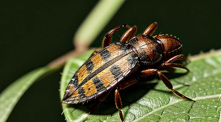
«The Anatomy of a Tick»
«Body Segmentation»
An unfed tick presents a distinct segmentation that aids identification. The arthropod’s body consists of two principal sections: the anterior capitulum, which houses the mouthparts, and the posterior idiosoma, containing the bulk of the organs. In the unfed state, the capitulum appears as a compact, dark‑colored structure distinct from the lighter, softer‑bodied idiosoma.
Key features of the segmentation include:
- Capitulum: small, rounded, often brown to black; visible as a separate unit from the main body.
- Idiosoma: oval, translucent to pale amber; surface may show fine granulation but lacks the engorged expansion seen after feeding.
- Leg attachment points: four pairs emerge from the ventral margin of the idiosoma, positioned symmetrically around the capitulum.
- Sclerotized plates: dorsal shield (scutum) covers part of the idiosoma in hard‑tick species, remaining visible and unaltered in the unfed condition.
These characteristics provide a reliable visual framework for distinguishing a starved tick from other arachnids and from engorged specimens. The clear demarcation between capitulum and idiosoma, combined with the consistent coloration and size, defines the appearance of an unfed tick.
«Legs and Mouthparts»
An unfed tick presents a compact, oval body covered by a smooth, pale exoskeleton. The legs and mouthparts are the most conspicuous external features that differentiate it from a engorged specimen.
The eight legs are arranged in four pairs, each emerging from a distinct segment of the idiosoma. Every leg consists of six articulated segments: coxa, trochanter, femur, patella, tibia and tarsus. The segments are slender, lightly sclerotized and uniformly light‑brown, giving the tick a delicate, hair‑free appearance. When the tick is not attached to a host, the legs extend outward at roughly right angles to the body, creating a characteristic “spider‑like” stance that facilitates locomotion across vegetation.
Mouthparts comprise the chelicerae, the hypostome and the palps. The chelicerae are short, curved structures positioned laterally, each ending in a sharp tip used for cutting the host’s skin. The hypostome, located centrally beneath the mouth opening, is a stout, barbed rod that projects forward; in an unfed state it appears as a dark, slightly ridged cylinder. The palps are a pair of elongated, sensory appendages situated anterior to the chelicerae; they are pale, tapering to fine tips and bear sensory setae that detect chemical cues. Together, these components form a compact, needle‑like apparatus designed for rapid penetration of host tissue when the tick initiates feeding.
«Key Characteristics of Unfed Ticks»
«Size and Shape»
Unfed ticks present a compact, dorsoventrally flattened body. The dorsal surface is smooth, lacking the swollen appearance seen after blood intake. Body size varies markedly among developmental stages.
- Larva: approximately 0.5 mm in length, 0.3 mm in width; oval shape, resembling a tiny seed.
- Nymph: 1.0–1.5 mm long, 0.5–0.8 mm wide; retains the elongated oval profile.
- Adult female: 2.0–3.5 mm long, 1.5–2.0 mm wide; slightly longer than male, still flattened.
- Adult male: 2.0–2.5 mm long, 1.2–1.5 mm wide; more rounded than female.
The overall silhouette is an elongated oval with a rounded anterior (capitulum) and a broader posterior (idiosoma). Legs extend laterally from the body, giving a slightly wider appearance when viewed from above. Coloration ranges from reddish‑brown to dark brown, but does not affect size or shape.
«Coloration»
Unfed ticks display a distinctive coloration that serves as a primary field‑identification cue. The external cuticle of a non‑engorged specimen generally appears pale, ranging from ivory to light brown, with a subtle reddish hue in many species. This baseline palette contrasts sharply with the darkened abdomen that develops after blood intake.
Typical coloration patterns by tick family:
- Hard ticks (Ixodidae) – dorsal shield (scutum) light brown to tan; ventral surface pale cream; legs and mouthparts often darker brown.
- Soft ticks (Argasidae) – overall body uniformly pale beige; dorsal surface may exhibit faint mottling; legs and gnathosoma slightly darker.
Species‑specific notes:
- Ixodes scapularis – shield dark brown, abdomen pale yellow‑white.
- Dermacentor variabilis – shield reddish‑brown, legs dark brown.
- Ornithodoros moubata – body uniformly light tan, minimal contrast.
Coloration remains stable until the tick begins feeding; the cuticle does not change until the engorgement phase, when the abdomen expands and darkens due to the presence of host blood. Consequently, observation of a tick’s coloration provides reliable information about its feeding status and assists in rapid species identification.
«Texture and Appearance»
An unfed tick presents a compact, oval body that appears smooth to the naked eye. The dorsal surface is covered by a hard, chitinous exoskeleton, giving a glossy sheen that reflects ambient light. Coloration ranges from light brown to reddish‑brown, depending on species and environmental factors, and lacks the engorged, gray‑blue hue seen after blood intake.
Key visual and tactile characteristics include:
- Size: typically 2–5 mm in length, expanding minimally after feeding.
- Shape: rounded anterior, tapering slightly toward the posterior.
- Surface texture: hard, slightly ridged scutum on the back; softer, softer‑looking ventral plates.
- Legs: eight slender, pale legs extending from the ventral side, each ending in tiny claws for anchoring to host fur or vegetation.
- Mouthparts: visible as a short, beak‑like structure (capitulum) positioned forward of the body.
When compared with a fed counterpart, the unfed specimen retains a uniform coloration, a compact silhouette, and a rigid, non‑distended exoskeleton. These attributes enable identification in field surveys and laboratory examinations.
«Comparing Unfed vs. Fed Ticks»
«Visual Distinctions»
An unfed tick presents a compact, oval body approximately 2–5 mm in length, depending on species. The dorsal surface is covered by a hard, shield‑like scutum that is lighter in color than the surrounding integument. Legs are long, slender, and extend outward in a characteristic “spider‑like” posture, each bearing a small claw at the tip.
Key visual distinctions include:
- Scutum color: pale tan to light brown, often uniform without visible blood stains.
- Body contour: smooth, unexpanded, with a clearly defined anterior‑posterior axis.
- Leg arrangement: eight legs uniformly spaced, each joint visible, contrasting with the shorter, less distinct legs of a fed specimen.
- Mouthparts: visible capitulum positioned forward, lacking the swollen appearance seen after engorgement.
Compared with a fed tick, an unfed individual lacks abdominal distension, retains a rigid, flattened profile, and exhibits a consistent coloration without the reddish or darkened patches that result from blood intake. These attributes enable reliable identification in field surveys and laboratory examinations.
«Behavioral Differences»
Unfed ticks exhibit distinct activity patterns that differentiate them from engorged specimens. Mobility remains limited; the organism relies on passive attachment to a host rather than active pursuit. Questing behavior, characterized by elevated front legs, persists until a suitable host contacts the vegetation. Environmental stimuli such as temperature and humidity modulate the frequency of questing, with optimal conditions prompting increased elevation and duration of the stance.
Key aspects of «Behavioral Differences» include:
- Reduced locomotion: unfed individuals conserve energy, moving only to reposition on vegetation.
- Extended questing intervals: periods of inactivity alternate with brief, heightened readiness to attach.
- Sensitivity to microclimate: low humidity triggers descent to more protected microhabitats, whereas favorable moisture encourages prolonged exposure.
- Absence of blood‑feeding drive: lack of engorgement eliminates the need for prolonged attachment, resulting in shorter attachment attempts.
These behavioral traits influence detection probability and control strategies, as unfed ticks rely primarily on opportunistic host contact rather than active searching.
«Common Types of Unfed Ticks»
«Deer Ticks (Blacklegged Ticks)»
Deer ticks, also known as blacklegged ticks, display a distinct morphology when they have not taken a blood meal. The unfed adult measures 2–3 mm in length, expanding to 5–6 mm after feeding. The body is reddish‑brown on the dorsal surface and lighter on the ventral side. Six legs extend from the anterior segment, each bearing a dark, scutum that covers the entire back in males and a portion in females. The mouthparts, including the hypostome, are visible as a small, pale projection at the front of the body.
Larval stage: approximately 0.5 mm long, uniformly golden‑brown, lacking a scutum.
Nymphal stage: 1–2 mm long, reddish‑brown, with a partial scutum and more pronounced legs than the larva.
Adult stage: as described above, with a fully developed scutum in males and a partially visible scutum in females.
Key visual identifiers for an unfed tick:
- Small, compact body shape; no swelling of the abdomen.
- Uniform coloration without the engorged, balloon‑like appearance seen after feeding.
- Visible scutum (hard shield) on the dorsal surface.
- Prominent, dark legs extending from the anterior region.
- Mouthparts positioned at the front, not enlarged.
These characteristics allow rapid identification of unfed deer ticks in field surveys and public health assessments.
«American Dog Ticks»
The «American Dog Tick» (Dermacentor variabilis) presents a distinct morphology before a blood meal. Adults measure 3–5 mm in length, expanding to 6 mm when engorged. The dorsal shield (scutum) is reddish‑brown, marked with lighter, irregular patches that form a mottled pattern. The ventral side is paler, often yellow‑white, with fine hairs covering the abdomen. Legs are long, robust, and visible from a dorsal view, each bearing a dark band near the tip.
- Size: 3–5 mm (unfed adult); 1 mm (nymph); 0.5 mm (larva)
- Body shape: oval, slightly flattened dorsoventrally
- Scutum: brown with pale mottling, smooth texture
- Coloration: dorsal reddish‑brown, ventral pale; legs dark‑banded
- Eyes: two simple eyes positioned laterally on the dorsal surface
Compared with other North American ticks, the unfed «American Dog Tick» exhibits a broader scutum and more pronounced mottling than the deer tick (Ixodes scapularis), while its leg length exceeds that of the lone star tick (Amblyomma americanum). Accurate visual identification relies on these combined traits.
«Lone Star Ticks»
The unfed stage of the tick species known as «Lone Star Ticks» presents a distinct set of visual characteristics that aid identification in the field.
Adult females exhibit a reddish‑brown dorsum with a single white spot on the anterior scutum, the feature that gives the species its common name. Males lack the spot, displaying a uniformly brown scutum. Both sexes possess a compact, oval body measuring approximately 3 mm in length and 2 mm in width when not engorged. The ventral surface is lighter, often pale yellow, and the legs are relatively short, each bearing a dark band near the tip.
Immature stages (larvae and nymphs) differ in size and coloration. Larvae are about 0.5 mm long, with a translucent amber hue and no discernible markings. Nymphs measure roughly 1.5 mm, showing a darker brown coloration and a faintly visible scutum without the characteristic white spot.
Key identification points for an unfed «Lone Star Tick» include:
- Size: 2–3 mm (adult), 0.5–1.5 mm (immature).
- Color: reddish‑brown dorsum, pale ventral side.
- Marking: solitary white spot on female scutum; absent in males and immature stages.
- Body shape: compact, oval, with short, banded legs.
These traits collectively define the appearance of an unfed lone star tick and support accurate recognition during surveillance or medical assessment.
«Brown Dog Ticks»
Unfed «Brown Dog Ticks» (Rhipicephalus sanguineus) exhibit a distinct morphology that allows rapid identification in the field.
The adult female measures 4–6 mm in length when not engorged, expanding to over 10 mm after feeding. The male is slightly smaller, 3–5 mm long. Both sexes possess a hard, oval‑shaped dorsal shield (scutum) that is uniformly reddish‑brown, lacking the lighter markings seen in many other tick species. The ventral surface is paler, with a smooth, glossy integument.
Key visual characteristics of an unfed specimen:
- Body shape: Compact, oval, and slightly flattened dorsally.
- Color: Uniform reddish‑brown on the scutum; lighter, creamy‑tan on the ventral side.
- Legs: Eight slender legs, each ending in a small claw; legs are pale and clearly visible against the darker body.
- Mouthparts: Prominent, forward‑projecting chelicerae and a short, robust hypostome used for piercing host skin.
- Sensory organs: Pair of small, dark eyes positioned laterally on the dorsal surface.
These features differentiate unfed «Brown Dog Ticks» from other ixodid ticks, facilitating accurate recognition before they attach to a host.
«Where to Find Unfed Ticks»
«Habitat Preferences»
Unfed ticks are typically flat, reddish‑brown to brown, and display a noticeable scutum covering the dorsal surface. Their compressed body enables them to remain concealed in environments where they await a host.
- Leaf litter in deciduous and mixed forests, where moisture and shade sustain larval and nymphal stages.
- Underbrush and low vegetation, providing contact points for passing mammals and birds.
- Rocky outcrops and soil crevices that retain humidity, essential for preventing desiccation.
- Grassy meadow edges adjacent to wooded areas, offering transitional zones for host movement.
- Peridomestic yards with tall grasses or mulch, especially where wildlife such as rodents frequent.
These habitats share high relative humidity, moderate temperature fluctuations, and abundant host traffic. The combination of physiological adaptation and habitat selection enhances the likelihood of encountering unfed ticks during field surveys or routine inspections.
«Seasonal Activity»
Unfed ticks are typically flat, reddish‑brown, and lack the engorged, balloon‑shaped abdomen seen after a blood meal. Their bodies measure 2–5 mm in length, with legs extending outward, giving a “spider‑like” silhouette.
Seasonal activity determines when unfed ticks are most likely to be encountered. Activity peaks correspond to temperature and humidity thresholds that support questing behavior.
- Spring (March‑May): Nymphs emerge, actively questing on low vegetation; prevalence of unfed ticks is highest.
- Summer (June‑August): Adult females increase questing; mid‑day heat reduces activity, prompting ticks to quest in cooler morning and evening periods.
- Autumn (September‑November): Declining temperatures shift activity to lower elevations; nymphs and adults seek shelter, reducing surface presence.
- Winter (December‑February): Activity drops sharply; ticks remain dormant in leaf litter, with only occasional questing during warm spells.
Understanding these seasonal patterns aids in recognizing unfed ticks in the field. During peak months, visual surveys should focus on low grass and leaf litter, where the flat, reddish‑brown form is most conspicuous. In colder periods, reduced activity limits detection opportunities, requiring targeted habitat sampling.
«Identifying Unfed Ticks on Pets and Humans»
«Inspection Techniques»
Unfed ticks present a smooth, flattened dorsum measuring 1–3 mm in length, depending on species. The body appears glossy, with a uniformly colored scutum that may be brown, reddish‑brown, or amber. Legs are clearly visible, extending laterally from the ventral side, and the mouthparts are positioned forward, giving the organism a “spider‑like” silhouette. The overall shape is oval when viewed from above and elongate when observed from the side.
Effective detection relies on systematic visual examination of the host’s skin, hair, and clothing. Inspection should occur in well‑lit environments to distinguish the tick’s subtle coloration from surrounding tissue. Use of magnification devices, such as handheld lenses or digital microscopes, enhances identification of the minute scutum and leg articulation, reducing the risk of misclassification.
Key inspection techniques include:
- Direct visual sweep: run gloved fingers over the entire body surface, paying special attention to warm, moist areas (neck, armpits, groin, scalp).
- Fine‑tooth comb: pass a fine comb through hair to dislodge hidden specimens; examine comb teeth after each pass.
- Dermatoscope examination: apply a dermatoscope to suspected spots for high‑resolution imaging of the dorsal shield and ventral capitulum.
- Photographic documentation: capture macro photographs for later analysis and record‑keeping.
- Tick‑specific adhesive strips: press adhesive pads onto skin to capture unattached ticks; inspect pads under magnification.
Prompt identification and removal of unfed ticks prevent attachment and subsequent pathogen transmission. Regular inspections, especially after outdoor exposure, constitute a critical component of personal and veterinary health protocols.
«Tools for Removal»
An unfed tick presents as a small, flat, oval organism, typically pale or reddish‑brown, with a smooth dorsal surface. Its mouthparts are hidden beneath the body, making visual identification straightforward when the creature has not yet engorged.
Effective removal relies on precise instruments that grasp the tick close to the skin without compressing its abdomen. Recommended tools include:
- Fine‑point tweezers or spring‑loaded tick‑removal devices designed to slide beneath the mouthparts.
- Curved forceps with a narrow tip for angled access on curved body surfaces.
- Single‑use disposable gloves to prevent direct contact and reduce contamination risk.
- A magnifying glass or handheld lens to enhance visibility of the tick’s attachment point.
- Antiseptic solution (e.g., iodine or alcohol) for post‑removal skin disinfection.
Procedure: grasp the tick as close to the skin as possible, apply steady upward pressure, and avoid twisting. After extraction, place the specimen in a sealed container for identification if needed, then clean the bite area with antiseptic. Proper tool selection minimizes the chance of leaving mouthparts embedded and reduces the risk of pathogen transmission.
