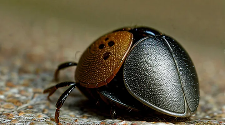
Understanding Tick Anatomy
External Features of a Tick
Ticks possess a compact body divided into two distinct regions: the anterior capitulum and the posterior idiosoma. The capitulum houses the mouthparts that penetrate host tissue; it consists of the hypostome, a barbed structure that anchors the parasite, two palps that guide attachment, and a pair of chelicerae that cut the skin. When a tick embeds, the capitulum remains partially exposed, allowing the host’s skin to envelope the hypostome while the palps and chelicerae stay visible on the surface.
The idiosoma includes several external structures essential for identification and survival:
- Dorsal scutum, a hard shield covering the back of unfed females and all males; it varies in color and pattern among species.
- Legs, eight in total, attached in four pairs to the ventral side; each leg ends in a pair of claws and sensory Haller’s organs that detect heat and carbon dioxide.
- Spiracular plates, openings on the ventral surface that facilitate respiration.
- Anal groove, a groove surrounding the anus that assists in waste elimination.
The external surface is covered by a thin, waxy cuticle that reduces water loss and protects against environmental hazards. Sensory setae distributed across the body detect movement and humidity. These features collectively enable the tick to locate hosts, attach securely, and endure prolonged feeding periods beneath the skin.
Internal Structures Involved in Feeding
The tick’s mouthparts embed within the host’s dermis, creating a compact feeding unit that remains invisible beneath the skin surface. The hypostome, a barbed, tube‑like structure, penetrates the tissue and anchors the parasite while providing a channel for blood flow. Chelicerae, positioned laterally to the hypostome, slice the skin to facilitate insertion and maintain a secure grip. Salivary glands, extending through the idiosoma to the mouth cavity, release anti‑coagulant and immunomodulatory substances that keep the blood fluid and suppress host defenses. A cement cone, composed of secreted proteins, hardens around the mouthparts, stabilizing the attachment and preventing dislodgement. Together, these components form a self‑contained apparatus that appears as a small, elongated silhouette beneath the epidermis, with the hypostome’s tip forming the deepest point of penetration.
- Hypostome: barbed feeding tube, anchors and channels blood.
- Chelicerae: lateral cutting appendages, assist insertion.
- Salivary glands: deliver pharmacologically active secretions.
- Cement cone: protein matrix, secures mouthparts in place.
The arrangement of these structures enables continuous ingestion of host blood while the tick remains concealed beneath the skin.
The Tick's Mouthparts Under the Skin
The Hypostome: The Primary Anchoring Structure
Barbs and Their Function
When a tick embeds itself, the anterior segment of its body, the capitulum, becomes visible just beneath the host’s epidermis. The capitulum is flattened, with a short, tapering projection that houses the mouthparts. Along this projection, microscopic barbs extend outward, giving the head a spiny, irregular contour.
The barbs consist of hardened cuticular material arranged in a staggered pattern. Each barb projects at an angle that resists backward motion, anchoring the tick firmly to the skin. Their primary functions are:
- Securing the feeding tube during blood extraction, preventing dislodgement by host movement.
- Maintaining a sealed channel that limits host immune detection.
- Facilitating the insertion of salivary secretions that contain anticoagulants and pathogen vectors.
The anchoring mechanism created by the barbs complicates removal; pulling directly on the capitulum can fracture the mouthparts, leaving fragments embedded in the tissue. Proper extraction requires grasping the tick’s body close to the skin and applying steady, upward traction, allowing the barbs to disengage without tearing.
Understanding the morphology of these barbs informs both clinical practice and public‑health strategies, as their effectiveness directly influences the duration of attachment and the likelihood of pathogen transmission.
Salivary Glands and Secretions
The tick’s capitulum becomes hidden beneath the host’s epidermis, while its salivary apparatus remains active within the feeding cavity. The salivary glands consist of a pair of paired acini that expand as blood intake proceeds, delivering a continuous stream of biologically active fluids directly into the wound.
Key components of the tick’s saliva include:
- Anticoagulants (e.g., apyrase, tick‑derived thrombin inhibitors) that prevent clot formation and maintain blood flow.
- Immunomodulators (e.g., histamine‑binding proteins, prostaglandin‑E2) that suppress host inflammatory responses and reduce detection.
- Cytolytic enzymes (e.g., metalloproteases) that degrade extracellular matrix, facilitating deeper insertion of the mouthparts.
- Anti‑hemostatic peptides (e.g., disintegrins) that interfere with platelet aggregation.
These secretions create a localized environment where the tick’s mouthparts can remain concealed, the feeding canal stays patent, and the host’s defense mechanisms are muted. Consequently, the visible portion of the tick’s head is limited to the small, darkened hypostome tip that anchors the organism, while the majority of its functional anatomy operates unseen beneath the skin.
Chelicerae: Cutting and Lacerating Tools
The tick’s head lies beneath the host’s epidermis, anchored by a pair of sharp, blade‑like chelicerae. These structures protrude just enough to breach the skin surface, creating a microscopic entry point that appears as a tiny, translucent puncture surrounded by a faint halo of blood. The chelicerae themselves are composed of hardened cuticle, giving them a glossy, ivory hue that contrasts with the reddish tissue they cut.
- Paired, asymmetrical blades; one functions as a guide, the other as a cutter.
- Length typically 0.1–0.2 mm, sufficient to slice through the stratum corneum.
- Jointed at the base, allowing a scissor‑like motion that lacerates skin and separates the epidermal layer from underlying dermis.
- Edges serrated at a microscopic scale, producing clean incisions that minimize host detection.
During attachment, the chelicerae work in concert with the hypostome, which secures the tick by embedding barbs into the tissue. The combined action creates a stable feeding portal while the host’s skin remains largely intact except for the narrow, linear wound visible only under magnification. This precise cutting mechanism enables the tick to remain concealed and feed for days without provoking a pronounced inflammatory response.
Palps: Sensory Organs
When a tick inserts its mouthparts into a host, the visible portion of the head consists of the capitulum, which includes the chelicerae and the palps. The palps are elongated, segmented appendages positioned laterally to the chelicerae. Under the skin they remain exposed, allowing direct contact with the surrounding tissue.
Palps serve as primary sensory structures. Their functions include:
- Detecting chemical cues from host skin, guiding the tick toward optimal feeding sites.
- Sensing temperature gradients, which help maintain attachment in warm environments.
- Providing mechanoreceptive feedback that assists in positioning the hypostome for secure penetration.
Morphologically, each palp is composed of four podomeres that taper toward the tip. The distal segment bears sensory sensilla—minute hair‑like structures that respond to volatile compounds and tactile stimuli. The palps move independently, sweeping the host surface to locate suitable insertion points.
During feeding, the palps remain static while the chelicerae cut the epidermis and the hypostome anchors the tick. Their sensory input continues throughout the engorgement phase, enabling the parasite to adjust its attachment as the host’s skin stretches. Consequently, the palps are essential for successful blood acquisition, even though they are not involved in the actual piercing of the skin.
Potential Dangers and Complications
Localized Tissue Reaction
When a tick penetrates the dermis, its capitulum remains anchored beneath the epidermal layer. The head appears as a small, pale, elongated structure, often slightly raised above the surrounding tissue. The surrounding skin forms a narrow, translucent ring that delineates the point of attachment.
The immediate tissue response includes:
- Localized erythema caused by vasodilation around the feeding site.
- A whitish, edematous halo that outlines the tick’s mouthparts.
- Mild hyperkeratosis as the epidermis attempts to seal the breach.
Microscopically, the area shows a concentration of neutrophils and macrophages surrounding the capitulum, with occasional eosinophils indicating an allergic component. The inflammatory infiltrate is confined to a radius of 2–3 mm from the attachment point, preserving surrounding structures.
Clinically, the visible head and its surrounding reaction aid in distinguishing a live attachment from a detached remnant. Prompt removal before the reaction expands reduces the risk of pathogen transmission and minimizes residual tissue changes.
Transmission of Pathogens
Bacterial Infections
When a tick embeds, its capitulum – a compact assembly of chelicerae, hypostome and palps – penetrates the epidermis and remains lodged in the dermal layer. The hypostome, a barbed, hollow tube, appears as a tiny, dark, filamentous projection beneath the skin surface, often indistinguishable from surrounding tissue without magnification. The surrounding tissue may show a slight raised, erythematous halo that marks the attachment site.
Bacterial pathogens transmitted during this attachment exploit the same breach. The most frequent agents include:
- Borrelia burgdorferi (Lyme disease) – presents with expanding erythema, fever, arthralgia.
- Anaplasma phagocytophilum (anaplasmosis) – produces fever, leukopenia, thrombocytopenia.
- Rickettsia rickettsii (Rocky Mountain spotted fever) – characterized by rash, headache, gastrointestinal distress.
- Ehrlichia chaffeensis (ehrlichiosis) – leads to fever, malaise, elevated liver enzymes.
Recognition of the tick’s head under the skin assists clinicians in evaluating infection risk. Key observations include:
- Visible hypostome protrusion or a small puncture scar.
- Persistent local inflammation beyond 24 hours.
- Systemic signs consistent with the pathogens listed above.
Prompt removal of the tick, followed by targeted antimicrobial therapy based on suspected organism, reduces the likelihood of severe complications. Empirical doxycycline remains the first‑line agent for most tick‑borne bacterial infections, adjusted according to patient age, allergy profile and regional resistance patterns.
Viral Infections
Ticks attach by inserting their hypostome, a barbed feeding tube, into the dermal layer. Beneath the epidermis the head region presents as a slender, translucent projection that may be partially visible as a faint ridge or puncture surrounded by a slight erythema. The hypostome remains anchored by cement proteins, creating a stable channel for blood intake and pathogen transfer.
Viral agents commonly associated with this mode of transmission include:
- Powassan virus: neuroinvasive, incubation 1‑4 weeks, symptoms range from fever to encephalitis.
- Tick-borne encephalitis virus: flavivirus, causes meningitis or encephalitis, prevalent in Eurasian tick populations.
- Heartland virus: phlebovirus, produces febrile illness with leukopenia and thrombocytopenia.
- Bourbon virus: orthomyxovirus, rare but severe febrile disease.
During feeding, the tick’s salivary glands release viral particles directly into the host’s bloodstream through the hypostome conduit. The microscopic size of the embedded head prevents immediate visual detection, allowing viruses to enter before the host recognizes the bite. Early removal of the tick, ideally within 24 hours, reduces the likelihood of viral transmission because the pathogen requires several hours of salivary secretion to reach effective concentrations.
Proper Tick Removal Techniques
Proper tick removal requires steady hands, appropriate tools, and prompt action. Use fine‑point tweezers or a specialized tick‑removal device; avoid blunt instruments that may crush the organism. Grasp the tick as close to the skin surface as possible, holding the mouthparts firmly without squeezing the abdomen. Apply a smooth, continuous force directed upward; jerking or twisting can cause the head to break off and remain embedded.
After extraction, clean the bite site with antiseptic solution and wash hands thoroughly. Inspect the wound for any retained fragments; a small, dark point may indicate a lingering mouthpart. If any part remains, repeat the removal process with fresh tweezers, ensuring the same close‑to‑skin grip. Persisting fragments can lead to localized irritation or infection.
Monitor the area for several days. Signs such as redness expanding beyond the bite, swelling, or fever warrant medical evaluation. Record the date of removal and, if possible, the tick’s appearance, as this information assists healthcare providers in assessing disease risk.
Key steps in concise form:
- Prepare fine‑point tweezers or a tick‑removal tool.
- Pinch the tick near the skin, avoiding pressure on the body.
- Pull upward with steady, even force.
- Disinfect the site and wash hands.
- Examine for retained mouthparts; repeat removal if necessary.
- Observe for adverse reactions; seek professional care if symptoms develop.
Visualizing the Subdermal Landscape
Microscopic Views
Microscopic examination of a feeding tick reveals a compact, dorsoventrally flattened capitulum protruding from the host’s dermis. The capitulum consists of a pair of chelicerae, a palpal organ, and the hypostome, each visible at magnifications of 40–100 ×. The chelicerae appear as slender, blade‑like structures that cut the epidermal surface, while the palps are short, curved appendages that guide the hypostome into the tissue.
The hypostome is the most conspicuous element; it presents as a barbed, cone‑shaped projection approximately 0.2–0.5 mm in length. Its rows of backward‑pointing denticles embed firmly in the dermal matrix, preventing detachment. Surrounding the capitulum, a thin sheath of saliva‑filled salivary glands can be observed, forming a translucent halo that spreads into the surrounding interstitial space.
Key microscopic features include:
- Dorsal shield (scutum) covering the anterior dorsal surface, visible as a smooth, sclerotized plate.
- Ventral basis capituli, a recessed platform supporting the mouthparts.
- Internal musculature visible as striated fibers attached to the chelicerae, enabling rhythmic movement during blood intake.
At higher magnification (400–1000 ×), cellular details emerge: hemocytes congregate around the feeding site, and host epidermal cells exhibit localized necrosis. These observations provide a precise visual account of the tick’s head morphology beneath the skin.
Imaging Techniques
Electron Microscopy
Electron microscopy provides the only resolution capable of revealing the morphology of a feeding tick’s anterior apparatus beneath the epidermis. Specimens are fixed in glutaraldehyde, post‑fixed in osmium tetroxide, dehydrated, and embedded in resin before ultrathin sectioning. Transmission electron beams pass through sections as thin as 70 nm, producing images that display internal and external features of the mouthparts against surrounding host cells.
The images show a compact arrangement of chelicerae that pierce the epidermal layer, a barbed hypostome that anchors the organism, and a network of salivary ducts that penetrate the dermal matrix. Cuticular layers of the mouthparts remain intact, while host fibroblasts and inflammatory cells appear displaced around the insertion site. The interface between tick cuticle and host tissue is marked by a thin, electron‑dense cement-like material that seals the feeding canal.
Key ultrastructural observations:
- Paired chelicerae with serrated tips, positioned laterally to the hypostome.
- Hypostomal tip bearing dorsal and ventral rows of backward‑pointing barbs.
- Salivary duct lumen surrounded by a multilayered basal membrane.
- Host dermal collagen fibers stretched and partially degraded near the attachment zone.
- Electron‑dense cement matrix filling gaps between tick cuticle and host extracellular matrix.
These details clarify how the tick secures itself, delivers saliva, and manipulates host tissue during prolonged feeding, informing strategies for interrupting attachment and pathogen transmission.
Histopathology
Histopathological examination of an embedded tick focuses on the cephalothorax, which remains lodged in the dermis after the abdomen detaches. Under routine hematoxylin‑eosin staining, the head appears as a dense, eosinophilic structure bounded by a tough chitinous cuticle. The cuticle exhibits layered lamellae that resist routine sectioning, often requiring decalcification or enzymatic digestion to obtain thin sections. Within the cuticle, the mouthparts—chelicerae and hypostome—are visible as slender, basophilic projections extending into the surrounding tissue.
Surrounding the tick head, the host’s dermal response includes:
- A rim of acute inflammatory cells (neutrophils, eosinophils) immediately adjacent to the cuticle.
- A zone of chronic inflammation containing lymphocytes, plasma cells, and macrophages.
- Granulomatous formations in prolonged cases, with multinucleated giant cells attempting to wall off the foreign material.
- Fibroblastic proliferation leading to collagen deposition and eventual scar formation.
Special stains enhance diagnostic clarity. Periodic acid‑Schiff (PAS) highlights glycoprotein layers on the hypostome, while Masson’s trichrome delineates collagen encasing the tick head. Immunohistochemistry for CD68 confirms macrophage involvement, and CD3/CD20 panels characterize the lymphoid infiltrate.
Electron microscopy reveals the ultrastructure of the cuticle: alternating electron‑dense and electron‑lucent laminae, a surface epicuticle with waxy hydrocarbons, and internal microtubules supporting the mouthparts. These features differentiate tick remnants from other foreign bodies such as plant spines or insect mandibles.
In summary, histopathology identifies the tick’s cephalothorax as a resilient, chitinous entity surrounded by a predictable sequence of host inflammatory reactions, which progress from acute cellular infiltrate to chronic granulomatous and fibrotic stages. Accurate recognition of these patterns guides appropriate clinical management and prevents misdiagnosis of neoplastic or infectious processes.
