Initial Appearance and Common Locations
How a Freshly Attached Tick Might Look
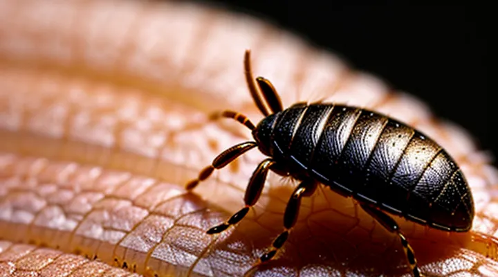
A freshly attached tick presents a distinctive visual profile that differs from an unattached specimen. The engorged mouthparts, known as the hypostome, pierce the epidermis and remain visible as a small, dark protrusion. The surrounding body often appears swollen, gray‑brown, and slightly raised above the skin surface. The dorsal shield (scutum) may be partially visible, while the abdomen expands as the tick begins to feed on blood.
Key visual indicators include:
- Small, dark puncture at the center of the attachment site.
- Slightly raised, rounded area surrounding the puncture.
- Visible scutum or partial shield, typically lighter in color than the abdomen.
- Absence of a clear blood pool; feeding fluid is absorbed internally, causing gradual abdominal enlargement.
Early identification relies on recognizing these features before the tick becomes fully engorged. Prompt removal reduces the risk of pathogen transmission and minimizes tissue irritation.
Preferred Body Areas for Tick Attachment
Ticks select attachment sites that provide easy access to thin epidermis, abundant blood flow, and protection from clothing friction. The preferred locations are typically concealed, moist, and have minimal hair, allowing the mouthparts to penetrate the skin with little resistance.
• Scalp and hairline, especially behind the ears
• Neck, particularly the posterior region
• Axillary folds (armpits)
• Groin and genital area
• Inguinal crease and inner thigh
• Behind the knees and popliteal fossa
• Wrist and ankle creases
These areas offer optimal conditions for engorgement and reduce the likelihood of early detection. Regular self‑examination of the listed regions after outdoor exposure aids early removal and minimizes the risk of disease transmission.
Distinguishing a Tick from Other Skin Conditions
Moles, Freckles, and Scabs
Moles are typically raised or flat, uniform in color ranging from light brown to black, and have well‑defined borders. The surface may be smooth or slightly textured, and pigmentation often remains stable over time. A subdermal tick appears as a small, dome‑shaped bump, often darker than surrounding skin, with a central punctum where the mouthparts attach. Unlike a mole, the bump may be slightly mobile and may elicit mild swelling or redness around the attachment site.
Freckles consist of clusters of melanin particles, presenting as flat, tan to light brown spots with irregular edges. They fade under strong lighting and do not change in size. A tick embedded beneath the skin rarely mimics this flat, diffuse pattern; instead, it forms a localized, raised nodule that may become inflamed, producing a noticeable halo of erythema.
Scabs are crusted lesions that form over a wound. They are typically rough, brownish‑red, and may detach as healing progresses. The core of a scab is composed of dried blood and plasma, not living tissue. In contrast, a tick under the skin maintains a firm, intact body that can be palpated as a distinct, rounded mass beneath the epidermis, often accompanied by a small puncture wound at the surface.
Key distinguishing features:
- Shape: rounded, dome‑shaped for a tick; flat or irregular for moles, freckles, and scabs.
- Mobility: slight movement possible with a tick; lesions are fixed in moles, freckles, scabs.
- Border: clear, defined edge for a tick; diffuse or irregular for freckles and scabs.
- Surrounding reaction: localized erythema or swelling common with ticks; absent or minimal with moles and freckles; crust formation typical of scabs.
Recognition of these characteristics enables accurate identification and appropriate removal of a tick without confusing it with benign skin markings.
Ingrown Hairs or Splinters
A sensation described as a tick moving beneath the skin often leads to concern about an actual parasite, yet two common non‑parasitic sources mimic this feeling.
Ingrown hairs develop when a shaved or broken hair re‑enters the epidermis. The resulting papule appears red, may contain a tiny visible hair tip, and can generate a fleeting, prick‑like motion that feels identical to a crawling tick. Typical locations include the beard area, legs and underarms, where friction encourages hair re‑entry.
Splinters introduce a foreign fragment—wood, glass, metal—into the dermis. The entry point is usually a pinpoint puncture surrounded by localized swelling. As the fragment shifts slightly with movement, a subtle crawling sensation emerges. Linear tracks or a faint line of discoloration often accompany the entry site, distinguishing splinters from live arthropods.
Key distinguishing features:
- Visible hair tip emerging from a papule → ingrown hair.
- Linear puncture with possible discoloration → splinter.
- Absence of a live organism or bite marks → non‑parasitic cause.
- Rapid onset after shaving or handling sharp objects → likely ingrown hair or splinter.
Management involves gentle extraction. For ingrown hairs, sterile tweezers can lift the exposed tip, followed by antiseptic application to prevent infection. Splinters require careful removal, often with a fine needle or forceps; thorough cleaning and a topical antibiotic reduce inflammation risk. Persistent redness, swelling, or pain warrants professional evaluation to exclude secondary infection or deeper tissue involvement.
Insect Bites (Mosquitoes, Spiders, Fleas)
A tick that has embedded its mouthparts beneath the epidermis creates a small, raised lesion often described as a papule. The central area may appear as a darkened punctum, sometimes resembling a tiny black dot, while the surrounding skin can be slightly erythematous and swollen. The lesion typically persists without the rapid spreading of redness seen in many other arthropod bites.
Mosquito, spider and flea bites present distinct visual patterns:
- Mosquito bite: round, itchy wheal, surface pale with a surrounding halo of redness that expands within minutes.
- Spider bite: variable; some species produce a necrotic ulcer with a central blister, others cause a localized, painful papule without extensive erythema.
- Flea bite: cluster of tiny, red punctate spots, often grouped in a line or “breakfast‑scrambled” pattern, each surrounded by a thin halo of inflammation.
Key identifiers for a tick bite include the presence of a firm, raised nodule, a central punctum that may exude a tiny amount of fluid, and the absence of immediate intense itching. Unlike mosquito bites, the reaction does not typically peak within an hour; instead, it remains relatively stable for several days. Spider bites may develop necrosis, a feature not associated with tick lesions. Flea bites appear as multiple adjacent points, whereas a tick bite is singular.
Recognition of these characteristics enables accurate differentiation and informs appropriate medical response, such as removal of the tick’s mouthparts and monitoring for potential pathogen transmission.
Identifying the Tick’s Body Parts When Embedded
The Head (Hypostome) and Mouthparts
A tick that has penetrated the skin presents a small, dark projection at the attachment site. The projection corresponds to the head region, which contains the feeding apparatus.
The head, or «hypostome», is a cone‑shaped structure covered with rows of backward‑pointing barbs. Barbs anchor the tick firmly in the host tissue, preventing easy removal. The surface of the hypostome appears matte and brown to black, matching the overall coloration of the engorged body.
Mouthparts associated with the hypostome include:
- Chelicerae: paired, needle‑like cutters that create a tiny incision in the skin.
- Palps: sensory appendages that guide the hypostome toward blood vessels.
- Hypostome: the primary anchoring organ, described above.
Visible signs of the head and mouthparts are:
- A slightly raised, point‑like tip protruding from the skin surface.
- A smooth transition from the tick’s dorsal shield to the hypostome, without obvious gaps.
- No visible segmentation; the head blends into the body’s oval outline.
Recognition of these features enables accurate identification of an embedded tick and informs appropriate removal techniques.
The Body (Engorged vs. Unengorged)
An engorged tick embedded in the skin appears as a swollen, balloon‑like structure. The abdomen expands to several millimetres, often reaching the size of a small pea. The surface becomes smooth and semi‑transparent, allowing underlying blood to be visible as a faint pink or reddish hue. The mouthparts may protrude slightly, forming a tiny dark point at the centre of the lesion.
An unengorged tick presents a flat, oval body about 2–4 mm long. The exoskeleton is hard, dark brown to black, with clearly defined segmentation. The legs are visible as tiny, pale extensions around the perimeter. The mouthparts are less apparent, recessed within the surrounding skin.
Key visual differences:
- Shape: balloon‑like versus flat oval
- Size: up to several millimetres when engorged, markedly smaller when unfed
- Colour: semi‑transparent with reddish tint versus solid dark brown/black
- Surface texture: smooth, glossy versus hard, chitinous
Recognition of these characteristics aids accurate identification and timely removal.
Legs and Size Variations
A tick that has penetrated the dermis appears as a small, rounded nodule whose surface may be slightly raised or flat. The skin over the body often shows a faint pink or reddish hue, while the embedded portion remains a darker brown, reflecting the tick’s exoskeleton. The head and mouthparts are usually hidden beneath the skin, leaving only the posterior abdomen visible.
The legs of an embedded tick retain their characteristic arrangement despite the concealment. Each adult tick possesses eight legs, grouped in four pairs. The legs are short, stout, and covered with tiny sensory hairs that enable the parasite to detect host movement. When the tick is partially embedded, the legs may be visible at the periphery of the nodule, appearing as tiny, pale protrusions.
Size variations depend on feeding status:
- Unfed adult: length ≈ 3–5 mm, width ≈ 2 mm.
- Partially engorged: length ≈ 5–10 mm, width ≈ 3–5 mm.
- Fully engorged: length ≈ 10–12 mm, width ≈ 6–8 mm.
The increase in size results from the expansion of the abdomen as the tick fills with blood. Leg length remains relatively constant, while the overall silhouette becomes more oval and prominent. Observing the combination of a raised nodule, darkened abdomen, and the characteristic eight‑leg pattern provides reliable visual confirmation of a tick embedded in the skin.
Signs of an Embedded Tick
Changes in Skin Color Around the Bite
A tick that has penetrated the dermis frequently produces a localized discoloration that serves as an early visual cue. The skin surrounding the attachment point may exhibit one or more of the following changes:
- A reddish‑pink halo extending a few millimeters from the bite site, caused by capillary dilation.
- A darker, purplish ring that develops as hemoglobin breaks down, indicating mild hemorrhage.
- A pale or ivory‑colored area directly under the tick, reflecting tissue compression and reduced blood flow.
The intensity and duration of each color shift depend on the tick’s feeding stage and the host’s vascular response. Initial redness typically appears within hours, fades over 24–48 hours, and may be replaced by a darker ring if inflammation persists. Persistent discoloration beyond several days warrants medical evaluation, as it can signal secondary infection or early signs of tick‑borne disease.
Swelling and Inflammation
A tick lodged beneath the epidermis initiates a localized inflammatory response. The skin around the attachment becomes swollen, forming a palpable, raised area that may feel firm or pliable depending on individual tissue reaction. Redness typically radiates outward, creating a concentric halo of erythema that contrasts with surrounding healthy tissue. The central point of the bite often appears as a small punctum, sometimes visible as a tiny dark spot where the mouthparts remain embedded.
Key visual indicators of swelling and inflammation include:
- Elevated, dome‑shaped nodule measuring 2–5 mm in diameter
- Surrounding erythema extending 5–10 mm from the core
- Warmth detectable by touch, indicating increased blood flow
- Possible secondary itching or tenderness, reflecting histamine release
In some cases, the inflammatory zone may enlarge within 24–48 hours, forming a more pronounced lump. Persistent or rapidly expanding swelling can signal secondary infection, requiring medical assessment. Absence of systemic symptoms such as fever does not exclude localized reaction; the primary presentation remains confined to the cutaneous area surrounding the embedded arthropod.
Itching, Pain, or Discomfort
An embedded tick appears as a small, dark, dome‑shaped body lodged just below the skin surface. The head may protrude slightly, creating a visible puncture or a tiny raised spot. The surrounding area often shows a faint halo of redness.
Symptoms commonly reported include:
- «Itching» that intensifies after the tick attaches, often localized around the bite site.
- «Pain» ranging from a mild prick at the moment of penetration to a persistent ache as the tick remains embedded.
- «Discomfort» manifested as swelling, a feeling of pressure, or a vague irritation that does not subside with typical skin soothing measures.
Persistent or worsening sensations, expanding redness, or the presence of a visible tick warrant prompt medical evaluation to reduce the risk of infection and other complications.
What to Do Upon Discovery
Safe Tick Removal Techniques
A tick lodged beneath the epidermis appears as a small, rounded swelling. The body may be expanded with blood, while the head and mouthparts remain hidden under the skin surface. The surrounding tissue often shows a slight reddening, but the tick’s silhouette is visible as a dark, dome‑shaped bump.
Safe removal requires precision and hygiene. Follow these steps:
1. Select fine‑pointed tweezers with smooth, non‑slipping jaws.
2. Grasp the tick as close to the skin as possible, securing the mouthparts without squeezing the body.
3. Apply steady, upward traction; avoid twisting or jerking motions that could fracture the hypostome.
4. Place the detached tick in a sealed container with alcohol for proper disposal.
5. Disinfect the bite area with an antiseptic solution.
6. Observe the site for several days; note any expanding rash, fever, or flu‑like symptoms and seek medical advice promptly.
Proper technique minimizes the risk of pathogen transmission and prevents residual mouthparts from remaining embedded in the tissue.
When to Seek Medical Attention
A tick that has become embedded beneath the skin may appear as a small, raised bump, often resembling a tiny, dark speck or a hollowed-out area where the mouthparts remain visible. Swelling, redness, or a palpable nodule can accompany the lesion, especially if the tick has been attached for several days.
Seek professional medical evaluation if any of the following conditions occur:
- Persistent or worsening erythema extending beyond the immediate bite site
- Development of a target‑shaped rash (often described as a “bull’s‑eye”)
- Fever, chills, fatigue, or muscle aches appearing within weeks of the bite
- Noticeable swelling of lymph nodes near the bite area
- Signs of secondary infection, such as pus, increasing pain, or foul odor
- Uncertainty about the duration of attachment or incomplete removal of the tick
Prompt assessment enables appropriate testing for tick‑borne diseases, administration of prophylactic antibiotics when indicated, and management of complications that may arise from delayed treatment.
Potential Complications and Symptoms to Monitor
Redness and Rash Development (Erythema Migrans)
A tick bite can trigger erythema migrans, the earliest cutaneous sign of Lyme disease. The rash typically begins as a small red macule at the attachment site and expands outward over several days.
Key visual features include:
- A circular or oval shape with a clear center and a peripheral erythematous halo.
- Diameter reaching 5 cm or more, often exceeding 10 cm in advanced cases.
- Uniform redness that may become slightly raised at the margins.
- Absence of vesicles, pus, or necrotic tissue.
The lesion progresses rapidly, enlarging at a rate of 2–3 cm per day. In some patients, multiple concentric rings develop, creating a “bull’s‑eye” appearance. The rash may be accompanied by mild itching or a warm sensation, but pain is uncommon.
Early identification of this expanding erythema is critical for prompt antimicrobial therapy, which reduces the risk of systemic complications. Medical assessment should occur as soon as the characteristic pattern is observed, regardless of the presence of systemic symptoms.
Flu-like Symptoms After a Tick Bite
A tick lodged beneath the epidermis appears as a small, dark, rounded mass. The head, or capitulum, may be visible as a tiny protrusion, while the body often resembles a pea‑sized bump. The surrounding skin can be slightly reddened, but the tick itself remains firmly attached, sometimes with its legs barely discernible.
After attachment, the host may develop systemic signs resembling a viral infection. These manifestations arise from the transmission of pathogens such as Borrelia burgdorferi or Anaplasma species. Early recognition of these symptoms can prevent progression to more severe disease stages.
Typical flu‑like manifestations include:
- Fever ranging from 38 °C to 40 °C
- Chills and sweats
- Generalized headache
- Muscle aches and joint pain
- Fatigue and malaise
- Mild nausea or loss of appetite
Medical evaluation is warranted when fever persists beyond 48 hours, when a rash emerges, or when neurological symptoms (e.g., facial palsy, meningitis signs) appear. Prompt antimicrobial therapy reduces the risk of chronic complications.
Long-Term Health Concerns
A tick that has migrated beneath the epidermis often appears as a small, raised nodule, sometimes surrounded by a faint erythema. The body of the arachnid may be partially visible through the skin, resembling a tiny, dark lump. Over time the lesion can enlarge as the tick feeds, producing localized swelling and a palpable firmness.
Long‑term health concerns arise when the parasite remains attached for several days, providing a conduit for pathogen transmission. Pathogens commonly associated with subdermal tick bites include spirochetes that cause Lyme disease, rickettsiae responsible for spotted fevers, and viruses such as Powassan. Delayed removal increases the probability of systemic infection, which may manifest weeks or months after the initial exposure.
Chronic complications can develop even after successful extraction. Potential outcomes include:
- Persistent arthritic pain affecting joints, especially the knees
- Neurological disturbances such as peripheral neuropathy or cognitive impairment
- Cardiac involvement presenting as conduction abnormalities or myocarditis
- Post‑treatment fatigue syndrome lasting several months
Early identification of the characteristic nodule and prompt medical evaluation reduce the risk of these sequelae. Monitoring for systemic symptoms—fever, rash, headache, or joint pain—should continue for at least six weeks after removal. Persistent or worsening signs warrant immediate laboratory testing and targeted antimicrobial therapy.
