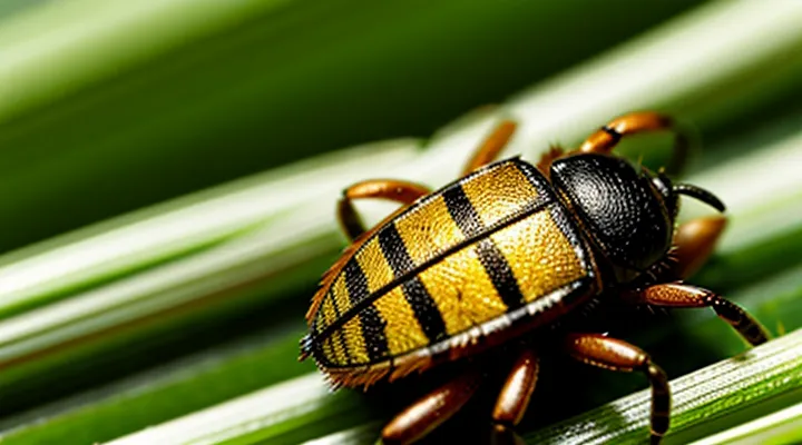
General Characteristics of Ticks
Size and Shape Variations
Ticks attached to human skin range from about 1 mm to over 15 mm in length, depending on species and feeding stage. Unfed larvae measure roughly 0.5 mm, nymphs 1–2 mm, and adult females 3–5 mm before blood intake. After engorgement, adult females can expand to 10–15 mm, while males remain near 3–5 mm.
Size differences reflect the tick’s developmental stage and the amount of blood consumed. Early‑stage ticks appear flat, oval, and light‑colored, resembling tiny specks. As they fill with blood, the body becomes rounded, markedly larger, and darkens to a deep brown or gray. Engorged females often assume a balloon‑like silhouette, with the dorsal surface swelling uniformly.
Shape variations arise from species‑specific anatomy. Hard ticks (Ixodidae) possess a shield‑like scutum covering part of the dorsal surface, giving a rectangular appearance. Soft ticks (Argasidae) lack a scutum, resulting in a more uniformly rounded form. Mouthparts may protrude forward in some species, creating a pointed tip that can be visible through the skin.
Typical size ranges by feeding status:
- Unfed: 0.5–3 mm, flat, oval
- Partially fed: 2–8 mm, semi‑rounded, slightly darkened
- Fully engorged: 10–15 mm, spherical, dark brown/gray
These dimensions and shapes assist clinicians in identifying tick species and assessing the duration of attachment.
Color and Texture
Ticks that have attached to human skin display a limited palette of hues and a distinctive surface quality. The organism’s coloration changes as it feeds, providing reliable visual cues for identification.
- Unfed or early‑stage ticks: reddish‑brown to dark brown, sometimes with a lighter “ornament” on the dorsal shield.
- Fully engorged specimens: deep crimson to purplish‑black, often appearing glossy due to stretched cuticle.
- Species‑specific variations: some Ixodes species exhibit a pale, almost ivory, scutum, while Amblyomma may show mottled patterns of tan and dark brown.
The texture of a feeding tick is equally characteristic. The outer exoskeleton feels firm when the tick is flat, then becomes supple as the abdomen expands. The mouthparts, including the hypostome, are rough and barbed, allowing firm attachment to skin. The engorged body feels soft, slightly gelatinous, and may appear swollen, while the dorsal shield remains comparatively rigid. These color and texture traits together enable rapid recognition of a tick on a person.
Identifying Ticks on the Body
Common Hiding Spots
Ticks attach to warm, protected skin areas where they remain unnoticed. Adult females are 3–5 mm long, expanding to 8–10 mm after feeding; they appear as dark, oval bodies that may become pale or engorged. Their small size and slow movement enable them to stay hidden for days.
- Scalp and hairline – dense hair shields the tick from visual inspection.
- Behind the ears – limited visibility and warmth attract attachment.
- Neck folds – skin creases retain moisture and conceal the parasite.
- Armpits – humid environment and limited exposure create a favorable niche.
- Groin and inner thigh – warm, moist skin folds provide optimal conditions.
- Waistband and belt line – friction‑resistant clothing regions allow ticks to cling unnoticed.
- Behind the knees – skin folds and reduced visibility protect the tick.
- Under fingernails or between toe webbing – tight spaces hinder detection during routine checks.
Regular skin examinations, especially in these locations, are essential for early removal and prevention of disease transmission.
Stages of Tick Attachment
A tick begins its interaction with a human host by climbing onto vegetation and waiting for a suitable passage of skin. The organism is small, reddish‑brown, and resembles a tiny seed. When it contacts a person, it climbs onto the skin surface and initiates the first stage of attachment.
- Questing and initial contact – The tick’s legs grasp the hair or clothing. At this point the body is flat, 2‑5 mm long, and its back appears smooth and matte. No visible mouthparts are exposed.
- Insertion of mouthparts – The tick inserts its hypostome, a barbed feeding tube, into the epidermis. The surrounding skin may show a pinpoint puncture, often unnoticed. The tick’s body lifts slightly, forming a small, raised “cap” that can be mistaken for a mole.
- Feeding phase (early) – Blood begins to flow into the tick’s foregut. The abdomen swells minimally, remaining a pale brown oval. The tick’s legs remain anchored, and a thin, translucent halo may appear around the attachment site due to slight irritation.
- Engorgement – Over several days, the tick’s abdomen expands dramatically, turning from brown to a grayish‑blue hue and increasing to 10‑15 mm in length. The skin around the tick may become reddened, and the tick’s shape changes from a flat disc to a balloon‑like silhouette.
- Detachment – Once fully engorged, the tick releases its grip and drops off. The empty exoskeleton left behind is a small, empty shell, typically 2‑3 mm, and the puncture site may remain as a tiny, healed scar.
Recognizing each visual stage aids rapid identification and removal, reducing the risk of pathogen transmission.
Unfed Tick Appearance
Unfed ticks that have attached to human skin are small, flat, and dark‑brown to reddish‑brown in color. Their bodies are oval and lack the rounded, balloon‑like appearance seen after feeding. The dorsal shield (scutum) covers the entire back in males and the anterior half in females, giving a smooth, hard surface. The mouthparts (capitulum) project forward, visible as a small, triangular structure near the front edge. Six legs extend from the ventral side, each ending in a claw that grips the skin.
Key visual characteristics:
- Length: 2–5 mm, depending on species; some are as small as 1 mm.
- Width: roughly half the length, creating a flattened profile.
- Color: uniform dark brown to reddish brown; occasional lighter patches on the scutum.
- Surface texture: hard, glossy exoskeleton without visible pores.
- Legs: thin, evenly spaced, visible as tiny protrusions when viewed from above.
These traits allow quick identification of a tick before it expands with blood. Recognizing the unfed form aids prompt removal and reduces the risk of disease transmission.
Engorged Tick Appearance
An engorged tick is a blood‑filled arachnid that has completed a feeding cycle on a person. After attachment, the tick expands dramatically, altering its appearance in ways that signal a potentially serious health risk.
The visible characteristics of an engorged tick include:
- Size increase from a few millimeters to as much as 1 – 2 cm in length and width, depending on species.
- A rounded, balloon‑like body that may appear glossy or translucent.
- A color shift from brown or reddish‑brown to a pale gray‑white or bluish hue as the abdomen fills with blood.
- A softened, less defined segmentation, making the legs appear tucked against the swollen abdomen.
- A clear demarcation between the engorged abdomen and the anterior mouthparts, which remain anchored to the skin.
Location on the body does not change the tick’s morphology, but areas with thin skin (e.g., scalp, armpits, groin) often reveal the swelling more prominently. The engorged tick remains attached for several days; as it detaches, the body contracts, returning to a smaller, darker form.
Prompt removal is advised because the enlarged size increases the likelihood of pathogen transmission. Visual identification of the described features enables rapid recognition and appropriate medical response.
Distinguishing Ticks from Other Skin Blemishes
Moles and Freckles
Moles and freckles are common pigmented skin lesions that can be mistaken for arachnid bites when they appear on the torso, limbs, or face. Both are composed of melanin, yet they differ in origin, texture, and clinical significance.
- Moles (nevus): usually raised, well‑defined borders, uniform or variegated color ranging from light brown to black, may contain hair follicles, and remain stable for years; occasional growth or change warrants dermatologic evaluation.
- Freckles (ephelides): flat, small (1–2 mm), uniformly tan or light brown, become more pronounced after sun exposure, and typically fade in low‑light conditions; they do not enlarge or develop new structures.
Ticks attached to skin present as a distinct, engorged arachnid body with a rounded or oval shape, often darker than surrounding tissue, and may be surrounded by a clear or hemorrhagic halo. Unlike moles and freckles, ticks are mobile, have visible legs, and can be removed without cutting into the lesion. Recognizing these visual criteria enables accurate differentiation and appropriate medical response.
Scabs and Insect Bites
Ticks embed themselves in the skin, creating a small, raised area that may be mistaken for a scab or bite mark. The attachment point is typically a reddish‑brown, dome‑shaped swelling about the size of a pencil eraser. When the tick’s mouthparts penetrate the epidermis, a tiny puncture hole appears at the center of the swelling, often surrounded by a faint halo of erythema.
Key visual differences between a tick attachment and a typical insect bite:
- Shape: Ticks form a rounded, slightly raised nodule; most bites are flat, irregular reddened patches.
- Color: The nodule is brown to dark brown, matching the engorged tick’s body; bite reactions are usually pink or light red.
- Surface texture: The area may feel firm or slightly gritty due to the tick’s hard exoskeleton; bites feel soft and may be itchy.
- Central puncture: A minute, often unnoticed opening exists where the tick’s hypostome anchors; bites lack a distinct central point.
- Duration: Tick attachment persists for hours to days, maintaining the raised nodule; ordinary bites fade within a few days.
If a scab forms, it typically covers the tick’s mouthparts after the insect detaches. The scab is a whitish, keratinized crust that may conceal the tick’s head. In contrast, a normal insect bite may crust over but usually retains the original pink-red coloration without a defined central hole.
Recognizing these characteristics enables prompt removal of the tick and reduces the risk of disease transmission. Immediate extraction with fine‑point tweezers, grasping the tick close to the skin, followed by cleaning the site with antiseptic, is the recommended response.
What to Do After Finding a Tick
Safe Tick Removal Techniques
Ticks attach with a small, engorged body that may appear as a dark, oval lump on the skin. Removing them safely prevents infection and minimizes skin damage.
- Use fine‑tipped tweezers or a specialized tick‑removal device.
- Grasp the tick as close to the skin as possible, avoiding the legs.
- Apply steady, gentle pressure to pull upward without twisting.
- After extraction, inspect the mouthparts; if any remain, repeat the process.
- Disinfect the bite area with alcohol or iodine.
- Wash hands thoroughly.
- Observe the site for redness, swelling, or rash over the next several days.
Do not squeeze the body, cut it off, or apply heat, chemicals, or petroleum products. Do not use folk remedies such as “painting” the tick with nail polish. If symptoms develop, seek medical evaluation promptly.
When to Seek Medical Attention
A tick attached to the skin may appear as a small, darkened bump, often resembling a tiny, engorged bead. After removal, the bite site can develop redness, swelling, or a rash. Seek professional evaluation promptly if any of the following occur:
- Redness expands beyond a few centimeters or forms a bull’s‑eye pattern.
- Fever, chills, headache, muscle aches, or fatigue develop within weeks of the bite.
- Joint pain or swelling appears, especially if it moves from one joint to another.
- The tick was attached for more than 24 hours, as longer attachment increases disease risk.
- The bite area becomes increasingly painful, ulcerated, or shows signs of infection such as pus or warmth.
- You have a weakened immune system, are pregnant, or have a history of Lyme disease or other tick‑borne illnesses.
Early medical assessment enables accurate diagnosis, appropriate antibiotic therapy, and prevention of complications associated with tick‑borne pathogens. If uncertainty exists about the tick’s identification or removal technique, contact a healthcare provider without delay.
Preventing Tick Bites
Protective Clothing and Repellents
Protective clothing forms the first barrier against tick attachment. Tight‑weave fabrics such as polyester or nylon reduce the ability of ticks to crawl through material. Long sleeves, long trousers, and closed shoes limit exposed skin. Tucking pants into socks and securing cuffs with elastic bands prevents ticks from reaching the lower legs. Light‑colored garments aid visual inspection after outdoor activity.
Repellents complement clothing by creating a chemical deterrent. Effective active ingredients include DEET (20‑30 % concentration), picaridin (10‑20 %), and permethrin (0.5 % for treated clothing). Apply skin repellents evenly, avoiding eyes and mucous membranes. Treat outer layers of clothing with permethrin and allow them to dry before wear. Reapply skin products every 4–6 hours or after swimming or heavy sweating.
Combined use of barrier clothing and repellents lowers the probability of tick bites, thereby reducing the chance of finding engorged ticks on the body. After exposure, conduct a systematic skin check: examine scalp, behind ears, underarms, groin, and between fingers. Prompt removal of any attached tick minimizes the risk of disease transmission.
Key practices
- Wear long, tightly woven garments; secure cuffs and pant legs.
- Choose light colors for easier visual detection.
- Apply DEET or picaridin to exposed skin; follow label instructions.
- Treat clothing with permethrin, refresh after multiple washes.
- Perform a full‑body tick inspection within 24 hours of outdoor activity.
Post-Outdoor Activity Checks
After returning from a hike, a wooded walk, or any activity in a tick‑infested area, a systematic body examination is essential. Ticks are typically flat, oval, and range from the size of a pinhead to a pea when engorged. Their bodies are reddish‑brown to dark brown, with a hard shield (scutum) covering the front half. Legs are six on each side and can be seen protruding from the sides of the abdomen.
The inspection routine should include:
- Remove clothing and wash hands with soap.
- Use a mirror or ask a partner to view hard‑to‑reach areas.
- Examine scalp, behind ears, neck, underarms, groin, waistline, behind knees, and between fingers.
- Look for attached organisms: a small, rounded shape attached to the skin, often near hair follicles or skin folds.
- If a tick is found, grasp it with fine‑pointed tweezers as close to the skin as possible, pull upward with steady pressure, avoiding crushing the body.
- Clean the bite site with antiseptic and store the tick in a sealed container for identification if needed.
- Document the date, location of the bite, and any symptoms that develop over the following weeks.
A thorough post‑activity check reduces the risk of disease transmission by ensuring early removal of any attached parasites. Regular practice of this protocol after each outdoor exposure maintains health safety.
