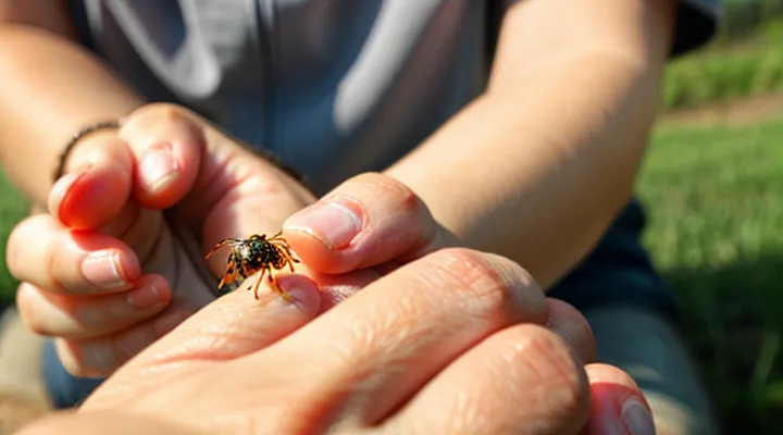
Identifying a Tick Bite on Your Hand
Initial Appearance and Symptoms
Immediate Reactions
A tick attachment on the hand usually presents as a tiny, red papule centered around the tick’s mouthparts. The skin may appear slightly raised, with a clear halo of erythema extending a few millimeters outward. Occasionally, a faint, white or pale spot marks the exact point of insertion, known as the feeding punctum.
Immediate reactions often include:
- Localized redness and swelling
- Mild to moderate itching
- Sharp or throbbing pain at the bite site
- Small blister formation in sensitive individuals
- Rapid onset of a rash if an allergic response occurs
These signs appear within minutes to a few hours after the tick attaches and may intensify before stabilizing. Prompt removal of the tick and cleaning of the area reduce the risk of complications.
Common Rash Characteristics
A tick bite on the hand often begins with a small, red papule centered on the site where the tick’s mouthparts remain attached. The lesion typically measures 2‑5 mm in diameter, though it may expand to 1 cm or more as inflammation progresses. The erythema is usually uniform in color, ranging from pink to deep crimson, and may develop a clear or slightly raised border.
The central punctum, sometimes called a “tick mouth‑mark,” appears as a pinpoint depression or dark spot. This feature distinguishes a tick‑related rash from other insect bites, which commonly lack a focal point. In the early stage, the surrounding skin may feel warm to the touch and exhibit mild tenderness.
As the reaction evolves, the rash can become:
- Slightly raised, forming a firm, dome‑shaped bump.
- Itchy, prompting scratching that may cause secondary irritation.
- Swollen, especially if an allergic response is present, leading to noticeable edema of the fingers or palm.
In some cases, a vesicular component emerges, creating small blisters that may rupture and ooze clear fluid. When the rash persists beyond a week or spreads beyond the bite site, it may indicate infection or transmission of a pathogen such as Borrelia burgdorferi. Prompt medical evaluation is advisable if the lesion enlarges, becomes necrotic, or is accompanied by fever, joint pain, or neurological symptoms.
Distinguishing from Other Insect Bites
A tick bite on the hand typically appears as a small, firm, raised area where the tick was attached. Unlike the shallow puncture left by a mosquito, the site may retain the tick’s mouthparts, creating a central dark spot surrounded by a red halo. The surrounding skin often remains relatively smooth, without the intense itching or welts common to flea or spider bites.
Key differences from other arthropod bites:
- Mosquito: superficial puncture, raised itchy bump, rapid swelling, no central dark point.
- Flea: multiple tiny red papules, intense itching, often in clusters; no central lesion.
- Spider: may produce a painful, necrotic ulcer or a raised blister; sometimes accompanied by a “target” pattern of concentric rings.
- Bee/wasp: immediate stinging pain, large swollen wheal, clear allergic reaction potential; no persistent attachment site.
Additional diagnostic clues for a tick bite:
- Presence of a tick’s engorged body or empty shell attached to skin.
- Minimal immediate pain; delayed onset of redness or swelling.
- Possible “bull’s-eye” erythema developing days after attachment, indicating potential infection.
When evaluating a hand lesion, verify whether a tick is still attached, note the size and firmness of the central point, and compare the reaction pattern with those typical of other insects. Prompt removal of the tick and monitoring for expanding rash are essential steps in management.
What to Do After a Tick Bite
Safe Tick Removal Techniques
Tools for Removal
When a tick attaches to the skin of the hand, prompt removal reduces the risk of pathogen transmission. Effective extraction depends on using the proper instruments and following a precise technique.
Fine‑point tweezers are the standard tool. Their narrow tips grasp the tick close to the skin surface, allowing steady upward traction without crushing the body. Choose stainless‑steel tweezers with a smooth grip to avoid slippage.
A dedicated tick‑removal device, often shaped like a small, curved hook, slides beneath the mouthparts. This design isolates the head, preventing compression of the abdomen, which can force infectious fluids into the wound.
If tweezers or a hook are unavailable, sterilized flat‑head forceps or a blunt‑ended needle can serve as alternatives, provided they allow firm contact with the tick’s head region.
Additional items that support safe removal include:
- Disposable nitrile gloves – protect the handler from direct contact.
- Antiseptic solution (e.g., 70 % isopropyl alcohol) – cleanse the bite area before and after extraction.
- Small container with a lid – store the tick for identification if needed.
- Clean gauze or cotton swab – apply pressure after removal to stop minor bleeding.
The removal process should be performed as follows:
- Don gloves and clean the bite site with antiseptic.
- Position the chosen tool so it grasps the tick’s head as close to the skin as possible.
- Apply steady, upward force without twisting or jerking.
- Once detached, place the tick in the container and seal it.
- Disinfect the bite area again and monitor for signs of infection.
Using these instruments correctly ensures the tick is extracted whole, minimizing tissue damage and the chance of disease transmission.
Step-by-Step Guide
Inspect the skin surface of the hand. Look for a small, raised spot that may be pink, red, or slightly darker than surrounding tissue. The spot often measures 2‑5 mm in diameter and may have a central puncture mark where the tick’s mouthparts entered.
Identify the attached tick. An engorged tick appears as a soft, oval body, usually brown or gray, gradually expanding as it feeds. The abdomen may be noticeably swollen, giving the tick a balloon‑like shape. The legs are short and positioned near the mouthparts, which are embedded in the skin.
Assess surrounding reaction. In many cases the bite site is surrounded by a faint halo of erythema, which can be smooth or irregular. Occasionally a small blister or a tiny ulcer may develop if the bite is irritated.
Document the findings. Record the size of the lesion, the color of the tick, and any changes in the skin’s appearance over time. Photographs can aid in monitoring progression.
Remove the tick if still attached. Use fine‑point tweezers to grasp the tick as close to the skin as possible, pulling upward with steady, even pressure. Avoid squeezing the body to prevent saliva release.
Clean the bite area. Apply antiseptic solution after removal, then cover with a sterile bandage if needed.
Monitor for signs of infection or disease transmission. Watch for expanding redness, warmth, swelling, fever, fatigue, or a rash resembling a bullseye. Seek medical evaluation promptly if any of these symptoms appear.
Repeat inspection after 24‑48 hours. Verify that the bite site is healing, that no new ticks have attached, and that any residual redness is diminishing. If the lesion persists or worsens, consult a healthcare professional.
Post-Removal Care
Cleaning the Bite Area
When a tick attaches to the skin of the hand, the surrounding area may appear red, swollen, or develop a small puncture mark. Prompt and proper cleaning reduces the risk of infection and helps identify any early signs of illness.
Begin cleaning immediately after removal:
- Wash hands thoroughly with soap and warm water.
- Apply a gentle antiseptic solution (e.g., povidone‑iodine or chlorhexidine) to the bite site using a clean cotton swab.
- Rinse with sterile saline to remove residual antiseptic.
- Pat the area dry with a sterile gauze pad; avoid rubbing.
After cleaning, monitor the site for:
- Persistent redness or expanding rash.
- Increasing pain, swelling, or warmth.
- Flu‑like symptoms such as fever, headache, or muscle aches.
If any of these develop, seek medical evaluation without delay. Maintaining a clean bite area and observing changes are essential steps in managing a tick encounter on the hand.
Monitoring for Symptoms
A tick bite on the hand often begins as a small, red puncture. Within hours to days, the site may swell, itch, or become painful. Monitoring the area and overall health is essential to detect early signs of infection or disease transmission.
Key symptoms to watch:
- Redness extending beyond the bite margin
- Increasing swelling or warmth
- Persistent itching or burning sensation
- Development of a target‑shaped rash (erythema migrans)
- Fever, chills, headache, or muscle aches
- Joint pain or swelling, especially in the knees or elbows
Observe the bite daily for at least two weeks. Record any changes in size, color, or sensation, and note systemic symptoms such as fever or fatigue. If a rash expands rapidly, reaches a diameter of 5 mm or more, or if flu‑like symptoms appear, seek medical evaluation promptly. Early treatment with appropriate antibiotics can prevent complications from tick‑borne illnesses.
When to Seek Medical Attention
Signs of Infection
Localized Infection
A tick attachment on the hand often progresses to a localized infection if the bite is left untreated. The skin around the feeding site becomes inflamed, producing a well‑defined, erythematous halo that may expand over several days. The central puncture point is usually small, sometimes covered by the engorged tick or a scab after removal. Common manifestations include:
- Redness extending 1–2 cm from the bite, with clear margins
- Swelling that feels firm to the touch
- Warmth and tenderness localized to the area
- Small vesicles or pustules that may develop within the erythema
If bacterial invasion occurs, the lesion may evolve into an abscess, characterized by a fluctuating center and possible purulent discharge. Systemic symptoms such as fever, chills, or malaise indicate that the infection is spreading beyond the initial site and require immediate medical evaluation. Prompt removal of the tick, thorough cleansing with antiseptic, and, when indicated, empirical antibiotic therapy reduce the risk of complications such as cellulitis, Lyme disease, or other tick‑borne illnesses.
Systemic Symptoms
A tick bite on the hand may appear as a small, red papule, often surrounded by a clear halo. While the local lesion is the most immediate sign, systemic manifestations can develop within hours to weeks, indicating possible infection or allergic reaction.
Common systemic symptoms include:
- Fever or chills
- Headache, often described as persistent or throbbing
- Muscle aches and joint pain, which may migrate between joints
- Fatigue or a general feeling of malaise
- Nausea, vomiting, or abdominal discomfort
- Swollen lymph nodes, particularly in the armpit or neck region
- Skin rash beyond the bite site, such as a diffuse red maculopapular eruption or a target‑shaped lesion
When these signs appear, they may signal diseases transmitted by ticks, such as Lyme disease, Rocky Mountain spotted fever, or ehrlichiosis. Prompt medical evaluation is essential; laboratory testing can confirm the pathogen, and early antibiotic therapy reduces the risk of severe complications.
Tick-Borne Illnesses
Common Diseases Associated with Tick Bites
Tick bites on the hand often serve as the entry point for a range of tick‑borne infections. The skin may show a small puncture mark, sometimes surrounded by a red halo that expands over days. This early visual cue can precede systemic illness caused by pathogens transmitted during feeding.
Common illnesses linked to hand‑acquired tick bites include:
- Lyme disease – caused by Borrelia burgdorferi; characterized by an expanding erythema migrans lesion, fever, fatigue, and joint pain.
- Rocky Mountain spotted fever – infection with Rickettsia rickettsii; presents with fever, headache, and a maculopapular rash that may spread from the bite site.
- Anaplasmosis – caused by Anaplasma phagocytophilum; symptoms include fever, chills, muscle aches, and sometimes a mild rash.
- Ehrlichiosis – caused by Ehrlichia chaffeensis; manifests as fever, leukopenia, and elevated liver enzymes, with occasional skin changes.
- Babesiosis – infection with Babesia microti; produces hemolytic anemia, fever, and fatigue, rarely accompanied by a rash.
- Tularemia – caused by Francisella tularensis; may cause ulceration at the bite site, swollen lymph nodes, and systemic illness.
Recognition of the initial bite mark, followed by any of the described clinical patterns, warrants prompt medical evaluation. Early laboratory testing and antibiotic therapy, especially for Lyme disease and spotted fever, reduce the risk of severe complications.
Early Warning Signs to Watch For
A tick attached to the hand often leaves a small, dark spot where its mouthparts have pierced the skin. The surrounding area may appear pink or reddish, and the bite site can be slightly raised. In many cases the tick’s body remains visible, attached to the skin for several hours or days.
Early warning signs that require attention include:
- Localized redness extending beyond the bite point
- Swelling or a raised bump that enlarges over time
- Persistent itching or burning sensation
- Pain that intensifies rather than subsides
- Fever, chills, or flu‑like symptoms within a few days
- Development of a target‑shaped rash (often called a “bullseye”) around the bite
If any of these signs appear, remove the tick promptly with fine‑tipped tweezers, clean the area with antiseptic, and seek medical evaluation to rule out infection or tick‑borne disease.
