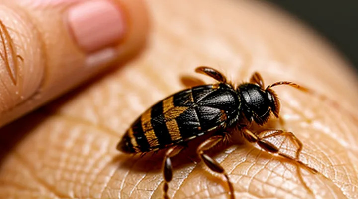
The Size and Appearance of Ticks
Tiny Ticks: Nymphs and Larvae
Tiny ticks that are in the nymph or larval stage present distinct visual cues. Nymphs measure about 0.5 mm in length, appear translucent to pale brown, and have a flattened, oval body that blends with the skin’s surface. Their legs are short, often difficult to see without magnification, and the scutum (shield) covers most of the dorsal surface, giving a smooth appearance.
Larvae, commonly called seed ticks, are even smaller—approximately 0.2 mm long. They are almost entirely transparent, allowing the underlying skin tone to show through. The body is spherical, lacking a hardened scutum, and the legs are proportionally longer relative to body size, sometimes visible as tiny, pale filaments.
Key identification points:
- Size: nymphs ≈ 0.5 mm; larvae ≈ 0.2 mm.
- Color: nymphs pale brown to amber; larvae nearly colorless.
- Body shape: nymphs oval, covered by scutum; larvae round, no scutum.
- Leg visibility: nymph legs short, often hidden; larval legs longer, sometimes discernible.
When examining skin, use a magnifying lens or dermatoscope to detect these minute structures. The attachment point is typically a small, punctate area where the mouthparts have pierced the epidermis, often without noticeable swelling. Recognizing these features enables accurate detection of early‑stage ticks.
Distinguishing Features: Legs and Body Shape
A tiny tick attached to the skin appears as a compact, rounded object, typically 1–3 mm in diameter. The body is oval to slightly elongated, with a smooth, glossy surface that may resemble a small pea or a grain of sand. Its coloration ranges from light brown to reddish‑brown, depending on species and engorgement stage.
- Legs: Eight slender legs emerge from the anterior margin. Each leg ends in a small claw that grips the skin, creating a subtle, radiating pattern when viewed under magnification. The legs are noticeably shorter than the body, causing the tick to look like a smooth dome rather than a multi‑legged insect.
- Body shape: The dorsal shield (scutum) covers most of the back in unfed specimens, giving a hard, shield‑like appearance. In partially fed ticks, the abdomen expands, forming a more rounded, balloon‑like silhouette while the scutum remains distinct. The overall profile is low‑profile and dome‑shaped, lacking obvious segmentation.
Color Variations: From Brown to Black
A small tick attached to the skin appears as a round, flat body roughly 2–5 mm in diameter before feeding. The organism’s mouthparts penetrate the epidermis, creating a tiny puncture that may be barely visible.
Color is the most reliable visual cue. Ticks exhibit a spectrum that progresses from light brown to deep black as they mature and ingest blood. Typical shades include:
- Light brown: newly attached, unfed stage.
- Medium brown: partially fed, beginning engorgement.
- Dark brown to reddish‑brown: moderate blood intake.
- Black: fully engorged adult or older specimen.
The transition reflects physiological changes rather than disease presence. Recognizing these hues aids rapid identification and timely removal.
Location and Common Attachment Sites
Preferred Areas for Tick Bites
A tiny tick on the skin often goes unnoticed because it resembles a speck of dirt. Recognizing the typical attachment sites helps locate the parasite before it enlarges.
- Scalp, especially behind the ears
- Neck and collarbone region
- Underarms
- Groin and inner thigh folds
- Behind the knees
- Around the waistline, particularly under clothing seams
These locations share thin, less‑hairy skin that offers easy penetration and a steady blood supply. The parasite also benefits from reduced friction, allowing it to remain attached longer without detection.
On the surface, the tick appears as a small, rounded, dark spot about the size of a pinhead. The surrounding skin may show a faint red halo, but inflammation is often minimal in the early stage. Prompt inspection of the listed areas can reveal the tick before it expands and increases the risk of disease transmission.
Hiding Spots: Hairline and Skin Folds
A small tick measures roughly 2–5 mm when unfed, appearing as a rounded, dark gray or brown speck that may be mistaken for a hair or a spot of dirt. Its body is flattened, with a pair of forward‑pointing mouthparts that can be seen as tiny protrusions if the insect is examined closely.
Hairlines and skin folds provide optimal concealment because the tick can align its body with fine hairs or creases, reducing its visibility. In these areas the insect often adopts a flatter profile, blending with the surrounding tissue and making detection difficult without magnification.
Typical indicators of a hidden tick in these zones include:
- A pinpoint, slightly raised spot that does not blanch under pressure.
- A faint, linear groove where the feeding apparatus penetrates the skin.
- A subtle change in skin texture, such as a tiny, firm bump amidst hair or a crease.
Immediate inspection of the scalp, behind the ears, under the arms, and within any abdominal or groin folds is essential for accurate identification. If a tick is suspected, gentle removal with fine‑point tweezers, grasping close to the skin surface, minimizes the risk of leaving mouthparts embedded.
What a Tick Might Look Like vs. Other Skin Conditions
Ticks vs. Moles or Freckles
A tiny tick attached to the skin appears as a raised, oval or round body measuring 2–5 mm in diameter. Its surface is rough, often resembling a miniature crustacean shell, and it may be gray, brown, or dark reddish. The creature’s legs are visible only when the tick is examined closely; they protrude from the edges of the body and may twitch. Ticks embed their mouthparts into the epidermis, creating a firm, sometimes painful anchor point that does not flatten under gentle pressure.
Moles and freckles differ markedly:
- Size: Moles range from a few millimeters to several centimeters; freckles are usually less than 2 mm.
- Surface: Moles have smooth, uniform texture; freckles are flat, matte, and lack a raised profile.
- Color: Moles present uniform brown, black, or tan tones; freckles are light brown to reddish and fade under sunlight.
- Attachment: Neither mole nor freckle adheres to the skin via a feeding apparatus; they are integral to epidermal cells.
- Mobility: Ticks remain fixed until removed; moles and freckles do not move or detach.
When a small, hard, raised spot is discovered, examine it for the characteristic rough shell and visible legs. If the lesion does not blanch when pressed, does not change color with sun exposure, or is accompanied by itching, swelling, or a rash, seek medical evaluation promptly. Early removal of a feeding tick reduces the risk of pathogen transmission.
Ticks vs. Scabs or Dirt
A small tick attached to the skin appears as a rounded, dome‑shaped body, often 2–5 mm in length before feeding. The surface is smooth, slightly glossy, and matches the surrounding skin tone, though it may become darker as it swells with blood. Ticks have six legs that are visible as tiny protrusions near the edge of the body, and the mouthparts form a pointed, pin‑like structure that penetrates the skin. The attachment site is usually firm; the tick does not easily detach when touched.
In contrast, a scab is a dry, crusty formation that develops after a superficial wound heals. Scabs are irregularly shaped, rough to the touch, and may be reddish, brown, or yellow. They lack legs or a distinct head, and they can be peeled away without causing pain. Dirt particles are granular, vary in color from gray to brown, and lie loosely on the skin surface. They can be brushed off and do not embed themselves or cause localized swelling.
Key distinguishing points:
- Shape: Tick – rounded, dome; scab – irregular, flaky; dirt – granular.
- Texture: Tick – smooth, slightly moist; scab – dry, crusty; dirt – gritty.
- Attachment: Tick – firmly anchored, mouthparts embedded; scab – loosely adherent, removable; dirt – surface‑level, easily brushed away.
- Presence of legs: Tick – six visible legs near the perimeter; scab and dirt – none.
- Reaction: Tick – may cause localized redness and mild swelling; scab – normal healing inflammation; dirt – no specific reaction unless irritated.
Observing these characteristics enables accurate identification and appropriate removal of a tick, while avoiding confusion with harmless scabs or environmental debris.
Ticks vs. Other Insect Bites
A tiny, attached tick appears as a round, flattened disc, usually 2‑5 mm in diameter, with a darker, leathery body and a lighter, translucent underside. The organism may be partially engorged, giving it a slightly bulging silhouette, and it often remains firmly anchored by its mouthparts, which are not visible on the skin surface.
Compared with bites from common insects, a tick can be distinguished by several observable traits:
- Size and shape: ticks are larger than most mosquito or flea bites and have a compact, oval silhouette; spider bites may be similar in size but lack the distinct, segmented body.
- Color and texture: a tick’s exoskeleton is brown to black and appears smooth, whereas mosquito bites are reddened, raised welts without a solid core.
- Attachment: ticks remain attached for hours to days, creating a localized, often painless puncture; most other bites are transient and detach immediately.
- Reaction: tick sites may develop a small, central puncture surrounded by a faint ring of erythema, while mosquito or sandfly bites typically produce an itchy, raised bump with a clear halo.
Recognizing these differences enables accurate identification of a small tick on the skin and informs appropriate removal and medical assessment.
Post-Bite Appearance: Engorged Ticks
Swelling and Discoloration Around the Bite
A small tick attached to the skin appears as a tiny, often dark, dome‑shaped object, sometimes resembling a speck of dirt. The immediate reaction around the attachment site typically includes localized swelling and a change in skin color.
- Swelling: mild to moderate edema develops within minutes to hours, forming a raised, firm area that may feel warm to the touch. The extent of the swelling correlates with the host’s immune response and the tick’s saliva composition.
- Discoloration: erythema or a reddish‑purple halo frequently surrounds the bite. In some cases, a central punctate spot remains, representing the tick’s mouthparts. The discoloration can spread outward, forming a target‑like pattern known as a “bull’s‑eye” lesion, especially with certain disease‑transmitting species.
Persistent or worsening edema, expanding discoloration, or the appearance of a rash beyond the immediate perimeter warrants medical evaluation to rule out secondary infection or tick‑borne illness. Early removal of the tick, followed by proper wound care, reduces the risk of complications.
The Changing Size of an Engorged Tick
A newly attached tick measures roughly 1–2 mm in length, resembling a tiny, flat speck. Its body is smooth, color ranging from pale beige to reddish‑brown, and legs are barely visible against the skin surface. The mouthparts protrude only a fraction of a millimeter, making the organism appear as a faint dot.
When the parasite begins to feed, its abdomen expands dramatically. Within 24 hours, the engorged stage reaches 5–7 mm, assuming a balloon‑like shape that swells outward from the original flat profile. By the end of the feeding period—typically 3–5 days—the tick can grow to 10–12 mm, sometimes exceeding 15 mm in the most voracious species. The enlarged body becomes noticeably rounded, darker in hue, and the legs spread more widely, creating a conspicuous, raised bump on the skin.
Key visual indicators of size progression:
- Initial stage: 1–2 mm, flat, light‑colored.
- Early engorgement (≈24 h): 5–7 mm, rounded, moderate darkening.
- Late engorgement (3–5 days): 10–15 mm+, fully swollen, deep brown to black.
Recognizing these changes enables prompt removal before the tick reaches maximal expansion, reducing the risk of pathogen transmission.
Next Steps After Identifying a Tick
Safe Tick Removal Techniques
A tiny, flat-bodied arachnid attached to the epidermis appears as a pinpoint or a small, dark speck, often resembling a pinhead or a tiny bruise. The body may be partially engorged after feeding, giving a slightly raised, rounded shape that can be difficult to see without magnification.
Safe removal follows a strict sequence:
- Grasp the tick as close to the skin surface as possible using fine‑point tweezers or a specialized tick‑removal tool.
- Apply steady, downward pressure to pull the mouthparts straight out without twisting or squeezing the body.
- Avoid crushing the tick; compression can release pathogen‑laden fluids.
- After extraction, cleanse the bite area with antiseptic solution and wash hands thoroughly.
- Dispose of the tick by submerging it in alcohol, sealing it in a container, or flushing it down the toilet; do not crush it with fingers.
Post‑removal observation includes monitoring the site for redness, swelling, or a rash over the next several weeks. If any signs of infection or illness appear, seek medical evaluation promptly. The described method minimizes the risk of pathogen transmission while ensuring complete removal of the parasite.
When to Seek Medical Attention
A tiny tick attached to the skin appears as a small, dark, round or oval spot, often resembling a speck of dirt. The body may be slightly raised, and the legs can be visible as fine hairs extending from the edges. When the tick swells after feeding, the lesion may become larger and more prominent.
Seek professional evaluation if any of the following conditions occur:
- The tick remains attached for more than 24 hours.
- The bite site becomes increasingly red, swollen, or painful.
- A rash develops that expands outward from the bite, especially with a bullseye pattern.
- Flu‑like symptoms appear, such as fever, chills, headache, muscle aches, or fatigue, within weeks of the bite.
- The individual has a compromised immune system, is pregnant, or is a child under ten years old.
Contact a healthcare provider promptly when the above signs are present. Early diagnosis and treatment reduce the risk of complications associated with tick‑borne infections. If uncertain, err on the side of caution and arrange an evaluation.
