Why Ticks Detach
Natural Lifecycle
The natural lifecycle of a tick proceeds through four distinct phases: egg, larva, nymph, and adult. Each phase culminates in a detachment event when the tick leaves its host, and the physical characteristics of the detached specimen provide clues to its developmental stage.
- Egg – microscopic, translucent, clustered on the ground; not encountered as a detached tick on a host.
- Larva – size 0.5–1 mm, reddish‑brown, six‑legged, smooth body; after feeding, it drops off appearing soft and engorged.
- Nymph – size 1–2 mm, darker brown, eight‑legged, slightly flattened; post‑feeding nymphs are noticeably swollen, with a glossy surface.
- Adult female – size 2–4 mm (unfed) to 6–12 mm (engorged), dark brown to gray, eight‑legged, rounded abdomen; an engorged female resembles a small, blood‑filled sac, often visible as a dark, oval lump.
- Adult male – size 2–3 mm, lighter brown, eight legs, less engorged; appears as a small, tapered body with a narrower abdomen.
During detachment, the tick’s cuticle hardens, color deepens, and the body becomes more rigid. Recognizing these visual cues assists in accurate identification and informs appropriate removal procedures.
Host Response
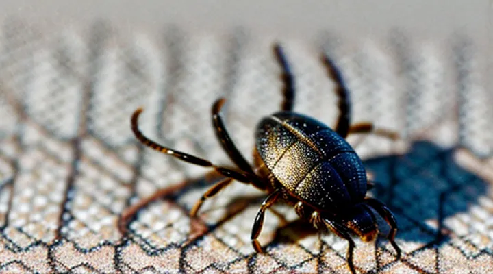
A detached tick typically appears as a small, flattened arthropod, ranging from 2 mm (unengorged) to 8 mm (partially engorged) in length, with a reddish‑brown dorsal surface and a lighter ventral side. The body may be swollen if recent feeding occurred, and legs are clearly visible.
When the parasite separates from the host, the following physiological responses are commonly observed:
- Local erythema at the bite site, often forming a pinpoint or circular rash.
- Mild swelling that peaks within 24 hours and subsides over several days.
- Pruritus that may intensify after the tick’s removal.
- Formation of a central punctum, sometimes surrounded by a halo of erythema.
- Transient fever or malaise in cases of pathogen transmission.
Immunologically, the host’s innate system deploys neutrophils and macrophages to the wound, releasing cytokines such as IL‑1β and TNF‑α. This initiates inflammation and promotes wound healing. Adaptive immunity generates specific IgG antibodies against tick salivary proteins, which can neutralize subsequent bites and reduce pathogen load.
Monitoring should include daily inspection of the bite area for expanding erythema, ulceration, or necrosis. Persistent symptoms beyond a week, systemic signs (headache, joint pain), or a history of exposure to disease‑endemic regions warrant medical evaluation and possible serologic testing.
General Appearance of a Detached Tick
Size and Shape
A detached tick presents a compact, oval body that tapers slightly at the ends. When unfed, the organism is flat and measures roughly 2–5 mm in length and 1.5–2 mm in width. After a blood meal, the abdomen expands dramatically, producing a rounded, engorged silhouette that can reach 6–10 mm long and 4–8 mm wide, depending on species and feeding duration.
Key dimensional characteristics:
- Unfed stage: 2–5 mm (length) × 1.5–2 mm (width); smooth, leathery cuticle.
- Partially engorged: 4–7 mm (length) × 3–5 mm (width); abdomen visibly swollen.
- Fully engorged: up to 10 mm (length) × 8 mm (width); pronounced, balloon‑like abdomen.
The overall shape remains roughly elliptical throughout, with a distinct, darker dorsal shield (scutum) that may cover only part of the back in females and the entire back in males. The ventral side is softer and more pliable, allowing the tick to detach easily after feeding.
Coloration
A detached tick’s coloration provides immediate clues about its feeding status, species, and time since removal.
Unfed ticks typically appear light‑brown to tan, with a smooth, uniform surface. Their legs and mouthparts may be a paler hue than the body, especially in larvae and nymphs.
Engorged ticks change dramatically after a blood meal. The abdomen swells and turns deep red, dark brown, or grayish‑black, while the dorsal shield (scutum) often remains a lighter shade. The overall impression is a markedly enlarged, mottled specimen.
Species differences influence color patterns:
- Black‑legged (deer) tick: dark, almost black legs; body ranges from reddish‑brown (unfed) to bright red (fully engorged).
- American dog tick: brown to reddish‑brown body; abdomen may become dark brown after feeding.
- Lone star tick: ivory or pale cream scutum with a distinctive white spot; abdomen shifts from pale to gray‑black when engorged.
After removal, the tick’s cuticle begins to dry, causing the colors to dull and darken. Within hours, the bright red of a fully fed specimen may turn brownish‑gray, while unfed ticks retain a more stable tan.
Observing these color cues enables rapid assessment of a tick’s recent activity and assists in accurate identification.
Leg Configuration
A detached tick can be identified by the arrangement of its legs, which is a reliable characteristic for species determination and stage identification.
Adult ticks possess eight legs arranged in four symmetrical pairs. Each leg consists of a coxae that attaches to the body, followed by a trochanter, femur, patella, tibia, and a tarsus ending in a small claw. The front pair (leg I) is generally longer and equipped with sensory organs that aid in host detection. The legs are positioned laterally, extending outward from the dorsal surface, and are clearly visible when the tick is removed from a host.
Nymphal ticks also have eight legs, but the segments are proportionally shorter and the overall body size is reduced. The legs retain the same segmental structure as adults, allowing easy differentiation from larvae.
Larval ticks differ markedly: they have only six legs, arranged in three pairs, with the same segmental composition but a noticeably smaller overall profile.
Key observable points for a detached tick:
- Number of legs (six in larvae, eight in nymphs and adults)
- Symmetrical pairing of legs
- Visible segmentation (coxa → trochanter → femur → patella → tibia → tarsus)
- Relative length of the front pair compared to the remaining legs
- Presence of claws at the distal end of each tarsus
These leg‑configuration details enable precise identification of a detached tick’s developmental stage and assist in subsequent taxonomic assessment.
Key Features to Observe
Distinguishing from Other Pests
A detached tick is a small, oval‑shaped arachnid measuring 2–5 mm when unfed and up to 10 mm after engorgement. Its body consists of a dorsal shield (scutum) and a ventral plate, both typically brown to reddish‑brown. The legs are clearly visible, eight in total, arranged in pairs and extending from the anterior edge. The mouthparts form a short, protruding beak (hypostome) that may appear as a tiny hook. Surface texture is smooth, not glossy, and the tick’s outline remains rounded rather than elongated.
- Fleas: laterally compressed, 1–3 mm, lack a scutum, possess strong hind legs for jumping, and exhibit a darker, shiny exoskeleton.
- Lice: elongated body, 2–4 mm, six legs only, no visible scutum, cling tightly to hair shafts, and have a more granular texture.
- Mites: often microscopic (<1 mm), round to oval, may lack distinct legs, and display a softer, translucent appearance.
- Beetles: hard, glossy elytra covering the abdomen, six legs, and a more rigid, angular shape.
- Cockroaches: larger (10–30 mm), flattened body, prominent wings, and a rough, brown exoskeleton.
These visual criteria enable rapid identification of a separated tick and prevent confusion with other common household pests.
Signs of Engorgement
A detached tick is typically a soft, rounded body that has expanded to several times its original size after feeding. The abdomen appears swollen, often resembling a small grape or a plump, translucent sphere. Color shifts from light brown to a darker, reddish or grayish hue as blood fills the gut. The legs remain clearly visible, but the mouthparts may be obscured by the engorged abdomen.
Key indicators of engorgement include:
- Marked increase in overall length and width compared to an unfed tick
- Bulging, dome‑shaped abdomen that dominates the body outline
- Darkened, semi‑transparent skin revealing internal blood meal
- Loss of distinct segmentation on the dorsal surface
- Slight flattening of the ventral side as the tick prepares to detach
These characteristics enable reliable identification of a tick that has completed its blood meal and is ready to fall off its host.
Post-Detachment Behavior and Survival
Locomotion
A tick that has been removed retains the anatomical features that enable its movement, making these structures essential for visual identification. The body is oval, slightly flattened, and ranges from 2 mm to 6 mm depending on species and feeding stage. The dorsal shield (scutum) is visible as a dark, hardened plate on the anterior half of the female; males display a complete scutum covering the entire back.
The legs, the primary locomotor organs, are six pairs of short, sturdy appendages. Each leg ends in a claw that can be seen as a tiny hook when the tick is examined under magnification. The legs are positioned symmetrically around the body, giving the tick a characteristic “spider‑like” silhouette. The articulation points are visible as pale, flexible joints, indicating the tick’s ability to pivot and cling to hosts.
Key visual cues for a detached tick:
- Oval, flattened body with clear segmentation
- Dark scutum on the anterior dorsal surface (partial in females, complete in males)
- Six pairs of short legs ending in hooked claws
- Pale, flexible joints at each leg segment
- Visible mouthparts (hypostome) projecting forward from the front, appearing as a set of barbed tubes
Recognizing these locomotion‑related structures enables accurate assessment of a tick’s species and feeding status, which is critical for evaluating disease risk.
Questing for a New Host
A detached tick presents a flat, oval body measuring 2–5 mm in length, depending on species and engorgement level. The dorsal shield (scutum) retains its brown to reddish hue, while the ventral side appears lighter. Legs extend outward, each ending in small claws that grip the surrounding surface. The mouthparts remain visible as a short, angled projection beneath the body.
When seeking a new host, the tick adopts a “questing” posture: it climbs onto vegetation, stretches its forelegs upward, and maintains a rigid stance. This stance maximizes contact with passing mammals, birds, or reptiles. The tick’s sensory organs detect heat, carbon‑dioxide, and movement, prompting it to latch onto a suitable carrier.
Key visual indicators of a questing, detached tick:
- Flattened, elongated shape without blood‑filled abdomen
- Visible scutum with consistent coloration
- Four pairs of legs spread in a forward‑raised position
- Minute, hooked claws gripping the substrate
- Absence of a swollen, engorged abdomen seen in feeding ticks
These characteristics enable reliable identification of a tick that has detached and is actively searching for another host.
Health Implications and What to Do
Immediate Actions After Finding a Tick
Finding a tick demands swift, precise action to lower the chance of pathogen transmission. Begin by preparing clean tools; sterilize fine‑tipped tweezers with alcohol or heat. Grasp the tick as close to the skin’s surface as possible, avoiding compression of the body. Apply steady, upward pressure to extract the entire organism without twisting. After removal, place the tick in a sealed container for identification if needed, then clean the bite area with antiseptic. Monitor the site for several weeks; any rash, fever, or flu‑like symptoms should trigger medical evaluation.
Immediate steps:
- Sterilize tweezers or a tick‑removal device.
- Pinch the tick’s head or mouthparts near the skin.
- Pull upward with constant force; do not jerk or squeeze the body.
- Transfer the tick to a labeled vial or zip‑lock bag.
- Disinfect the bite site with iodine or alcohol.
- Wash hands thoroughly.
- Record the date and location of the bite.
- Observe for symptoms; seek professional care if they appear.
When to Seek Medical Advice
A detached tick can be small, dark, and flattened, often resembling a speck of dirt. After removal, the bite site may appear pink or red and can itch. Recognizing when the situation requires professional evaluation prevents complications such as tick‑borne illnesses.
Seek medical advice if any of the following occur:
- Fever, chills, or fatigue develop within two weeks of the bite.
- A rash emerges, especially a circular, expanding lesion with a clear center (often described as a “bull’s‑eye” pattern).
- Joint pain, swelling, or stiffness appear, particularly in large joints.
- Neurological symptoms arise, including headache, facial weakness, or confusion.
- The tick was attached for more than 24 hours, as longer attachment increases infection risk.
- The bite occurred in a region where Lyme disease or other tick‑borne diseases are common.
- The individual has a weakened immune system, chronic illness, or is pregnant.
Prompt evaluation enables appropriate testing and, if necessary, early treatment, reducing the likelihood of severe outcomes.
Preventing Future Tick Encounters
Personal Protective Measures
A detached tick appears as a tiny, flattened oval, usually 2–5 mm in length, with a pale, reddish‑brown body and a round, engorged abdomen if it has fed. The mouthparts may still be visible as tiny hooks at one end. When the tick is no longer attached, it often loses its glossy sheen and becomes more matte.
Personal protective measures focus on preventing attachment and ensuring rapid removal. Effective actions include:
- Wearing long sleeves, long trousers, and closed shoes; tuck pants into socks to create a barrier.
- Applying EPA‑registered repellents containing DEET, picaridin, or IR3535 to exposed skin and clothing.
- Conducting thorough body checks after outdoor activities, paying special attention to scalp, behind ears, underarms, and groin.
- Using fine‑tipped tweezers to grasp the tick as close to the skin as possible; pull upward with steady pressure, avoiding crushing the body.
- Disinfecting the bite site and hands with alcohol or iodine after removal; store the tick in a sealed container for identification if needed.
- Maintaining yard hygiene by trimming grass, removing leaf litter, and creating a barrier of wood chips between lawn and wooded areas.
Consistent application of these practices reduces the likelihood of tick bites and limits the risk of disease transmission.
Environmental Management
A detached tick is a small, oval arthropod typically ranging from 2 mm to 10 mm in length, depending on its stage of development and whether it has fed. The body is flat when unfed, becoming rounded and slightly swollen after a blood meal. Color varies from pale beige in larvae to dark brown or reddish‑black in adults. Six legs are clearly visible, each ending in a tiny claw. The mouthparts, especially the hypostome, may remain attached to the skin after removal; when the tick is fully separated, the hypostome is absent, leaving a smooth dorsal surface.
Accurate identification of a removed tick supports environmental management by informing surveillance, habitat modification, and public‑health interventions. Key practices include:
- Systematic collection of detached specimens for species verification and pathogen testing.
- Mapping of tick occurrence to pinpoint high‑risk habitats such as tall grasses, leaf litter, and edge zones between forest and pasture.
- Reducing tick density through targeted vegetation management, controlled burns, or grazing strategies that limit host wildlife congregation.
- Educating landowners and outdoor workers on proper removal techniques to prevent pathogen transmission and to ensure the tick is intact for laboratory analysis.
- Integrating tick data into broader ecosystem monitoring to assess the impact of climate change, land‑use alterations, and biodiversity shifts on vector populations.
By linking the physical characteristics of a separated tick with precise environmental management actions, agencies can enhance early detection, mitigate disease risk, and maintain ecological balance.
