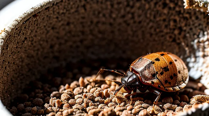
The Macabre Reality of Crushed Bed Bugs
Initial Appearance: A Bloody Smudge
Size and Shape of the Stain
When a bedbug is compressed, the resulting residue forms a distinct mark on the surface. The mark’s dimensions typically range from 2 mm to 5 mm in diameter, reflecting the insect’s body length of roughly 4 mm to 5 mm. Lengthwise, the imprint may appear slightly elongated, measuring up to 6 mm, because the abdomen can spread under pressure.
Key characteristics of the imprint:
- Shape: irregular oval with a central dark core and a lighter peripheral halo.
- Edge definition: blurred, often with fine radiating lines caused by fluid dispersion.
- Color: deep reddish‑brown at the center, fading to pale yellow toward the margins as hemolymph mixes with surrounding debris.
Surface texture influences the exact outline. Smooth, non‑porous surfaces retain sharper edges, while textured fabrics produce a more diffuse pattern. The stain’s size and form provide reliable visual cues for identifying recent bedbug activity.
Color Variation Based on Feeding Status
When a bedbug is flattened, the exoskeleton becomes translucent, revealing the internal tissues. The overall silhouette remains oval, with the head, thorax, and abdomen compressed into a single, flattened mass.
Color differences correspond to the insect’s recent feeding activity:
- Unfed individuals display a uniform pale‑tan or light brown hue; crushing exposes a whitish hemolymph that lacks strong pigmentation.
- Recently fed specimens exhibit a darker, reddish‑brown abdomen; crushing releases hemolymph tinged with a deep orange‑red color, highlighting the presence of digested blood.
- Long‑term post‑prandial bugs may show a mottled pattern of dark brown and black zones, reflecting partial digestion and the accumulation of waste pigments.
These variations allow rapid visual assessment of feeding status from a crushed sample.
Internal Contents Exposed
Digested Blood: The Primary Component
When a bedbug is flattened, the dominant visual element is the ingested blood that fills the abdominal cavity. This substance emerges as a dense, reddish‑brown mass, often spreading outward from the crushed exoskeleton.
The blood‑derived material exhibits several distinct characteristics:
- Color: deep rust hue, ranging from dark crimson to brownish‑black as oxidation progresses.
- Consistency: semi‑fluid, thick enough to cling to cuticle fragments yet capable of seeping onto surrounding surfaces.
- Distribution: concentrates around the ventral side where the gut resides, creating a noticeable stain that contrasts with the pale exoskeleton.
The presence of this digested fluid accounts for the majority of the residue observed after crushing, masking finer anatomical details and providing the most immediate indication of the insect’s recent feeding activity.
Undigested Blood: Fresher Meals
A crushed bedbug presents a flattened exoskeleton with a translucent, reddish hue. The abdomen ruptures, releasing a pool of liquid that retains the coloration of a recent blood meal. The fluid appears glossy and uncoagulated, indicating that digestion has not progressed far beyond ingestion.
Undigested blood within the crushed specimen exhibits several distinct characteristics:
- Coloration matches fresh human or animal blood, ranging from bright scarlet to deep crimson.
- Viscosity remains low, allowing it to spread thinly across the ruptured surface.
- Lack of hemolysis signs; cells retain their original shape, confirming minimal enzymatic breakdown.
- Surface tension creates a smooth, reflective film that contrasts with the matte texture of the insect’s cuticle.
These observations confirm that the liquid released from a flattened bedbug is essentially a recent, unprocessed meal, offering a visual cue of the insect’s feeding status at the moment of impact.
Bodily Fluids and Hemolymph
When a bed bug is subjected to crushing pressure, its exoskeleton ruptures and releases internal contents. The primary liquid that escapes is hemolymph, the insect equivalent of blood. Hemolymph appears as a translucent to slightly amber fluid, often tinged with a faint reddish hue due to the presence of hemocyanin or pigment cells. The fluid spreads rapidly across the surface, creating a glossy sheen that highlights the flattened body.
Additional bodily fluids, such as digestive residues, may be observed. These residues are typically darker, ranging from brown to black, reflecting the insect’s recent blood meals. The mixture of hemolymph and digestive waste can produce a mottled appearance, with contrasting light and dark patches on the crushed specimen.
Key visual indicators of the released substances:
- Clear to amber hemolymph, occasionally exhibiting a pinkish tint
- Dark brown or black digestive material
- Shiny, wet surface coating the flattened exoskeleton
The combined effect results in a visibly moist, discolored outline that distinguishes a crushed bed bug from an intact one.
Residual Evidence and Identification
Exoskeletal Fragments and Debris
When a bedbug is subjected to crushing pressure, the protective outer shell disintegrates into a collection of hard, translucent pieces and organic residue. The exoskeleton, composed primarily of chitin, fractures along predetermined weak points, producing shards that retain the original curvature of the insect’s dorsal surface. These fragments are typically amber‑to‑brown in hue, reflecting the natural pigmentation of the cuticle.
The debris includes:
- Small, irregularly shaped chitinous fragments, ranging from a fraction of a millimeter to several millimeters in length.
- Minute splinters of the thoracic shield, often visible as glossy, semi‑transparent plates.
- Residual abdominal membrane, which appears as thin, parchment‑like sheets.
- Traces of digested blood, manifesting as reddish or rust‑colored stains adhering to the broken pieces.
The overall appearance resembles a scattered mosaic of brittle plates interspersed with dark, wet spots where hemolymph has leaked from ruptured internal vessels. The fragments retain a glossy sheen, while the blood stains gradually darken as oxidation proceeds.
Antennas, Legs, and Other Appendages
A crushed Cimex lectularius loses its delicate sensory structures. The antennae, normally slender and segmented, become compressed into short, dark fragments that often remain attached to the body’s flattened mass. Leg segments, originally thin and jointed, collapse into irregular, flattened shards that may appear as faint, pale lines against the surrounding exoskeleton. Additional appendages—such as the mouthparts, cerci, and any residual setae—are reduced to indistinct, mottled smears. The overall impression is a flattened, reddish‑brown silhouette with scattered, broken remnants of the original appendages.
- Antennae: compressed, darkened, may retain partial segmentation.
- Legs: flattened, jagged pieces, often faintly visible as light streaks.
- Other appendages (mouthparts, cerci, setae): reduced to irregular, mottled remnants.
Potential for DNA Analysis
Crushed bedbugs leave a flattened, darkened exoskeleton with visible internal contents. The remnants contain sufficient cellular material for molecular extraction, making the specimen a viable source for genetic profiling. DNA integrity remains largely intact despite mechanical disruption, allowing standard polymerase chain reaction (PCR) protocols to amplify target regions such as mitochondrial COI or nuclear ribosomal genes.
Key considerations for successful analysis include:
- Prompt preservation of the crushed sample in ethanol or a silica‑gel desiccant to prevent nucleic acid degradation.
- Removal of external contaminants by rinsing with sterile buffer before tissue lysis.
- Utilization of bead‑beating or enzymatic digestion to release DNA from the compacted cuticle.
- Application of quantitative PCR to assess copy number and detect possible inhibitors introduced during crushing.
Forensic laboratories exploit these attributes to confirm species identification, trace infestation sources, and link specimens to crime scenes. The ability to recover reliable genetic data from a flattened bug expands investigative capabilities beyond visual assessment alone.
Factors Influencing the Crushing Outcome
Force Applied and Surface Type
When a bed‑bug is subjected to compression, the visual outcome depends primarily on the magnitude of force and the characteristics of the surface receiving the impact. Greater force produces more extensive rupture of the exoskeleton, exposing internal tissues and resulting in a flattened, mottled appearance. Lower force may leave the body partially intact, with the dorsal shield still recognizable but distorted.
Key variables influencing the post‑crush morphology:
- Force intensity – low, moderate, high; each tier correlates with increasing deformation and loss of structural integrity.
- Surface hardness – soft (fabric, carpet), medium (wood, laminate), hard (glass, metal); harder surfaces transmit force more efficiently, accelerating exoskeletal failure.
- Surface texture – smooth versus rough; rough textures introduce additional shear, producing irregular tear patterns and fragmented remnants.
On a soft, pliable substrate, even moderate pressure can spread the bug’s body, yielding a spread‑out, gelatinous smear. On a hard, smooth surface, high pressure concentrates the impact, creating a crisp, flattened silhouette with visible internal organs leaking onto the surface. The combination of force level and surface type determines whether the crushed specimen appears as a cohesive, flattened outline or a disintegrated mass of fragmented parts.
Bed Bug's Life Stage: Nymph vs. Adult
When a bed bug is compressed, the exposed body reveals the insect’s cuticle, hemolymph and any residual egg material. The observable characteristics differ markedly between immature individuals and fully developed ones.
The primary distinctions are:
- Size: crushed nymphs measure between 1 mm and 3 mm in length; adults range from 4 mm to 7 mm, producing a larger splatter of fluid.
- Color of hemolymph: nymphal hemolymph appears pale amber, while adult hemolymph is a deeper reddish‑brown due to higher concentration of pigments.
- Cuticle integrity: the exoskeleton of a nymph is thinner, resulting in a more fluid‑rich smear; the adult’s thicker cuticle yields a firmer, less dispersed residue.
- Presence of egg remnants: only nymphal crushes may contain fragments of the embryonic membrane, visible as translucent specks within the spill.
These factors enable identification of the life stage from the aftermath of a crush, assisting in accurate assessment of infestation severity.
Time Since Last Feeding
A crushed bedbug reveals physical changes that correlate with the interval since its last blood meal. Freshly fed insects retain a swollen abdomen filled with blood, which appears as a dark, glossy mass when the exoskeleton is broken. The surrounding cuticle often shows a reddish tint from the ingested hemoglobin.
As time passes without feeding, the abdomen gradually contracts. After 24–48 hours, the blood begins to digest, producing a duller, brownish coloration and a softer consistency. By the end of a week, the abdomen shrinks further, the internal contents become pale and gelatinous, and the crushed remains display a mottled, almost translucent appearance.
Key visual indicators linked to feeding interval:
- 0–12 hours: pronounced bulge, deep red‑black core, shiny surface.
- 12–48 hours: reduced bulge, brownish hue, semi‑fluid interior.
- 48 hours–7 days: minimal bulge, pale gelatinous mass, matte exterior.
- Beyond 7 days: small, dry remnants, faint coloration, brittle texture.
These characteristics allow accurate estimation of the last feeding period from a single crushed specimen.
Implications for Pest Control
Identifying the Problem: A Visual Cue
Confirming an Infestation Through Crushed Bugs
Crushed bed bugs provide unmistakable visual evidence of an infestation. The flattened insect appears as a dark, oval silhouette roughly 4–5 mm long. The exoskeleton collapses, exposing a glossy, reddish‑brown interior where blood‑filled gut contents are visible. The head, antennae, and legs become indistinct, leaving a smooth, mottled surface that contrasts sharply with surrounding debris.
Key diagnostic features observable in a squashed specimen:
- Dark, flattened body outline matching the size of an adult bed bug.
- Shiny, reddish interior indicating recent blood meals.
- Absence of distinct appendages, replaced by a uniform smear.
- Presence of small, dark spots that correspond to the insect’s eyes and mouthparts before compression.
When these characteristics are identified on furniture, bedding, or floor surfaces, they confirm the presence of the pest. Collecting multiple crushed examples strengthens the diagnosis, allowing targeted treatment decisions.
Differentiating from Other Pests' Remains
Crushed bedbug remains appear as a flattened, reddish‑brown mass. The exoskeleton fragments retain the characteristic oval shape and visible dorsal segmentation. Antennae and leg stubs often remain attached, giving a mottled texture.
Differentiation from other pest remains relies on several observable traits:
- «Bedbug»: oval, 4–5 mm length before crushing; after compression, retains segmented outline and dark reddish hue; antennae and leg remnants are distinct.
- «Cockroach»: larger (10–30 mm), glossy dark brown to black exoskeleton; wing pads or full wings may be visible; crushing yields a more uniform, shiny smear without clear segmentation.
- «Flea»: elongated, 2–4 mm, deep black coloration; after crushing, body collapses into a smooth, featureless dark spot; leg fragments are short and less defined.
- «Tick»: rounded, 3–5 mm, often engorged with blood; crushing produces a glossy, reddish‑orange mass with a smooth surface; leg and mouthpart remnants are minimal.
- «Mite»: microscopic (≤ 1 mm), translucent or pale; crushing results in a barely visible residue, lacking distinct body parts or coloration.
Key identifiers include size range, color intensity, presence of segmented outlines, and visibility of antennae or leg fragments. These factors enable reliable separation of crushed bedbug remains from those of other common household pests.
Cleaning and Sanitation Considerations
Removing Stains and Biological Residues
Crushed bedbugs leave dark reddish‑brown macules that spread outward from the point of impact, often accompanied by a thin oily sheen and fragmented cuticle fragments. The macules contain hemolymph, which oxidizes quickly, creating a deep rust‑colored stain that adheres to fibers and hard surfaces.
Effective removal follows a three‑stage protocol:
- Blot the fresh macule with a clean, absorbent cloth to eliminate excess fluid; avoid rubbing, which embeds particles deeper.
- Apply a mild detergent solution (approximately 0.5 % surfactant) to the affected area, allowing a brief dwell time of 5–10 minutes before gentle agitation with a soft brush.
- Rinse thoroughly with lukewarm water; for persistent «stain» or «biological residue», introduce an enzymatic cleaner containing protease and lipase, following manufacturer‑specified concentration and contact time.
Precautions include wearing disposable gloves, testing cleaners on an inconspicuous fabric segment, and ensuring complete drying to prevent mold growth. Heat treatment (≥ 60 °C) on washable items further degrades residual proteins, eliminating potential allergenic effects.
Preventing Secondary Infestations
Crushing a bedbug releases internal fluids that can contaminate surrounding surfaces. Those fluids contain viable eggs and live insects, creating a risk of secondary infestation if not managed promptly.
Immediate actions reduce spread:
- Place the crushed specimen in a sealed plastic bag; dispose of the bag in an outdoor trash container.
- Vacuum the area thoroughly, focusing on cracks, seams, and mattress edges; empty the vacuum canister into a sealed bag and discard it.
- Wash all bedding, clothing, and nearby fabrics at a minimum temperature of 60 °C; dry on high heat for at least 30 minutes.
- Apply a residual insecticide to the treated zone, following label instructions; repeat application after 7 days to target emerging survivors.
- Inspect adjacent rooms and furniture for additional signs; repeat cleaning and treatment steps where evidence appears.
Continual monitoring involves weekly visual checks for live bugs or fresh stains. Documentation of findings supports timely escalation to professional pest‑control services if recurrence persists.
Psychological Impact on Occupants
The Unpleasantness of Crushed Pests
Crushed bedbugs reveal a flattened, mottled exoskeleton tinged with dark brown to black coloration. The pressure splits the dorsal shield, exposing a glossy interior filled with hemolymph that spreads rapidly across the surface.
The aftermath presents several unpleasant qualities:
- Viscous, reddish‑brown fluid that stains fabrics and hard surfaces.
- A faint, musty odor produced by the release of defensive chemicals.
- Microscopic fragments that can become airborne, irritating respiratory passages.
Contact with the residual fluid may trigger skin irritation or allergic reactions in sensitive individuals. The presence of crushed insects also signals a larger infestation, increasing the likelihood of further bites and potential disease transmission. Prompt removal and thorough cleaning are essential to mitigate these risks.
Reinforcing the Need for Eradication
A crushed bed bug appears as a flattened, darkened body with a ruptured exoskeleton. The abdomen often spreads into a reddish‑brown smear, while the head and legs become indistinct. Blood remnants may pool around the collapsed form, creating a glossy patch on the surface.
Visible remnants serve as undeniable proof of an active infestation. Their presence confirms that the insects have accessed sleeping areas, increasing the risk of allergic reactions, secondary skin infections, and psychological distress. Immediate response prevents population growth and limits exposure to pathogens.
Key actions to eliminate the problem:
- Conduct thorough inspections of bedding, seams, and furniture.
- Engage licensed pest‑control professionals for targeted treatment.
- Apply heat or steam to eradicate hidden stages.
- Seal cracks and install protective mattress encasements.
- Maintain regular laundering of linens at high temperatures.
Prompt eradication eliminates the source of the described remnants, restores sanitary conditions, and averts further health complications. «The flattened insect reveals the urgency of decisive intervention».
