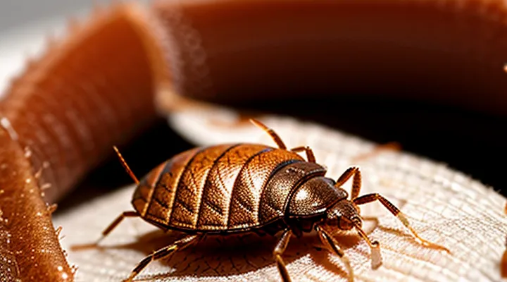
Initial Appearance
Immediate Reactions
When a bedbug penetrates the skin, the first visible sign is a small, raised spot that often appears as a red or pink papule. The lesion typically measures 2–5 mm in diameter and may develop a halo of lighter skin around its edge.
- Intense itching begins within minutes to a few hours after the bite.
- Swelling can accompany the papule, producing a slightly raised, firm bump.
- A thin, clear fluid may leak from the site if the skin is scratched.
- In some individuals, the reaction is delayed, and the bite remains unnoticed for up to 24 hours.
The initial response is mediated by the insect’s saliva, which contains anticoagulants and anesthetic compounds. These substances trigger histamine release, leading to the characteristic redness, swelling, and pruritus. Immediate scratching can exacerbate inflammation and increase the risk of secondary infection. Prompt washing of the area with mild soap and cool water can reduce irritation and limit the severity of the reaction.
Delayed Onset
Bedbug (Cimex lectularius) bites frequently exhibit a latency period before visible signs appear. The skin reaction often emerges 12–48 hours after the feed, sometimes extending to several days, which can mislead patients into attributing the lesions to other sources.
When the delayed response becomes evident, the lesions typically present as small, raised, erythematous papules. The central area may be slightly pale or exhibit a faint wheal, surrounded by a halo of redness that expands outward over time. In many cases, the rash remains confined to a few millimeters in diameter; however, pruritus can intensify as the inflammation progresses.
The onset timeline varies:
- 12–24 hours: faint redness, mild itching.
- 24–48 hours: pronounced papule, increased swelling, stronger itch.
- 48 hours–7 days: possible secondary inflammation, occasional vesicle formation.
Factors that influence the delay include:
- Individual hypersensitivity to bedbug saliva.
- Bite location (thin skin shows symptoms sooner).
- Number of bites delivered during a feeding episode.
- Prior exposure, which may sensitize or desensitize the host.
Delayed manifestation distinguishes bedbug bites from immediate reactions caused by mosquitoes or fleas, which typically appear within minutes. The characteristic linear or clustered arrangement of lesions, combined with the described latency, assists clinicians in recognizing bedbug infestations despite the postponed skin response.
Distinguishing Bed Bug Bites from Other Conditions
Mosquito Bites
Bedbug bites typically appear as small, red, raised welts that often group in linear or clustered patterns. The lesions may develop a central puncture point and can become itchy or swollen within a few hours. In many cases the reaction intensifies over 24–48 hours, leaving a darker, sometimes crusted spot that can persist for several days.
Mosquito bites share some visual features with those of bedbugs but differ in distribution and morphology. Common characteristics of mosquito-induced lesions include:
- A single, round, raised bump about 2–5 mm in diameter.
- A clear, pale halo surrounding the central red spot, giving a target‑like appearance.
- Pronounced itching that usually peaks within a few minutes and subsides within a day.
- Isolation of lesions; bites are rarely found in straight lines or tight clusters.
Key distinctions between the two types of bites are:
- Arrangement – bedbug bites often form rows or clusters, while mosquito bites appear singly.
- Size – mosquito welts are generally smaller than the larger, more inflamed bedbug lesions.
- Duration of discoloration – bedbug marks may darken and linger longer than the transient redness of mosquito bites.
Understanding these visual cues assists in accurate identification of the culprit insect and informs appropriate treatment decisions.
Flea Bites
Fleas leave tiny, pinpoint‑shaped lesions that turn pink or red within minutes of feeding. The puncture is usually less than 2 mm in diameter, surrounded by a thin halo of inflammation. Intense pruritus appears rapidly, often prompting a single raised bump or a small cluster of bumps when several insects bite the same area. Occasionally, a central punctum is visible where the flea’s mouthparts penetrated the epidermis. The reaction may persist for several days, with the erythema fading to a lighter pink before disappearing.
Bed‑bug bites differ in size and pattern. They are typically 3–5 mm, slightly raised, and may develop a dark center surrounded by a larger, erythematous halo. The lesions often appear in linear or zig‑zag formations, reflecting the insect’s movement along a host’s skin. The itching is moderate to severe, and secondary inflammation may develop if the bite is scratched.
Key points for distinguishing flea bites from bed‑bug bites:
- Size: flea ≤2 mm; bed‑bug 3–5 mm.
- Shape: flea punctate, often single; bed‑bug round with central dark spot.
- Distribution: flea isolated or small cluster; bed‑bug line or group.
- Onset of itching: flea immediate; bed‑bug within hours.
Accurate identification assists clinicians in selecting appropriate treatment and in advising patients on environmental control measures.
Allergic Reactions
Bedbug bites generally appear as small, red, raised punctures that may cluster in a line or a zig‑zag pattern. In individuals with heightened sensitivity, the skin’s response can extend beyond the basic lesion.
- pronounced swelling around the bite
- intense, persistent itching
- formation of larger, flat‑topped welts (hives)
- occasional blistering or secondary infection from scratching
The allergic component may manifest within minutes of the bite or develop several hours later, lasting from a few days to a week. Early signs include rapid enlargement of the initial redness and a spreading rash that does not conform to the typical linear arrangement.
Treatment focuses on controlling the immune response. Oral antihistamines reduce itching and swelling; topical corticosteroid creams limit inflammation. If symptoms progress to severe swelling, difficulty breathing, or widespread hives, immediate medical evaluation is required. Persistent or recurrent reactions warrant allergy testing to confirm bedbug sensitivity and guide long‑term prevention strategies.
Common Locations of Bites
Exposed Skin Areas
Bedbugs preferentially target skin that is uncovered during sleep, because the insects locate heat and carbon‑dioxide emissions without the barrier of clothing. The most frequently affected regions are:
- Face, especially around the eyes, cheekbones, and jawline.
- Neck and upper chest, where shirts are often rolled up or absent.
- Arms, particularly forearms and wrists that lie on pillow edges.
- Hands and fingers that rest on bedding.
- Legs and ankles when shorts or skirts are worn.
On these exposed areas, bites appear as small, raised papules 2–5 mm in diameter. The central point may be slightly erythematous, surrounded by a pale halo that expands as the reaction develops. In many cases, a linear or clustered pattern emerges, reflecting the bedbug’s feeding behavior of probing multiple nearby sites. The lesions often become itchy within a few hours, and the surrounding skin may swell modestly, especially on thinner, more vascular regions such as the eyelids or the inner forearm.
Patterns of Bites
Bedbug bites manifest as tiny, red, raised welts that may itch or swell. The spatial arrangement of the lesions helps distinguish them from other insect bites.
- Linear rows of 2‑5 puncta, often aligned along a straight line or slight curve, reflecting the insect’s feeding path.
- Clustered groups of 3‑6 spots, tightly packed, indicating multiple probes in a confined area.
- “Zig‑zag” formations where bites alternate direction, suggesting the bug’s movement across the skin.
- Isolated single puncta, less common, appearing when only one feeding event occurs.
The lesions typically develop within 24 hours, reaching peak redness after 48 hours. Uniform size, smooth edges, and the absence of a central puncture mark differentiate them from mosquito or flea bites.
Symptoms Accompanying Bed Bug Bites
Itching and Discomfort
Bedbug bites typically produce small, red welts that become intensely itchy within a few hours. The itching results from the insect’s saliva, which contains anticoagulants and anesthetic agents that trigger an inflammatory response. This reaction causes histamine release, leading to a burning sensation and persistent urge to scratch.
The discomfort may evolve as follows:
- Initial phase (0‑4 hours): Red papules appear, often clustered in a linear or zig‑zag pattern. Mild swelling accompanies the itch.
- Peak phase (12‑24 hours): Intensity of itch increases; lesions may enlarge and become raised.
- Resolution phase (2‑7 days): Swelling subsides, but itching can persist, especially if the skin is scratched, risking secondary infection.
Repeated scratching can break the skin barrier, introducing bacteria and prolonging irritation. Antihistamines, topical corticosteroids, and cold compresses reduce histamine activity and alleviate the sensation. Maintaining clean bedding and applying insect‑proof covers prevent further bites and limit cumulative discomfort.
Swelling and Redness
Bedbug bites typically produce a localized reaction characterized by swelling and redness. The affected area expands within minutes to a few hours, forming a raised, firm papule that may reach a diameter of 3–5 mm. The surrounding skin often turns pink to deep red, reflecting increased blood flow and inflammation.
Key features of the swelling and redness include:
- Rapid onset – the lesion becomes noticeable shortly after the bite, unlike some arthropod bites that develop more slowly.
- Symmetrical pattern – multiple bites frequently appear in a linear or clustered arrangement, each with similar size and coloration.
- Persistent erythema – the red hue may persist for several days, gradually fading as the body resolves the inflammatory response.
- Variable intensity – swelling can be mild in some individuals and pronounced in others, depending on personal sensitivity and prior exposure.
The swelling may be accompanied by a clear or slightly cloudy fluid accumulation under the skin, giving the papule a slightly translucent appearance. In contrast to mosquito bites, which often present as a single, isolated bump, bedbug bites commonly involve several adjacent lesions, each exhibiting the described swelling and redness.
Resolution typically occurs within one to two weeks without medical intervention, though residual discoloration can linger longer. Persistent or worsening swelling may indicate secondary infection and warrants clinical assessment.
Potential Secondary Infections
Bedbug bites create small, red, raised lesions that often appear in clusters of three or more. When the skin is repeatedly scratched, the disrupted epidermis provides an entry point for bacteria normally present on the surface or in the environment. This secondary colonization can develop into a localized infection that may progress rapidly if left untreated.
Typical signs of bacterial involvement include:
- Increased redness extending beyond the original bite margin
- Warmth and swelling of the surrounding tissue
- Purulent discharge or crust formation
- Pain that intensifies rather than diminishes
- Systemic symptoms such as fever or malaise in severe cases
Common pathogens associated with these complications are Staphylococcus aureus and Streptococcus pyogenes. Individuals with compromised immunity, diabetes, or peripheral vascular disease are at higher risk for deeper tissue infection and delayed healing. Prompt cleansing of the area with mild antiseptic, avoidance of further trauma, and early medical evaluation for antibiotics are essential measures to prevent escalation.
Factors Influencing Bite Appearance
Individual Skin Sensitivity
Bedbug bites manifest as small, raised lesions that may appear red, pink, or flesh‑colored. The exact presentation depends largely on a person’s cutaneous reactivity. Individuals with heightened sensitivity often develop pronounced erythema, swelling, and intense pruritus within a few hours of the bite. Those with low sensitivity may notice only faint discoloration or a barely perceptible spot that resolves quickly.
Key factors influencing the visual outcome include:
- Immune response strength – strong histamine release produces larger, more inflamed welts; weak response yields minimal changes.
- Skin thickness – thinner epidermis allows deeper vascular exposure, resulting in brighter red marks.
- Age – children and the elderly typically exhibit more noticeable reactions due to immature or compromised immune regulation.
- Medications – antihistamines or immunosuppressants can blunt typical swelling and redness.
Typical patterns observed across sensitivity levels:
- High sensitivity – clusters of 2–5 mm papules, vivid red, surrounded by a halo of swelling; may coalesce into larger plaques.
- Moderate sensitivity – isolated papules, pink to light red, moderate itching, limited peripheral edema.
- Low sensitivity – single, flat macules, faint pink or tan, negligible itching, rapid fading.
Understanding these variations helps clinicians differentiate bedbug bites from other arthropod reactions and guides appropriate management based on the patient’s cutaneous profile.
Number of Bites
Bedbug infestations usually produce several bites rather than a single lesion. The count of bites depends on the severity of the infestation, the duration of exposure, and the individual’s sleep habits.
Typical patterns include:
- Clustered groups: three to five bites appearing close together, often in a line or “breakfast‑lunch‑dinner” arrangement.
- Multiple clusters: several groups of three to five bites spread across exposed skin, such as the arms, neck, or face.
- Isolated bites: occasional single lesions when only one insect feeds before being disturbed.
A light infestation may result in only one or two bites per night, while heavy infestations can generate dozens of lesions over several nights. The total number of bites often correlates with the length of time the host remains exposed to active insects.
Time Since Bite
Bedbug bites evolve noticeably as the hours and days pass after the insect feeds. The progression provides clues for identifying the source of an eruption.
- First few hours: A small, raised red spot appears at the feeding site. The center may be a pinpoint puncture, surrounded by a faint halo of erythema. It often feels itchy or mildly painful.
- 12–24 hours: The lesion expands slightly, becoming a larger, more pronounced welt. The surrounding skin may turn darker red, and swelling can increase. Intense itching is common during this interval.
- 48–72 hours: Some bites develop a central blister or a shallow ulcer, especially in individuals with heightened sensitivity. The surrounding redness may peak, and the area can become warm to the touch.
- 4–7 days: The inflammatory response begins to subside. Redness fades, while the bite may leave a pink or brown macule. Persistent itching may continue, and secondary bacterial infection can appear if the skin is scratched excessively.
- 1–2 weeks: Most lesions resolve completely, leaving only a faint post‑inflammatory hyperpigmentation that disappears over several weeks. Rarely, a scar may remain if the bite was severely irritated.
Variations depend on personal immunity, skin type, and the number of bites clustered together. Rapid onset of intense swelling or prolonged ulceration may indicate an allergic reaction and warrants medical evaluation. Understanding the temporal pattern helps differentiate bedbug bites from other arthropod or allergic skin lesions.
When to Seek Medical Attention
Severe Allergic Reactions
Bedbug bites normally present as small, red, raised spots, often grouped in a line or cluster. In most individuals the reaction is mild and resolves within a few days.
In a minority of cases the immune response escalates to a severe allergic reaction. Characteristics include:
- Large, intensely pruritic welts that exceed the size of a typical bite.
- Marked swelling that spreads beyond the immediate puncture area.
- Erythema with a deep, violaceous hue.
- Blister formation or necrotic centers in extreme cases.
- Systemic signs such as hives, difficulty breathing, rapid heartbeat, or low blood pressure.
These manifestations may appear within minutes to several hours after exposure and can persist for weeks if untreated. Immediate medical evaluation is warranted when any of the following occur: widespread edema, respiratory distress, dizziness, or rapid progression of skin lesions. Antihistamines, topical corticosteroids, and, in severe cases, epinephrine are standard interventions to mitigate the reaction and prevent complications.
Signs of Infection
Bedbug bites typically appear as small, red, raised spots that may develop a darker center. When a bite becomes infected, the surrounding skin exhibits additional, clinically significant changes.
- Redness that expands beyond the immediate bite area, forming an ill‑defined, erythematous halo.
- Swelling that increases in size, feels firm to the touch, and may be accompanied by a sensation of heat.
- Presence of pus or a yellowish discharge, indicating bacterial colonisation.
- Development of a painful, tender nodule that does not resolve within a few days.
- Fever, chills, or malaise, suggesting systemic involvement.
- Enlarged, tender lymph nodes near the bite, reflecting regional immune response.
If any of these signs appear, medical evaluation is warranted to prevent complications such as cellulitis or abscess formation. Prompt treatment may involve topical or oral antibiotics, wound cleaning, and monitoring for worsening systemic symptoms.
Persistent Symptoms
Bedbug bites typically manifest as small, red papules that may develop a central punctum. While the initial reaction often subsides within a few days, certain symptoms can persist and require attention.
Persistent itching is the most common lasting complaint. Repeated scratching can exacerbate inflammation and lead to secondary bacterial infection, indicated by increasing warmth, pus formation, or spreading erythema. Even after the bite heals, some individuals experience lingering hyperpigmentation or discoloration that may last weeks or months, especially on darker skin tones.
Other enduring effects include:
- Localized swelling that remains for several days beyond the acute phase.
- Persistent tenderness or a dull ache at the bite site.
- Delayed hypersensitivity reactions, presenting as larger, raised welts that resist standard antihistamine treatment.
- Post‑inflammatory scarring when the skin barrier is compromised by intense scratching.
When symptoms do not resolve within two weeks, or if signs of infection appear, medical evaluation is advised. Prompt treatment with topical antibiotics, corticosteroids, or oral antihistamines can mitigate complications and reduce the duration of lingering discomfort.
