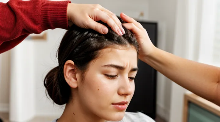
«Understanding Head Lice and Their Bites»
«The Nature of Head Lice Infestations»
«Life Cycle of Head Lice»
Head lice (Pediculus humanus capitis) progress through three distinct stages: egg, nymph, and adult. Eggs, called nits, are attached firmly to hair shafts near the scalp, typically within a half‑inch of the skin. After about seven days, each nit hatches into a nymph, which resembles a miniature adult but lacks fully developed reproductive organs. Nymphs mature through three molts over a period of nine to twelve days, emerging as fertile adults capable of laying up to eight eggs per day.
Adult lice survive on the host for approximately three weeks. They feed several times daily, inserting a short, painless proboscis into the skin to draw blood. Repeated feeding on the neck region produces small, red, flat or slightly raised spots. These lesions often appear in clusters where the hairline meets the skin, reflecting the lice’s preference for warm, protected areas.
Key points of the life cycle:
- Egg (nit): 5–7 days; firmly glued to hair, near scalp.
- Nymph: 3–4 days per molt; three molts total.
- Adult: 9–12 days to maturity; up to three weeks of activity.
Understanding each phase clarifies why bite marks are most noticeable on the neck: mature lice concentrate feeding there after establishing a population, and the skin’s thinness makes the inflammatory response more visible. Effective control targets all stages, eliminating both the visible bites and the underlying infestation.
«How Lice Spread»
Lice bites on the neck typically appear as tiny, red, raised spots that may itch or develop a slight swelling. The lesions often form in clusters where the insect has repeatedly fed, and they may be surrounded by a faint halo of irritation.
The spread of head lice follows predictable pathways that determine where bites are likely to occur. Key mechanisms include:
- Direct head‑to‑head contact, the most efficient route for transfer of live insects.
- Sharing of personal items such as hats, hairbrushes, headphones, or scarves that have come into contact with an infested scalp.
- Contact with contaminated surfaces, for example, pillowcases, helmets, or upholstered furniture, where lice can survive briefly before relocating to a new host.
Once transferred, lice crawl to the hairline and scalp, where they lay eggs. As the insects move across the scalp, they may reach the neck region, especially if hair is long or if the host frequently brushes or scratches the area. Repeated feeding in this zone produces the characteristic bite pattern described above.
Preventing the spread requires interrupting these pathways: avoid sharing headgear, maintain personal items separate, and wash or disinfect bedding and clothing at temperatures of at least 130 °F (54 °C). Regular inspection of the hairline and neck can catch early infestations before bites become widespread.
«Identifying Lice Bites on the Neck»
«Visual Characteristics of Lice Bites»
«Common Appearance and Size»
Lice bites on the neck usually appear as tiny, reddish papules. The lesions are round to slightly oval, with a well‑defined border and a central punctum where the insect’s mouthparts penetrated the skin. Typical diameter ranges from 1 mm to 3 mm; occasional larger reactions may reach 5 mm, especially in individuals with heightened sensitivity.
- Color: pink to bright red, may darken to a purplish hue if inflammation intensifies.
- Surface: smooth, sometimes slightly raised; a faint wheal may be felt.
- Itchiness: moderate to strong pruritus, often prompting scratching.
- Distribution: clustered in a linear or irregular pattern along the hairline and posterior neck region.
- Duration: lesions persist 3–7 days, fading as the bite heals and inflammation resolves.
«Coloration and Texture Changes»
Lice bites on the neck typically appear as small, raised spots that differ in color and surface feel from the surrounding skin. The initial reaction is often a pink or reddish hue caused by localized inflammation. Within a few hours, the area may darken to a purplish tone as blood vessels constrict and hemoglobin breaks down. In some cases, the bite may turn a brownish shade during the healing phase, indicating the presence of hemosiderin deposits.
Texture changes accompany the color shift. Fresh bites feel firm and slightly raised, resembling a tiny papule. As the inflammatory response subsides, the raised edge softens and the center may become flat or develop a shallow crater. Prolonged scratching can lead to a rough, scaly surface or even a crusted lesion, signaling secondary irritation or infection.
Key observations:
- Early stage: Pink‑red, firm papule; clear borders.
- Mid stage (12–24 hours): Purplish or brownish discoloration; edges begin to soften.
- Late stage (48 hours+): Flattened or cratered center; possible scaling or crust formation if irritated.
Monitoring these coloration and texture patterns helps differentiate lice bites from other dermatological conditions and guides appropriate treatment.
«Distinguishing Lice Bites from Other Conditions»
«Comparison with Allergic Reactions»
Lice bites on the neck typically appear as small, red papules clustered in a line or irregular pattern. The lesions are often pinpoint, measuring 1–2 mm, and may develop a tiny punctum at the center where the insect’s mouthparts penetrated the skin. Swelling is usually mild, and the surrounding tissue remains relatively flat. Itching develops within hours and can intensify over the next day.
Allergic skin reactions, such as those caused by contact allergens or insect venom, share some visual features but differ in key aspects.
- Size: allergic hives (urticaria) commonly exceed 5 mm and can enlarge rapidly, whereas lice bites stay confined to a few millimeters.
- Distribution: allergic eruptions often spread widely across the body in a symmetric fashion; lice bites concentrate on hair‑bearing areas, especially the neck, scalp, and behind the ears.
- Shape: allergic lesions are typically raised, edematous wheals with well‑defined borders; lice bites remain flat or slightly raised papules with a central punctum.
- Duration: allergic swelling may persist for several hours to days before fading, while lice bite inflammation usually subsides within 24–48 hours if the infestation is removed.
When evaluating a neck rash, note the presence of a linear or grouped arrangement, the tiny central puncture, and limited size. These characteristics favor a parasitic bite over an allergic response. Absence of widespread, raised wheals and rapid resolution further supports the diagnosis of lice infestation.
«Differentiation from Other Insect Bites»
Lice bites on the neck appear as small, red papules, typically 1–2 mm in diameter. The lesions are often grouped in linear or clustered patterns that follow the hairline, and they may be surrounded by a faint halo of erythema. Intense pruritus develops within hours and can persist for several days. Unlike many insect bites, lice bites rarely produce a central punctum or a raised welt that swells noticeably.
Key distinguishing features compared with other common arthropod bites:
- Size and shape – Lice bites are uniformly tiny and dome‑shaped; mosquito bites are larger, raised, and often form a single raised bump.
- Distribution – Lice concentrate on the neck and scalp, creating rows or patches that align with hair shafts; flea bites appear as scattered spots on exposed skin such as ankles or legs.
- Timing of itch – Lice irritation begins quickly after the bite, whereas tick bites may remain painless for several hours before a delayed reaction.
- Associated signs – Presence of nits or live lice on hair confirms infestation; other insects leave no such evidence.
- Duration of lesions – Lice lesions resolve within a week with proper treatment; spider bites can linger longer and may develop necrotic centers.
Recognition of these criteria enables accurate identification of neck lesions caused by lice and prevents misdiagnosis with mosquito, flea, or tick bites.
«Exclusion of Skin Rashes and Infections»
Lice bites on the neck appear as small, red papules, often grouped in clusters of two to three. The lesions are pruritic and may develop a central punctum where the insect’s mouthparts penetrated the skin. Swelling is minimal; surrounding tissue remains flat, and the color does not progress beyond a light to moderate erythema.
To rule out other dermatological conditions, consider the following distinguishing factors:
- Absence of vesicles or pustules: Viral exanthems and bacterial infections typically produce fluid‑filled lesions or purulent collections, which are not seen with lice bites.
- Lack of spreading pattern: Contact dermatitis and fungal infections often spread outward from the point of contact, whereas lice bites remain localized near hair shafts.
- No systemic symptoms: Fever, malaise, or lymphadenopathy accompany many infections; these signs are absent in isolated lice bites.
- Negative laboratory tests: Cultures or rapid antigen assays return negative when lice are the sole cause.
Clinical evaluation should include a thorough inspection of hair and scalp for live lice or nits, as their presence confirms the diagnosis. If the described lesions meet the criteria above and no alternative etiology is identified, the condition can be classified as lice‑induced dermal irritation, with other rashes and infections effectively excluded.
«Symptoms Associated with Bites»
«Itching and Irritation Levels»
Lice bites on the neck typically produce a localized, red papule surrounded by a thin halo of inflammation. The primary symptom is a sharp, persistent itch that intensifies several hours after the bite and may persist for up to 48 hours. Intensity varies with individual skin sensitivity, but most reactions fall within a moderate range: the skin feels tender to the touch, and scratching can lead to secondary erythema or tiny blisters.
Key factors influencing irritation levels:
- Individual hypersensitivity – people with atopic dermatitis or allergic tendencies experience stronger itching and larger wheals.
- Number of bites – clusters of bites increase cumulative irritation, often creating a line or “scratch” pattern along the neck.
- Duration of exposure – prolonged contact with lice prolongs the inflammatory response, extending the itch cycle.
- Secondary infection – excessive scratching can introduce bacteria, converting a simple bite into a painful, inflamed lesion.
Management focuses on reducing itch and preventing infection. Topical antihistamines or corticosteroid creams lower histamine‑mediated inflammation within 15–30 minutes. Oral antihistamines provide systemic relief for severe cases. Keeping the area clean, applying cool compresses, and avoiding further scratching limit tissue damage and accelerate healing. Most lesions resolve without scarring within three to five days if proper care is maintained.
«Secondary Skin Reactions»
Lice bites on the neck commonly produce a small, red papule surrounded by a halo of erythema. The initial lesion may be pruritic and develop within minutes to a few hours after contact. Repeated scratching can trigger secondary skin reactions that extend beyond the primary bite site.
Typical secondary responses include:
- Erythematous wheals (urticaria) appearing days after the initial bite
- Vesicular eruptions that may coalesce into larger blisters
- Eczematous patches with dry, scaly surfaces
- Excoriated papules or crusted lesions caused by mechanical trauma
- Localized bacterial infection, presenting as pustules, purulent drainage, and increased warmth
- Post‑inflammatory hyperpigmentation or hypopigmentation persisting for weeks to months
- Hypertrophic scarring in cases of deep tissue damage or prolonged inflammation
These reactions often intensify the visual impact of the original bite, creating a pattern of clustered, inflamed spots that may spread across the cervical region if scratching continues. Prompt topical anti‑inflammatory or antiseptic treatment can limit progression and reduce residual discoloration.
«Potential for Scratch Marks and Sores»
Lice bites on the neck appear as small, red papules, often clustered near the hairline. The skin may feel itchy, prompting repeated scratching. When scratching persists, the following changes can develop:
- Linear or irregular scratch marks that overlay the original bite lesions.
- Crusty scabs formed from broken skin.
- Erosions that enlarge the initial papule, sometimes exposing underlying tissue.
- Secondary bacterial infection, indicated by increased redness, warmth, swelling, or purulent discharge.
These secondary lesions may persist longer than the primary bite reaction and can lead to noticeable scarring if left untreated. Prompt hygiene measures, such as washing the affected area with antiseptic soap and applying topical antibiotic ointment, reduce the risk of progression from simple bites to lasting sores.
«Areas on the Neck Prone to Bites»
«Hairline and Nape Involvement»
Lice bites on the neck typically present as small, red papules clustered around the hairline and the nape. The lesions are often pruritic and may develop a central punctum where the insect’s mouthparts penetrated the skin. Swelling is usually limited to a few millimeters, and the surrounding skin remains intact.
- Hairline: Bites concentrate at the front edge of the scalp, following the natural line of hair growth. Lesions appear in a linear or irregular pattern, matching the direction of hair shafts. Redness may be more pronounced where hair tension pulls the skin taut.
- Nape: Bites form on the lower back of the neck, often in groups of two to four. The area may show slight edema, and scratching can cause secondary irritation or crusting. The nape’s skin is thinner, so discoloration can be more visible.
The overall appearance includes raised, erythematous spots, occasional tiny vesicles, and a persistent itch that intensifies after exposure to heat or sweating. Absence of pus or necrosis distinguishes these bites from bacterial infections.
«Behind the Ears and Upper Neck»
Lice bites located behind the ears and on the upper neck appear as tiny, red, raised spots. Each spot typically measures 1–3 mm in diameter and may show a central puncture point where the insect’s mouthparts pierced the skin. The lesions are often grouped in clusters or arranged in short linear rows that follow the hairline. Intense itching accompanies the lesions, and scratching can cause the spots to become swollen or develop a darker, bruised center.
Typical visual cues include:
- Uniform redness with a well‑defined edge
- Central pinpoint or tiny white dot indicating the bite entry
- Slight elevation above surrounding skin
- Grouping of two to six lesions in close proximity
- Absence of a surrounding halo, which differentiates them from mosquito bites
In contrast, allergic reactions to other insects may produce larger, more diffuse welts, while contact dermatitis often shows a spreading rash with clear borders. Recognizing the specific pattern behind the ears and on the upper neck helps differentiate lice bites from other dermatological conditions.
«When to Suspect Lice and Seek Confirmation»
«Recognizing Other Signs of Lice»
«Presence of Nits and Live Lice»
Lice bites on the neck appear as small, red, pruritic papules that may develop a central punctum where the insect pierced the skin. The lesions are often grouped closely together, reflecting the feeding pattern of the insect. When nits and live lice are present, the visual clues become more definitive.
- Nits: oval, whitish‑cream structures firmly attached to the hair shaft, usually 1–2 mm from the scalp. Their smooth surface distinguishes them from dandruff; they do not detach easily when combed.
- Live lice: translucent to brownish bodies about 2–4 mm long, with six legs. Active movement can be observed when the hair is examined under good lighting. They tend to cluster near the bite sites, especially on the neck and behind the ears.
- Bite marks: erythematous, raised bumps, sometimes with a tiny central dot. The surrounding skin may show slight swelling or a halo of irritation. Repeated scratching can cause secondary erythema or crusting.
The coexistence of nits adhered to hair and mobile lice on the scalp confirms an infestation and explains the characteristic neck lesions. Absence of these indicators suggests alternative causes for similar skin changes.
«Movement Sensation»
Lice bites on the neck appear as small, red punctate lesions, often grouped in a linear or irregular pattern. The surface may be slightly raised, with a central point of discoloration surrounded by a faint halo. The lesions are typically less than 2 mm in diameter and may develop a tiny crust if scratched.
The primary sensory cue associated with these bites is a distinct movement sensation. Victims report a subtle, intermittent crawling feeling that seems to travel across the skin. This sensation precedes or accompanies the itch, creating an impression of live organisms moving beneath the epidermis. The perception is localized to the bite site and may extend a few millimeters beyond the visible marks.
Key aspects of the movement sensation:
- Tingling or prickling that emerges shortly after the bite occurs.
- A fleeting sensation of tiny organisms shifting direction.
- Amplified awareness when the skin is warm or moist.
- Diminished intensity after the bite area becomes inflamed or scabbed.
Understanding the visual characteristics and the accompanying movement perception aids in distinguishing lice bites from other dermal irritations.
«Importance of Professional Diagnosis»
Lice bites on the neck typically appear as small, red papules clustered near the hairline. The lesions may be slightly raised, sometimes surrounded by a faint halo of irritation, and can itch intensely. In some cases, a tiny puncture mark is visible at the center of each papule, indicating where the insect pierced the skin. The pattern of bites often follows the distribution of hair, distinguishing them from other dermatological conditions.
Professional assessment is essential for several reasons:
- Accurate identification distinguishes lice bites from allergic reactions, fungal infections, or other parasitic infestations.
- Laboratory confirmation (e.g., microscopic examination of lice or eggs) eliminates uncertainty and guides appropriate treatment.
- Prescription‑strength topical or oral agents may be required, and a clinician can select the most effective medication based on resistance patterns.
- A health‑care provider can evaluate secondary complications such as bacterial superinfection and recommend wound care or antibiotics if needed.
- Counseling on environmental decontamination and preventive measures reduces recurrence and limits spread within households or communal settings.
Relying on self‑diagnosis risks misinterpretation of symptoms, delayed therapy, and unnecessary exposure to ineffective over‑the‑counter products. A qualified practitioner ensures a definitive diagnosis and a targeted management plan.
