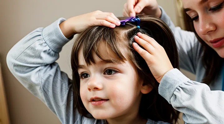
Understanding Head Lice
What Are Lice?
The Appearance of Adult Lice
Adult head lice are small, wing‑less insects measuring 2–3 mm in length. Their bodies are flattened laterally, allowing them to move easily through hair shafts. The head is broader than the thorax, giving a teardrop silhouette when viewed from the side. Color varies from pale gray to brown, becoming darker after a blood meal. Antennae are short and segmented, ending in a sensory tip. Legs are six, each ending in sharp claws that grasp hair strands tightly; the claws are visible as tiny, curved hooks at the end of the legs.
The abdomen consists of three visible segments covered by fine, translucent scales that give a slightly glossy appearance under magnification. The ventral side shows a pair of small, pale lateral plates. When engorged with blood, the abdomen expands and appears reddish or blackish, making the louse appear larger and more opaque.
Key visual cues for identifying an adult louse include:
- Size around 2–3 mm, visible to the naked eye as a moving speck.
- Oval, elongated body with a wider head.
- Darkening after feeding.
- Six legs equipped with clawed tarsi that cling to hair.
- Absence of wings or visible wings pads.
The Life Cycle of Lice
Lice develop through a predictable sequence of stages that can be observed directly on a child’s scalp. The cycle begins with the egg, commonly called a nit, which is firmly attached to a hair shaft near the scalp. Nits appear as tiny, oval, creamy‑white or yellowish bodies, often mistaken for dandruff; their size ranges from 0.5 to 1 mm. Because they are glued in place, they remain immobile until hatching.
- Egg (nit): 7–10 days to hatch; attached at a 45° angle to the hair; translucent to white in color.
- Nymph: Newly emerged insects are called nymphs; they are pale, translucent, and measure about 1 mm. Nymphs feed immediately and undergo three molts.
- Adult: After three molts, lasting roughly 9–12 days, the insect reaches adulthood. Adult lice are about 2–3 mm long, gray‑brown, and visible as moving specks on the scalp or hair.
The entire life cycle—from egg to reproductive adult—takes approximately 2–3 weeks under normal temperature and humidity. Adult females lay 5–10 eggs per day, depositing them close to the scalp where warmth accelerates development. Continuous reproduction means that, without intervention, a visible infestation can persist and expand rapidly.
Understanding each stage’s visual characteristics enables early detection. Nits are the most stationary and easiest to spot as a crusty coating at the base of hair shafts. Nymphs and adults are mobile; they may be seen crawling slowly or moving between hairs. Prompt identification of these forms is essential for effective treatment and prevention of further spread.
What Are Nits?
Distinguishing Nits from Dandruff
Nits are the eggs of head lice. They appear as tiny, oval bodies about 0.8 mm long. The shell is usually off‑white, tan, or yellow‑brown and has a slightly translucent quality. Nits are glued firmly to the hair shaft, typically within a half‑inch of the scalp. Because of the adhesive, they do not move when the hair is brushed and they remain attached even after a wash.
Dandruff consists of dead skin flakes that shed from the scalp. Flakes are usually larger, irregularly shaped, and range from white to gray. They are loosely attached to hair and scalp, so a gentle comb or a single brush stroke can dislodge them. Dandruff flakes are crumbly, break apart easily, and often appear in clusters rather than as isolated, single items.
Key visual distinctions:
- Attachment: Nits are cemented to each hair strand; dandruff slides off.
- Shape and size: Nits are uniform, oval, and <1 mm; dandruff is irregular and often larger.
- Color: Nits may show a slight yellowish hue; dandruff is typically white or gray.
- Texture: Nits feel hard and smooth; dandruff feels soft and flaky.
- Location: Nits cluster close to the scalp; dandruff can be found throughout the hair and on the shoulders.
To confirm the presence of nits, use a fine‑tooth nit comb on dry hair and examine each captured specimen under adequate lighting. If the material remains attached to the comb and displays the described characteristics, it is likely a nit rather than dandruff.
Nits vs. Empty Egg Shells
Nits are the eggs of head‑lice, firmly glued to the hair shaft close to the scalp. They appear as oval, ivory‑white or yellowish bodies about 0.8 mm long. The surface is smooth and reflective; the shell is opaque, and the interior may show a tiny dark spot where the embryo develops. Nits remain attached for 7–10 days until the hatchling emerges.
Empty egg shells are the remnants left after the nymph hatches. They retain the same oval shape and size but become translucent and slightly crinkled. The shell loses its opacity, allowing light to pass through, and the dark spot disappears. Because the shell no longer adheres strongly, it may shift or fall off the hair.
Key visual cues for differentiation:
- Color: live nits – opaque white/yellow; empty shells – translucent.
- Surface texture: nits – smooth, glossy; shells – dull, often with a subtle crease.
- Attachment: nits – tightly glued near the scalp; shells – loosely attached, may slide down the shaft.
- Movement: nits – remain stationary when hair is brushed; shells – may move or detach.
Accurate identification prevents unnecessary treatment and ensures appropriate response to a confirmed infestation.
How to Identify Lice and Nits
Visual Inspection Techniques
Tools for Examination
To identify the tiny insects and their eggs on a child’s scalp, specific examination instruments are required. A fine‑tooth louse comb, typically with 0.2‑mm spacing, separates lice and nits from hair shafts when drawn from the root toward the tip. A magnifying lens of 2‑3× power enlarges the visual field, allowing distinction between live insects, translucent eggs, and debris. Portable LED headlamps provide uniform illumination, reducing shadows that can conceal specimens. Handheld digital microscopes, offering up to 200× magnification, capture detailed images for documentation or remote consultation. Disposable nitrile gloves protect the examiner from potential allergic reactions and maintain hygiene. A clean, flat surface such as a white towel or tray serves as a background for collecting combed material, facilitating inspection and counting. Each tool enhances accuracy, speed, and safety during the assessment of lice infestations and their attached nits.
Areas to Focus On
Lice infestations on children present specific visual cues that require systematic observation. Accurate identification depends on concentrating on several key aspects of the head and hair.
- Adult lice: body length 2–4 mm, translucent to gray‑brown, six legs visible when the insect is alive; rapid, erratic movement across hair shafts.
- Nits (eggs): oval shape, 0.8 mm long, color ranging from white to yellow‑brown; firmly attached to the base of the hair shaft at a 45‑degree angle, resistant to sliding when a comb is run through the hair.
- Attachment sites: most common near the scalp on the nape, behind the ears, and at the crown; nits are often clustered in these regions because they require warmth for development.
- Hair type considerations: thick or curly hair can conceal nits, while fine hair may allow easier visibility of both lice and eggs.
- Timing of inspection: conduct examinations in daylight and after a warm shower, when lice are less active and nits are more apparent.
- Differentiation from debris: dandruff flakes are easily removable and lack a firm attachment; nits do not detach with a gentle pull, whereas hair particles do.
- Diagnostic tools: use a fine‑toothed lice comb on a well‑lit surface; magnification (hand lens or smartphone camera) improves detection of small, translucent nits.
- Signs of recent activity: presence of live lice, fresh nits (lighter color), and irritated scalp lesions indicate an ongoing infestation.
Focusing on these areas enables precise recognition of lice and their eggs, facilitating prompt treatment and preventing further spread.
Common Misidentifications
Dandruff
Dandruff appears as small, white to gray flakes that detach easily from the scalp and fall onto hair and shoulders. The flakes are dry, often irregularly shaped, and do not cling to hair shafts. Unlike lice or their eggs, dandruff does not move, does not cause itching that intensifies after scratching, and does not leave behind a visible brown or black residue.
Key visual differences between dandruff and head‑lice infestations:
- Dandruff: loose, powdery flakes; no attached particles; scalp may show mild scaling but no live insects.
- Lice: live insects about the size of sesame seeds, visible moving across hair; may be seen crawling.
- Nits: oval, yellow‑brown or white cemented eggs attached firmly to the hair shaft, often within ¼ inch of the scalp; cannot be brushed away easily.
When examining a child’s head, the presence of freely falling flakes without attached bodies indicates flaking rather than an infestation. Persistent itching, visible insects, or cemented eggs require separate assessment.
Hair Casts
Hair casts are thin, tube‑like sheaths that encircle the hair shaft. They are composed of keratin debris and remain firmly attached to the hair, sliding down as the hair grows. Unlike living parasites, casts contain no moving parts and are not attached to the scalp.
Adult lice appear as gray‑brown insects about 2–3 mm long. Their bodies are flattened, with six legs and a visible head. Nits are oval, tan‑white structures measuring 0.8 mm, cemented to the hair close to the scalp. Both lice and nits are typically found near the hairline, behind the ears, and at the nape of the neck.
Hair casts can be confused with nits because of their similar size and location. The following points differentiate them:
- Attachment: Casts encircle the entire hair shaft and can be moved up or down the hair; nits are glued to the hair surface and cannot be displaced without breaking the cement.
- Shape: Casts are cylindrical and translucent; nits are oval, opaque, and often display a pointed end where the embryo develops.
- Mobility: Casts slide freely when the hair is brushed; nits remain fixed even after vigorous combing.
- Surface texture: Casts have a smooth, silky feel; nits feel rough and may show a white, chalky coating.
When examining a child’s scalp, isolate a single hair and gently slide it between the fingertips. If the structure moves along the shaft without resistance, it is a cast. If it stays attached despite manipulation, it is a nit. Accurate identification prevents unnecessary treatment and focuses appropriate interventions on actual infestations.
Product Residue
Product residue left on a child’s hair can alter the visual cues used to identify head‑lice infestations. Residues from shampoos, conditioners, styling gels, or leave‑in treatments often create a glossy or sticky film that obscures the tiny, translucent bodies of lice and the oval, brownish‑gray shells of nits attached to hair shafts.
Live lice appear as 2–4 mm, wingless insects with a flattened body and six legs; they move quickly when the scalp is disturbed. Nits are firmly cemented to the hair near the scalp, measuring 0.8 mm in length, with a smooth, slightly curved surface that darkens as the embryo develops.
When a coating of product remains on the hair, the following effects occur:
- Light reflection from the film can make nits appear lighter, blending them with the hair shaft.
- Viscous residues can trap debris, producing specks that mimic nits in size and color.
- The film may immobilize live lice temporarily, reducing their movement and making detection more difficult.
To eliminate residue before an inspection, follow these steps:
- Rinse the child’s hair with warm water for at least one minute to loosen surface film.
- Apply a clarifying shampoo formulated to break down silicone and oil‑based ingredients; lather thoroughly from scalp to tips.
- Rinse completely, ensuring no suds remain.
- Condition only the lengths, avoiding the scalp to prevent re‑deposition of residue.
- Dry hair with a clean towel; avoid applying additional styling products until after the examination.
Removing product residue restores the natural texture of hair and scalp, allowing the characteristic size, shape, and attachment points of lice and nits to be observed accurately.
Signs and Symptoms of Infestation
Behavioral Cues
Excessive Scratching
Lice and their eggs (nits) are visible as tiny, gray‑white oval or teardrop shapes attached firmly to hair shafts near the scalp. Adult lice appear as six‑mm, crab‑shaped insects, reddish‑brown when unfed and darker after a blood meal. Excessive scratching often accompanies an infestation and creates additional diagnostic clues.
- Red, irritated skin on the neck, ears, and forehead indicates repeated trauma from biting insects.
- Crusty or scabbed patches suggest prolonged scratching, which can obscure nits but also reveal their locations when hair is pulled apart.
- Small blood spots on the hair or pillowcase result from lice bites and are frequently uncovered during vigorous scratching.
Frequent scratching may also cause secondary infection, characterized by swelling, pus, or foul odor. Identifying these signs alongside the visual appearance of lice and nits enables accurate assessment and timely treatment.
Irritability
Irritability often signals an active infestation of head lice. The discomfort caused by crawling insects and the itching from their bites triggers a child’s mood to shift quickly from calm to restless. Parents may notice sudden crying, difficulty concentrating on tasks, and frequent requests to scratch the scalp. These behaviors typically intensify after a period of unnoticed exposure, because the sensory irritation accumulates as more lice and nits become established.
Visible signs of the parasites reinforce the irritability response. Adult lice appear as small, tan‑brown insects about the size of a sesame seed, moving quickly across hair shafts. Nits, the oval eggs, cling firmly to the base of each strand and resemble tiny, white or yellowish specks. When a child’s scalp is examined, the combination of moving insects and attached nits creates a constant tactile stimulus that fuels agitation.
Key indicators of irritability linked to a lice infestation include:
- Frequent, unexplained crying episodes
- Persistent head scratching despite clean hair
- Resistance to bedtime or other routine activities
- Reduced attention span during school or play
- Complaints of a “tingling” or “crawling” sensation on the scalp
Assessing these behaviors alongside a careful visual inspection helps differentiate lice‑related irritability from other causes, such as allergies or stress. Prompt identification of the insects and their eggs allows timely treatment, which typically reduces the sensory irritation and restores the child’s usual temperament.
Physical Manifestations
Rash or Sores
Lice and their eggs present a distinct visual pattern that differs from dermatological lesions such as rashes or sores. Live lice appear as small, tan‑brown insects about the size of a sesame seed, moving quickly across hair shafts. Nits are oval, firmly attached to the base of a strand, measuring roughly 1 mm in length and resembling translucent or whitish specks. In contrast, a rash manifests as clusters of erythematous patches, while sores appear as localized breaks in the skin surface, often with surrounding inflammation.
Key distinguishing characteristics:
- Mobility – Lice crawl; rash and sores remain static.
- Attachment – Nits are glued to hair; rashes and sores are not attached to hair fibers.
- Color and texture – Lice are pigmented and three‑dimensional; nits are smooth, glossy, and may appear grayish. Rashes are flat, reddish, and may be raised. Sores exhibit ulcerated or crusted tissue.
- Location – Lice concentrate near the scalp, behind ears, and at the nape; rashes can occur anywhere on the skin, and sores typically develop in areas of irritation or trauma.
- Response to treatment – Pediculicidal products eliminate live insects and nits; topical corticosteroids or antiseptics reduce rash or sore symptoms but do not affect lice.
Accurate visual assessment, combined with these criteria, enables practitioners and caregivers to differentiate parasitic infestation from cutaneous conditions, ensuring appropriate intervention.
Swollen Lymph Nodes
Swollen lymph nodes are enlarged, often tender, masses of lymphatic tissue that can be felt in the neck, behind the ears, and under the jaw. They appear when the immune system reacts to a local or systemic stimulus.
Lice infestation presents as small, grayish insects moving quickly through hair and as oval, whitish‑brown eggs (nits) firmly attached to hair shafts near the scalp. The presence of these parasites can trigger an inflammatory response, causing nearby lymph nodes to enlarge as immune cells accumulate.
Typical characteristics of lymph node enlargement linked to a head‑lice problem include:
- Size up to 1 cm in diameter, sometimes larger if infection is severe
- Mild tenderness when pressed
- Mobility, allowing the node to shift under the skin
- Development within a few days of noticing live lice or nits
Factors that help differentiate lice‑related swelling from other causes:
- Recent observation of live insects or attached eggs on the scalp
- Absence of fever, sore throat, or respiratory symptoms that accompany viral infections
- Lack of purulent discharge or skin ulceration, which suggests bacterial involvement
- Nodes that remain localized to the cervical region rather than being widespread
Management steps:
- Perform a thorough scalp examination to confirm the presence of lice and nits
- Apply an appropriate pediculicide and remove nits with a fine‑toothed comb
- Re‑examine the neck after 7‑10 days; if nodes persist, increase in size, become markedly painful, or are accompanied by systemic signs, seek pediatric evaluation
- Document node size and tenderness to assist healthcare providers in assessing the need for further investigation or treatment.
