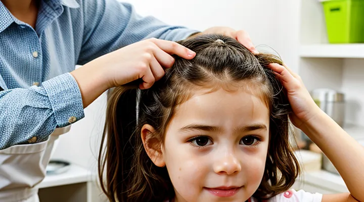
The Primary Diet of Head Lice
The Essential Nutrient: Blood
Source of Blood Meal: The Human Scalp
Head lice (Pediculus humanus capitis) are obligate ectoparasites that survive exclusively by ingesting human blood. The sole source of their nourishment is the vascular network of the scalp, accessed through the skin surface beneath hair shafts.
Feeding occurs as follows:
- The louse inserts its specialized mouthparts into a capillary near the epidermal surface.
- Saliva containing anticoagulants is released to prevent clotting.
- Blood is drawn in small volumes, typically 0.5–1 µL per feeding episode.
- Each adult louse feeds multiple times per day, while nymphs feed less frequently but follow the same mechanism.
The scalp provides a constant, warm environment rich in capillaries, enabling rapid replenishment of the louse’s blood supply. Absence of this specific host tissue halts development and leads to mortality.
Frequency and Volume of Feeding
Head lice survive by extracting blood from the scalp. An adult louse initiates a feeding episode roughly every three to four hours, which translates to five to six meals within a 24‑hour period. Nymphs follow a similar schedule, though younger stages may feed slightly less frequently because of smaller metabolic demands.
During each bout, the insect inserts its proboscis into a hair follicle and ingests a minute quantity of blood. The volume per feed ranges from 0.5 to 1.0 microliters, sufficient to sustain the louse’s rapid growth and egg production. Over the course of a day, an adult consumes approximately 3 to 6 microliters of blood, a fraction of the host’s total scalp blood supply but enough to cause noticeable irritation.
Key points:
- Feeding interval: 3–4 hours per episode.
- Daily frequency: 5–6 feeds per adult; slightly fewer for nymphs.
- Volume per feed: 0.5–1.0 µL.
- Total daily intake: 3–6 µL for an adult.
These parameters explain why infestations can develop quickly: frequent, low‑volume meals enable rapid population expansion without depleting the host’s resources.
The Mechanism of Feeding
Anatomy of the Louse Mouthparts
Piercing Stylets
Piercing stylets are the specialized mouthparts head lice use to obtain nutrients from their host. Each louse possesses a pair of elongated, needle‑like structures that penetrate the scalp skin to reach capillary blood vessels. The stylets are composed of a hardened cuticular sheath surrounding a flexible inner tube, allowing precise insertion and withdrawal without causing extensive tissue damage.
Functionally, the stylets perform three coordinated actions:
- Insertion: Muscular contraction drives the stylets through the epidermis and into the dermal layer where blood vessels lie.
- Suction: A negative pressure generated by the louse’s pharyngeal pump draws blood up the internal tube.
- Repositioning: Rapid retraction and re‑insertion enable the parasite to locate fresh feeding sites as the host’s skin thickens around previous wounds.
Morphological adaptations include serrated tips that facilitate entry through keratinized tissue and sensory receptors that detect temperature and chemical cues indicating blood flow. These features collectively enable head lice to sustain themselves exclusively on human blood throughout their life cycle.
Salivary Glands and Anticoagulants
Head lice obtain nourishment exclusively from human blood. The insect inserts its mandibles into the scalp, pierces a capillary, and draws fluid directly into its alimentary canal. Blood intake provides proteins, lipids, and carbohydrates required for development and reproduction.
During the piercing process, the louse injects saliva that contains a cocktail of anticoagulant substances. These compounds prevent clot formation at the wound site, ensuring uninterrupted flow of blood. Salivary glands located in the thorax synthesize and store the anticoagulants, releasing them with each bite.
Key anticoagulant agents identified in head‑lice saliva include:
- Apyrase, which hydrolyzes ADP and ATP, reducing platelet aggregation.
- Antithrombin‑like proteins that bind and inactivate thrombin.
- Serine protease inhibitors that interfere with the coagulation cascade.
- Histamine‑binding proteins that modulate host inflammatory responses.
The presence of these molecules extends the bleeding period, allowing the louse to ingest larger volumes of blood before the wound seals. Consequently, the efficiency of blood acquisition depends directly on the functionality of the salivary gland secretions.
The Feeding Process
Locating a Capillary
Head lice obtain nourishment by piercing the scalp’s epidermis and sucking blood from the underlying capillary network. Accurate identification of the specific capillary accessed by a louse is essential for understanding feeding behavior, assessing infestation severity, and developing targeted treatments.
Microscopic examination of plucked hair shafts reveals a minute puncture site surrounded by a halo of hemoglobin residue. High‑resolution optical microscopy, combined with differential interference contrast, highlights the translucent vessel wall and the louse’s stylet insertion point. Dermoscopy, using a polarized light source at 10–20 × magnification, visualizes the feeding channel as a linear dark line extending from the hair follicle to a superficial capillary. Transillumination of the scalp, achieved with a fiber‑optic light probe, produces a shadow pattern that pinpoints the vessel’s location beneath the louse’s attachment site.
Practical steps for locating the feeding capillary:
- Clean the scalp area with alcohol to remove debris.
- Apply a drop of saline to the hair shaft to enhance optical clarity.
- Use a dermatoscope to scan the region, focusing on the louse’s head and mouthparts.
- Record the position of the visible puncture relative to the follicle margin.
- Confirm the vessel’s depth with ultrasonography if needed for research purposes.
These methods provide reproducible data on the exact blood source exploited by head lice, facilitating precise epidemiological studies and informing the design of interventions that disrupt feeding pathways.
Ingesting Blood
Head lice survive by extracting human blood from the scalp. Their mouthparts form a specialized piercing‑suction apparatus that penetrates the epidermis to reach capillaries. Once the cuticle is breached, the louse draws a small volume of plasma and red blood cells, providing the proteins, iron, and nutrients required for growth and reproduction.
- Average intake per feeding: 0.5–1.0 µL of blood.
- Feeding frequency: every 4–6 hours during daylight.
- Duration of each meal: 5–10 minutes before the louse retreats to the hair shaft.
Blood ingestion supports the lice life cycle. Nymphs require multiple meals to molt, while adult females need a steady supply to produce up to 8 eggs per day. The constant removal of blood can cause scalp irritation, itching, and secondary bacterial infection if the skin is broken. Detection often relies on visual identification of live insects or nits attached near the base of hair shafts.
Consequences and Related Factors
Impact of Feeding on the Host
Itching and Irritation
Head lice survive by ingesting small amounts of human blood through their piercing mouthparts. During each feeding session, the insect injects saliva that contains anticoagulant compounds, which provoke a localized immune response in the scalp.
The immune reaction releases histamine, producing an intense pruritic sensation. Repeated bites amplify the sensation, leading to persistent scratching.
Typical manifestations of the irritation include:
- Red, inflamed patches surrounding bite sites
- Swollen papules or pustules caused by the inflammatory response
- Secondary bacterial infection when skin integrity is compromised by scratching
Continuous exposure to these symptoms can exacerbate discomfort and may require medical intervention to control both the infestation and the associated dermatological effects.
Allergic Reactions
Head lice obtain nutrients by piercing the scalp and ingesting small amounts of blood. The bite introduces saliva that contains proteins capable of triggering the body’s immune response. In some individuals, this exposure leads to allergic reactions ranging from mild irritation to more pronounced dermatitis.
Typical manifestations include:
- Red, inflamed patches around the bite sites
- Intense itching that may persist for several days
- Swelling or papular eruptions that can coalesce into larger lesions
- Secondary bacterial infection if scratching breaches the skin barrier
The allergic response originates from IgE‑mediated hypersensitivity to louse saliva. Repeated bites can sensitize the immune system, increasing reaction severity over time. Diagnosis relies on clinical observation of characteristic scalp lesions coupled with a history of infestation.
Management strategies focus on eliminating the parasites and mitigating inflammation:
- Apply approved pediculicidal agents to eradicate lice and nits.
- Use topical corticosteroids or antihistamine creams to reduce itching and swelling.
- For extensive reactions, oral antihistamines or short courses of systemic steroids may be prescribed.
- Maintain scalp hygiene, avoid sharing personal items, and conduct regular inspections to prevent re‑infestation.
Understanding the link between louse feeding and immune activation enables targeted treatment, limits discomfort, and reduces the risk of complications.
Survival Without a Blood Meal
Environmental Factors
Head lice survive by extracting blood from the human scalp; however, their feeding activity varies with external conditions that affect both the parasite and its host.
- Ambient temperature above 20 °C accelerates lice metabolism, prompting more frequent bites.
- Relative humidity between 50 % and 70 % prevents desiccation, allowing lice to remain active longer between meals.
- Hair length and density create a micro‑environment that retains heat and moisture, facilitating sustained feeding.
- Seasonal shifts that alter indoor climate—warmer summer months or heated winter interiors—modify lice feeding cycles.
- Crowded living spaces increase host contact rates, raising the likelihood of lice transfer and feeding opportunities.
- Frequent use of shampoos or conditioners containing insecticidal agents can suppress feeding by irritating lice.
- Exposure to strong light or ultraviolet radiation reduces lice activity, limiting blood intake.
These environmental parameters collectively shape the frequency and efficiency of head‑lice blood consumption.
Duration of Starvation
Head lice (Pediculus humanus capitis) obtain nutrients exclusively from human blood; without this source they enter a state of starvation. Laboratory observations show that adult lice survive up to 48 hours without a blood meal when kept at ambient room temperature (20‑22 °C) and relative humidity above 50 %. Under cooler conditions (10‑15 °C) survival extends to approximately 72 hours, while exposure to temperatures above 30 °C reduces the starvation limit to 24 hours. Nymphal stages are less tolerant, typically lasting 12‑24 hours without feeding.
- Adults at 22 °C, 55 % RH: 48 hours maximum
- Adults at 12 °C, 70 % RH: 72 hours maximum
- Adults at 33 °C, 40 % RH: 24 hours maximum
- Nymphs (any condition): 12‑24 hours maximum
These survival thresholds influence control strategies: rapid removal of infested individuals and maintenance of environmental conditions unfavorable to lice prolongation of starvation can significantly reduce population persistence. Understanding the limited duration of starvation clarifies why prompt treatment is essential for effective eradication.
