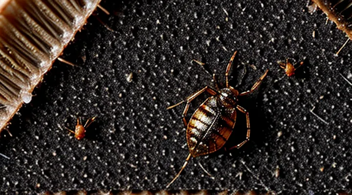
The Unseen Clues of Bed Bug Infestations
Understanding Bed Bug Fecal Marks
Why Fecal Marks are Important
Bed bug fecal marks serve as a primary visual cue for confirming an infestation. Their dark, elongated spots, often found on bedding, walls, and furniture, differ from other insect droppings by size and color, allowing accurate species identification without laboratory analysis.
Key reasons for their significance:
- Detection – Visible stains reveal the presence of hidden insects, enabling early intervention before populations expand.
- Mapping – Concentration patterns indicate where bugs hide, guiding targeted pesticide application and reducing unnecessary chemical use.
- Progress monitoring – Changes in the number and distribution of marks over time provide measurable feedback on treatment effectiveness.
- Legal documentation – Photographic records of fecal stains support tenant‑landlord disputes and insurance claims, establishing objective proof of infestation.
Because fecal deposits persist after the insects have moved, they remain reliable evidence even when live bugs are not observed. Accurate interpretation of these marks shortens response time, improves control strategies, and prevents further spread.
Location of Fecal Spots
Bedbug fecal spots appear as tiny, dark deposits that resemble pepper specks or a grain of rice. Their location provides reliable evidence of an infestation.
Inspect the following areas for these marks:
- Mattress and box‑spring seams, especially where stitching is visible.
- Headboard, footboard, and bed frame joints.
- Upholstered furniture, including sofa cushions and chair armrests.
- Cracks and crevices in wall baseboards, floorboards, and around electrical outlets.
- Behind picture frames, mirrors, and wall hangings.
- Luggage compartments, suitcase seams, and travel bags.
- Underneath or behind nightstands, dressers, and other bedroom furniture.
- Pet bedding and cages placed near sleeping areas.
Spotting clusters of black‑brown specks in these locations confirms the presence of bedbug excrement and warrants immediate pest‑control measures.
Differentiating from Other Stains
Bed‑bug fecal spots are tiny, dark‑brown to black specks, typically 0.5–2 mm in diameter. They appear as a linear trail when insects feed in a confined area, often along mattress seams, headboards, or behind baseboards. The deposits are dry, powdery, and may smudge into a reddish‑brown stain if they become saturated with blood from a recent bite.
Key visual and contextual cues that separate these marks from other household stains include:
- Color and texture – Bed‑bug droppings are uniformly dark and granular; mold spores are fuzzy, greenish or black, while dust is lighter and loosely attached.
- Location – Concentrations are found near hiding places: mattress folds, furniture crevices, and wall cracks. Pet urine stains spread over larger areas and emit a distinct ammonia odor.
- Pattern – Linear or clustered arrangements suggest insect movement; random splatters are typical of spilled liquids or food stains.
- Odor – Fresh fecal spots may emit a faint, sweet, musty scent; chemical cleaners or mildew produce stronger, recognizable smells.
- Associated evidence – Presence of shed exoskeletons, live insects, or blood‑tinged spots reinforces the diagnosis; none of these accompany ordinary dirt or water stains.
When uncertainty remains, a simple test can confirm identity: dampen a cotton swab with isopropyl alcohol and rub a suspect speck. Bed‑bug feces dissolve readily, leaving a faint brown residue, whereas mold or paint particles retain their structure. Microscopic examination reveals characteristic digested blood cells in confirmed droppings.
Identifying Bed Bug Feces
Visual Characteristics
Color and Appearance
Bedbug feces are typically tiny, dark‑colored spots that resemble specks of ink or pepper. The pigment ranges from deep brown to almost black, depending on the insect’s recent blood meal; after a full feeding, the waste may appear slightly lighter, showing a reddish‑brown hue.
The deposits are granular and dry, measuring less than a millimeter across. They often accumulate in cracks, seams, mattress tags, and along baseboards. When disturbed, the particles may smudge, leaving a faint, wet streak that quickly dries and darkens again.
- Color: dark brown to black; occasional reddish‑brown after recent feeding.
- Form: minute, dry granules, less than 1 mm in size.
- Distribution: seams, folds, cracks, and edges of furniture.
- Reaction to pressure: smears into a temporary wet line that reverts to dry specks.
Texture and Consistency
Bed bug feces consist of digested human blood that has been dehydrated during excretion. The material appears as minute, dark particles that settle on bedding, mattress seams, or walls.
- Color: deep brown to black, sometimes with a reddish tint from recent meals.
- Size: 0.5–2 mm in length, comparable to coarse sand grains.
- Shape: irregular, often angular, lacking a uniform silhouette.
- Surface: matte, non‑shiny, with a slightly powdery feel when touched.
When fresh, the droplets may retain a thin film of moisture, giving the deposits a tacky quality that hardens within minutes. As they age, the moisture evaporates, leaving a dry, crumbly consistency that can be brushed away easily. The transition from moist to dry is rapid; within an hour, the excrement usually becomes a fine, sand‑like residue.
Size and Shape
Bed bug feces are microscopic to the naked eye, typically measuring 0.5–1 mm in length. The deposits are solid, not liquid, and their shape varies slightly depending on the feeding state of the insect.
- Dimensions: roughly the size of a pinhead; length rarely exceeds 1 mm, width about 0.2–0.3 mm.
- Form: generally oval or slightly elongated; when multiple droplets coalesce, they may appear as irregular specks.
- Color: dark brown to black, resembling pepper grains or tiny ink stains.
These characteristics enable reliable identification on mattress seams, bed frames, and other surfaces frequented by the pest.
Common Misidentifications
Distinguishing from Mold
Bedbug droppings appear as tiny, dark‑brown to black specks, roughly the size of a pinhead, often grouped in a line or scattered near harborages. They lack any surface texture and do not grow outward.
Mold colonies differ markedly. Key distinguishing characteristics include:
- Texture: mold presents a fuzzy, velvety, or powdery surface; bedbug feces remain smooth and dry.
- Color range: mold exhibits a spectrum of hues—green, white, gray, or black—while bedbug excrement stays within a uniform dark brown to black palette.
- Growth pattern: mold expands outward in irregular patches, often with visible hyphae; droppings remain isolated or in short trails and do not spread.
- Location: mold favors damp, poorly ventilated areas such as walls, ceilings, or bathroom fixtures; bedbug waste is typically found on mattress seams, headboards, baseboards, and furniture crevices.
- Odor: mold emits a musty, earthy smell; bedbug feces have no distinct aroma.
When inspecting a suspected infestation, focus on these visual and contextual cues to separate bedbug excrement from mold growth accurately.
Separating from Dirt and Dust
Bed‑bug feces are small, dark specks that differ markedly from ordinary household dust or soil particles. The distinction relies on color, shape, texture, and location.
- Color: Fecal spots range from deep mahogany to black; dust is typically gray, beige, or light brown.
- Shape: Excrement appears as elongated or oval dots, often with a tapered end; granules of dirt are irregular and rounded.
- Texture: When brushed lightly, bed‑bug droppings crumble into fine powder; mineral dust remains gritty and does not disintegrate.
- Location: Fecal deposits concentrate near hiding places—mattress seams, bed frames, and cracks; dirt accumulates on floor surfaces, windowsills, and high‑traffic areas.
Microscopic examination confirms the difference: bed‑bug feces contain digested blood pigments, giving a reddish‑brown hue under magnification, whereas dust contains silicates, pollen, or fibers without pigment. Cleaning methods should target each material separately; vacuuming with a HEPA filter removes dust, while a steam treatment or targeted pesticide application eliminates fecal residues.
Comparing with Other Insect Droppings
Bed bug feces appear as tiny, dark specks roughly the size of a pinhead. The deposits are matte, often black or dark brown, and may be found along seams, mattress edges, or in cracks where the insects hide. When freshly excreted, the spots may be slightly glossy; after exposure to air they become dull and may smudge, leaving a faint, rust‑colored stain.
Compared with droppings of other household pests, several visual cues aid identification:
- Cockroach droppings – elongated, cylindrical pellets about 1 mm long, light brown to black, often resembling coffee grounds. They are typically found near food sources, drains, or behind appliances.
- Flea feces – minute, black specks measuring 0.2–0.5 mm, resembling pepper grains. They accumulate on pet bedding, carpets, or upholstery and may be mixed with blood stains from bite sites.
- Carpet beetle frass – fine, white or pale yellow powder composed of larval exuviae and feces. It gathers on floor surfaces, under furniture, or in closets where larvae feed on natural fibers.
- Silverfish droppings – small, dark, granular particles similar to bed bug feces but generally larger (0.5–1 mm) and often accompanied by silvery scales or shed skin fragments.
Key differentiators for bed bug excrement are its pinpoint size, matte darkness, and frequent placement along harboring crevices rather than on food surfaces. Recognizing these traits alongside the contrasting shapes, colors, and locations of other insect droppings streamlines accurate pest diagnosis.
Confirmation Methods
The Smear Test
Bedbug feces appear as tiny, dark specks resembling pepper grains or black‑brown dots. When deposited on fabric, mattress seams, or walls, the stains often form a linear trail following the insect’s movement. Fresh deposits are matte and may smear when touched; older spots become slightly glossy and can be lifted with a damp cloth.
The smear test provides a rapid method to confirm the presence of these droppings. A sterile cotton swab is moistened with distilled water, then gently brushed over the suspected stain. The swab is rolled onto a glass slide, air‑dried, and examined under a microscope at 10–40× magnification. Characteristic features include:
- Oval to elongated particles, 0.5–2 mm in length
- Uniform dark coloration with a rough surface texture
- Absence of cellular structures typical of mold or blood
If the microscopic view matches these criteria, the sample is classified as bedbug excrement, supporting infestation verification.
Magnification Techniques
Magnification is essential for confirming the characteristic dark specks left by bedbugs. A simple 10‑20× hand lens can reveal the grainy texture and size, typically 0.5–1 mm, resembling coffee grounds. For greater detail, a stereomicroscope at 40–100× shows the irregular edges and occasional reddish tint that distinguishes fecal pellets from dust or fabric lint.
Digital microscopes with built‑in illumination allow direct imaging on a screen, facilitating comparison with reference photos. When using a digital system, set the resolution to at least 2 µm per pixel and adjust the light angle to reduce glare on fabric surfaces.
Higher‑resolution analysis, such as scanning electron microscopy (SEM), provides surface morphology at 500–1 000×. SEM images display the rough outer shell of the pellet and any embedded particulate matter, confirming its biological origin.
Effective sample preparation includes:
- Gently lifting a single speck with fine tweezers to avoid crushing.
- Placing the specimen on a clean glass slide for hand lens or stereomicroscope examination.
- Mounting on an adhesive stub for SEM to prevent movement under vacuum.
Consistent lighting, preferably diffuse white light, enhances contrast and reduces false positives when inspecting bedding, mattress seams, or furniture crevices.
Professional Identification
Bedbug feces appear as minute, dark specks resembling dried blood or pepper grains. Individual droplets measure approximately 0.1–0.2 mm in diameter, are matte black or deep brown, and may coalesce into linear streaks along seams, mattress folds, or wall cracks. When fresh, the deposits retain a faint, rusty sheen; after drying they become powdery and may flake off under light pressure.
Professional identification relies on several objective techniques:
- Direct visual inspection with a 10–30× hand lens to confirm size, color, and texture.
- Microscopic examination of collected particles to verify the characteristic oval shape and surface granulation.
- UV illumination (365 nm) to highlight fecal residues, which fluoresce faintly compared to surrounding material.
- DNA extraction from swabbed spots for laboratory confirmation when morphological assessment is inconclusive.
- Trained detection dogs that alert to the specific odor of bedbug excrement.
Accurate recognition of these signs enables timely pest‑management response and prevents infestation escalation.
