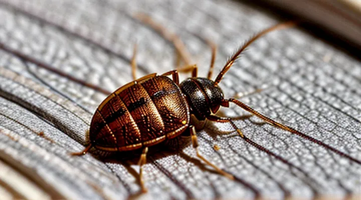
Key Characteristics of Bed Bug Bites
Size and Shape
Bedbug bite marks appear as small, raised lesions whose dimensions rarely exceed five millimetres. Typical measurements range from two to four millimetres in diameter, with occasional enlargement to six millimetres when secondary inflammation occurs.
The lesions are generally circular, exhibiting a smooth, well‑defined perimeter. In some photographs, a series of adjacent bites forms a short linear or clustered pattern, reflecting the insect’s feeding behaviour. The central area often presents a pale, slightly raised core surrounded by a reddened halo, creating a target‑like appearance.
Key visual indicators of size and shape:
- Diameter: 2–5 mm (occasionally up to 6 mm)
- Outline: round, with a distinct edge; occasional short linear arrangements
- Elevation: slight swelling above skin surface
- Colour contrast: pale centre with a surrounding erythematous ring
Recognition of these dimensional and morphological traits assists in differentiating bedbug bites from other arthropod reactions in photographic evidence.
Color and Appearance
Bedbug bite lesions appear as small, red to pink macules. Color ranges from light pink to deep reddish‑purple, reflecting individual skin response and the age of the bite.
The central area often stays pale while a darker halo surrounds it. A slightly raised, erythematous border may be visible around the macule.
Typical diameter measures 2–5 mm. Multiple bites frequently create linear rows or clustered groups, especially on exposed skin.
- Color spectrum: light pink, pink, reddish‑purple, occasional bruised hue.
- Shape: round or oval macule with peripheral erythema.
- Border: faintly raised, sometimes papular.
- Distribution: linear rows, clusters, or “breakfast‑n‑eggs” pattern.
- Duration: color fades from red to brownish over several days.
Pattern of Bites («Breakfast, Lunch, and Dinner»)
Bedbug bites commonly appear as small, erythematous papules ranging from 2 mm to 5 mm in diameter. The lesions often develop in groups rather than isolated points, a pattern that aids visual diagnosis.
The arrangement described as «Breakfast, Lunch, and Dinner» consists of three punctate lesions aligned linearly or in a shallow V‑shape. Key characteristics include:
- Three bites positioned within a 2 cm span.
- Uniform redness and similar size across the trio.
- Slightly raised edges with a central punctum, reflecting the feeding site.
Photographic evidence typically shows the trio against surrounding skin, with a clear separation from solitary or random clusters. The linear configuration distinguishes bedbug activity from other arthropod bites, which more often present as scattered or circular groupings.
Location on the Body
Bedbug bites frequently appear on exposed skin that is accessible during sleep. Typical sites include the face, neck, arms, and hands, as well as the torso and legs when clothing provides limited coverage. The distribution often reflects the position of the sleeper and the proximity of the insect to the body surface.
Common locations:
- Face and neck – small, clustered red welts, sometimes forming a linear pattern.
- Upper arms and forearms – isolated or grouped papules, often with a central punctum.
- Hands and fingers – discrete, itchy bumps, occasionally accompanied by a faint halo.
- Torso (chest, abdomen, back) – larger clusters of raised lesions, sometimes arranged in a zig‑zag line.
- Lower legs and ankles – solitary or grouped spots, typically less numerous than on upper limbs.
In photographic documentation, bites on the face and neck display clearer contrast against lighter skin tones, while lesions on the torso may be partially obscured by clothing. The size of each bite remains consistent across body regions, ranging from 2 mm to 5 mm in diameter, with erythema that may intensify after 24 hours. The presence of a central punctum is a reliable indicator regardless of location.
Distinguishing Bed Bug Bites from Other Insect Bites
Flea Bites
Flea bites appear as small, red papules, typically 1–3 mm in diameter. The central punctum may be slightly raised, and surrounding erythema is often faint. In photographic documentation, lesions present as isolated points or clusters of three to five spots, frequently located on the lower legs, ankles, and feet.
Key visual markers include:
- Uniform size across lesions;
- Sharp, well‑defined edges;
- Absence of a linear or “break‑fast‑nuggets” pattern;
- Presence of a central puncture point, sometimes with a tiny black dot.
Contrast with bedbug bites reveals distinct differences. Bedbug lesions often form a zigzag or linear arrangement, exhibit larger swollen welts, and display a darker central macule. Flea bites remain discrete, lacking the characteristic “breakfast‑cluster” distribution noted in bedbug infestations.
For accurate photographic identification, capture images under natural lighting, focus on the skin surface without flash glare, and include a scale reference such as a ruler. Document the anatomical location and any clustering pattern to facilitate differentiation from other arthropod bites.
Mosquito Bites
Mosquito bites appear as isolated, raised welts measuring 2‑5 mm in diameter. The center is often a pale pink spot surrounded by a red halo that may expand slightly over several hours. In photographs the surrounding skin shows uniform erythema without clustering, and the lesion typically resolves within a few days without leaving a mark.
Bedbug bites differ in visual presentation. Photographic evidence shows multiple, linearly arranged lesions, often grouped in rows of three to five. Each bite is a small, red papule with a darker central punctum, and the surrounding area may exhibit a mild swelling that persists longer than mosquito reactions. The pattern reflects the feeding behavior of the insect, which tends to bite sequentially along a host’s skin.
Key visual criteria for distinguishing the two types of bites:
- Arrangement: solitary (mosquito) versus linear or clustered (bedbug).
- Size: mosquito welts 2‑5 mm; bedbug papules generally 1‑3 mm.
- Central point: mosquito lesions lack a distinct punctum; bedbug bites display a dark core.
- Duration of erythema: mosquito redness fades within 48 hours; bedbug inflammation may last up to a week.
Recognizing these differences in photographs enables accurate identification of the biting insect.
Spider Bites
Spider bites appear as isolated or paired lesions, often centered on a single puncture site. The surrounding skin typically shows a raised, erythematous halo that may be smooth or slightly raised. In many photographs, the central punctum is visible as a tiny dot, sometimes surrounded by a darker ring of bruising. Swelling can develop rapidly, creating a dome‑shaped elevation that may persist for several days.
Key visual differences from other arthropod bites include:
- Single or paired lesions rather than the linear or clustered pattern common with other insects.
- Presence of a distinct central punctum, often absent in bedbug bite images.
- Uniform redness surrounding the punctum, without the “breakfast‑plate” arrangement of multiple, aligned spots.
- Occasional development of a central vesicle or ulceration, a feature rarely seen in photographs of other bite types.
When evaluating photographic evidence, focus on the lesion’s size, the clarity of the punctum, and the symmetry of the surrounding erythema. These characteristics provide reliable indicators that the bite originated from a spider rather than from other common pests.
Rash and Allergic Reactions
Bed‑bug bites often appear as small, red or pink macules that may develop a raised border. The lesions typically cluster in linear or zig‑zag patterns, reflecting the insect’s feeding behavior. Central puncta can be slightly darker, indicating the point of puncture.
Allergic responses vary among individuals. Common manifestations include:
- Erythema extending beyond the immediate bite site
- Pruritus that intensifies several hours after the initial exposure
- Swelling that may persist for days, sometimes forming a wel‑defined wheal
- Secondary excoriation resulting from scratching, which can produce crusted or ulcerated areas
In photographs, the rash may be confused with other arthropod bites, yet the combination of grouped lesions, pronounced itching, and occasional delayed swelling distinguishes it from most alternative causes. Recognizing these visual cues aids accurate identification and appropriate management.
Recognizing the Stages of Bed Bug Bites
Early Stages
Early-stage bedbug bites appear as tiny, reddish spots measuring approximately one to three millimeters in diameter. The lesions are usually flat or only minimally raised, lacking the pronounced swelling seen in later reactions. Color ranges from faint pink to bright red, depending on individual skin sensitivity and the interval since the bite occurred.
In photographs, the following features are typical:
- Small, discrete punctate lesions without surrounding edema.
- Arrangement in linear or clustered patterns, reflecting the insect’s feeding habit of moving along the skin.
- Absence of central crust or ulceration; the central point may be a subtle punctum where the proboscis pierced the skin.
- Minimal or no visible inflammation in the first 24‑48 hours; the redness may be faint and difficult to discern without close inspection.
«The early visual signature of a bedbug bite is a modest, isolated erythema that may be mistaken for a mosquito or flea bite, but its size, arrangement, and lack of swelling distinguish it.»
Photographic documentation often requires macro lenses or close-up focus to capture these minute details. Lighting should be even to avoid exaggerating shadows that could obscure the flat nature of the lesions.
Developed Bites
Bedbug bites that have progressed beyond the initial reaction display distinct visual cues in photographs. The lesions are typically raised, forming small papules that range from 2 mm to 5 mm in diameter. Redness surrounds the central puncture point, creating a halo of erythema that may darken to a brownish hue as the bite ages. The skin surface often shows a slight swelling, and the edges of the lesion can appear slightly raised compared to the surrounding tissue.
Key characteristics of «developed bites» observable in images:
- Central punctum or tiny dot indicating the feeding site.
- Uniform red or pink halo that expands outward over time.
- Mild edema giving the bite a raised, dome‑shaped profile.
- Possible discoloration to a darker shade after several days.
Photographic documentation frequently captures clusters of these lesions aligned in linear or zig‑zag patterns, reflecting the bedbug’s feeding behavior. The consistency of size, coloration, and arrangement aids in differentiating mature bedbug bites from other arthropod reactions.
Healed Bites
Healed bedbug bite lesions display a predictable progression in photographic documentation. Initial redness fades within a few days, leaving a flat, pale‑to‑pink macule that may persist for weeks. The macule often exhibits a subtle brownish hue due to post‑inflammatory hyperpigmentation, especially on individuals with darker skin tones.
Typical dimensions range from 2 mm to 5 mm in diameter. The outline is usually circular or slightly oval, lacking the raised central punctum seen in fresh bites. Occasionally, several healed spots appear in close proximity, forming a linear or clustered pattern that mirrors the insect’s feeding behavior.
Common anatomical sites include exposed areas such as the face, neck, forearms, hands, and lower legs. The lesions seldom occur on covered regions unless clothing is loose enough to expose the skin.
Key visual indicators of healed bedbug bites:
- Flat macule, 2–5 mm across
- Color transition from pink to light brown
- Absence of swelling or central puncture
- Arrangement in a line, zig‑zag, or small group
These characteristics help differentiate healed bedbug bites from other arthropod bite remnants, such as mosquito or flea bites, which often retain a raised center or exhibit a distinct circular pattern without linear clustering.
When to Seek Medical Attention
Severe Itching
Severe itching is a hallmark reaction to bites from the tiny nocturnal parasite that feeds on human blood. The sensation often begins within a few hours after the bite and can intensify rapidly, persisting for several days. Photographic evidence typically shows the following features associated with intense pruritus:
- Red, raised welts that may coalesce into larger patches when the skin is scratched repeatedly.
- Central puncture marks surrounded by a halo of inflammation, indicating repeated irritation.
- Evidence of secondary skin changes such as excoriations, crusts, or hyperpigmentation resulting from vigorous scratching.
The distribution pattern helps differentiate these bites from other arthropod reactions. Bites frequently appear in linear or clustered arrangements on exposed areas such as the arms, shoulders, neck, and face. When severe itching drives persistent scratching, the lesions become more pronounced, and photographs may capture signs of infection, including swelling, pus, or spreading redness.
Management focuses on alleviating the itch to prevent further skin damage. Topical corticosteroids, antihistamine creams, or oral antihistamines reduce inflammation and interrupt the itch‑scratch cycle. In cases where lesions become infected, medical evaluation and appropriate antimicrobial therapy are warranted. Prompt treatment minimizes the risk of long‑term skin discoloration and scarring.
Signs of Infection
Bedbug bites can progress to a secondary bacterial infection, and photographs often reveal characteristic changes that differentiate a simple reaction from an infected wound.
Typical visual indicators of infection include:
- Redness spreading beyond the original bite margin, forming a diffuse halo.
- Swelling that increases in size rather than subsiding.
- Pus or yellow‑white discharge emerging from the puncture site.
- Crusting or ulceration with a foul odor.
- Warmth felt when the area is touched, sometimes accompanied by a palpable induration.
Presence of fever, rapid pulse, or enlarged lymph nodes near the affected region signals systemic involvement. Immediate medical evaluation is advised when any of these signs appear, as prompt antimicrobial therapy reduces the risk of complications.
Allergic Reactions
Bedbug bites commonly appear as small, round welts surrounded by a reddish halo; when the host’s immune system reacts strongly, the lesions become markedly inflamed.
Typical visual indicators of an allergic response include:
- pronounced erythema extending several millimeters beyond the bite margin;
- noticeable edema that may cause the surrounding skin to swell;
- formation of a raised, itchy papule that can persist for days;
- occasional development of a central punctate area that may exude clear fluid.
Severity of these signs varies with individual hypersensitivity; some persons exhibit only faint discoloration, while others develop extensive, confluent patches. High‑sensitivity reactions often present with multiple bites clustered together, creating a linear or “breakfast‑cereal” pattern that intensifies the overall inflammatory appearance.
Distinguishing allergic bedbug reactions from other arthropod bites relies on pattern recognition and symptom chronology. Bedbug lesions typically appear in groups of three to five, aligned in a row, whereas solitary welts suggest alternative sources. The presence of intense itching, rapid swelling, and a pronounced red halo strongly points toward an immunologic response rather than a simple mechanical puncture.
Recognizing these photographic characteristics assists clinicians and lay observers in identifying allergic bedbug bites, facilitating appropriate treatment and infestation control measures.
Photographic Evidence and Identification
Taking Clear Photos
Clear, detailed photographs are essential for reliable identification of bite marks that may be caused by bedbugs. Proper technique reduces ambiguity and assists professionals in distinguishing these lesions from other skin conditions.
To achieve optimal image quality, follow these steps:
- Use natural daylight or a diffused white light source; avoid harsh shadows and colored lighting.
- Position the camera at a distance that allows the bite area to fill the frame while preserving surrounding skin for context.
- Employ a macro lens or the camera’s close‑up mode to capture fine details such as the central puncture and surrounding erythema.
- Stabilize the device with a tripod or steady surface; eliminate motion blur by using a short exposure time.
- Adjust focus manually on the bite’s center; verify sharpness by zooming in on the preview.
- Include a reference object of known size (e.g., a ruler or coin) placed beside the lesion to provide scale.
- Capture multiple angles: straight‑on view, oblique view, and a side profile to reveal depth and swelling.
- Store images in high‑resolution format (minimum 300 dpi) and keep original files without compression.
Consistent application of these practices yields photographs that accurately represent the appearance of bedbug bites, facilitating effective analysis and diagnosis.
Angles and Lighting
Accurate photographic documentation of bedbug bite marks requires careful control of camera angle and illumination. Improper positioning obscures characteristic patterns, leading to misidentification.
Angles that capture the bite from directly above minimize distortion and reveal the concentric redness typical of recent lesions. Oblique perspectives introduce shadows that can exaggerate swelling or mask peripheral inflammation. Side‑view shots highlight raised papules and enable assessment of depth, but should be supplemented by a perpendicular view for a complete record.
Lighting influences color fidelity and contrast. Diffused natural light reduces harsh shadows, preserving true hue and allowing subtle erythema to be seen. Direct flash creates glare, washes out red tones, and may produce reflective artifacts on skin. Warm‑temperature bulbs shift coloration toward orange, potentially mimicking bite redness; neutral‑white sources maintain accurate color balance. Consistent exposure settings prevent over‑ or under‑exposure that hides fine details.
Optimal capture protocol:
- Position camera perpendicular to the skin surface for primary image.
- Add a secondary oblique shot if swelling assessment is needed.
- Use soft, indirect lighting; avoid direct flash.
- Prefer daylight or daylight‑balanced artificial light (≈5,500 K).
- Maintain uniform exposure across all images.
Common Mistakes in Identification
Bedbug bite identification often relies on photographic evidence, yet several recurring errors undermine accuracy. Misinterpreting the visual characteristics of bites can lead to false conclusions about infestation status.
Common errors include:
- Assuming any red, itchy spot originates from bedbugs without considering alternative causes such as allergic reactions or other insects.
- Overlooking the typical arrangement of bites; bedbug marks frequently appear in linear or clustered patterns, while isolated lesions are less indicative.
- Ignoring the time frame of lesion development; fresh bites may appear as small, pale papules, whereas older bites can darken and swell, altering their appearance.
- Misreading skin tone variations; dark‑skinned individuals may exhibit less visible redness, causing underestimation of bite presence.
- Relying on a single photograph; lighting, focus, and angle affect color and size perception, making a series of images essential for reliable assessment.
Accurate identification demands comparison of multiple images taken under consistent conditions, awareness of differential diagnoses, and recognition of the characteristic bite distribution. Proper documentation reduces the risk of misidentification and supports effective pest‑control decisions.
