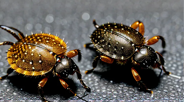
The General Anatomy of a Tick
The Main Body Sections
The Capitulum (Head)
The capitulum, or head, forms the anterior feeding apparatus of every tick species. It consists of a compact bundle of mouthparts that protrude from the dorsal surface when the tick is attached to a host. All ticks share the same basic arrangement, although the size and proportion of individual components differ among families.
Key elements of the capitulum include:
- Palps – paired sensory structures that locate the host’s skin and guide the feeding tube.
- Chelicerae – cutting appendages that pierce the epidermis.
- Hypostome – a central, barbed tube that anchors the tick within the host’s tissue and delivers saliva.
- Basis capituli – the sclerotized base that supports the other parts and connects to the gnathosoma.
The overall shape of the capitulum is typically rounded to oval, with the basis capituli forming a shield‑like plate. The hypostome’s barbs are present in all species, providing a universal mechanism for secure attachment. Palps are uniformly equipped with chemosensory organs, enabling detection of host cues across the entire tick clade.
Variations among species manifest as differences in:
- Length of the hypostome, ranging from short in some soft ticks to elongated in many hard ticks.
- Size of the palps, which may be proportionally larger in species that feed on small hosts.
- Degree of sclerotization of the basis capituli, influencing durability during prolonged feeding.
Despite these modifications, the capitulum’s fundamental architecture remains constant, offering a reliable morphological indicator for identifying ticks at the generic level.
The Idiosoma (Body)
The idiosoma constitutes the bulk of a tick’s body, situated posterior to the gnathosoma and bearing the eight legs. Across all tick species it presents as a compact, oval structure that is dorsoventrally flattened to facilitate attachment to hosts. The dorsal surface is covered by a hardened cuticle; in hard ticks a scutum occupies the anterior portion, while soft ticks lack a scutum, leaving the cuticle more flexible. The ventral side contains the following consistent elements:
- Anal groove positioned anterior to the anus, directing waste away from the ventral surface.
- Spiracular plates (stigmata) opening laterally, each surrounded by a sclerotized rim.
- A series of setae and sensory pits distributed along the margins, providing tactile feedback.
- Internal musculature linked to the leg bases, enabling precise locomotion and host‑seeking movements.
The idiosoma houses the digestive tract, reproductive organs, and the excretory system, all encapsulated within the cuticular envelope. Its uniform shape and cuticular architecture constitute the primary visual characteristic shared by every tick species, regardless of taxonomic family.
External Features Common to Most Ticks
Legs and Their Structure
Ticks belong to the order Ixodida and share a distinctive leg arrangement that contributes to their overall morphology. Each individual possesses four pairs of legs, numbered I–IV from front to rear. The legs are relatively short compared to the body, giving ticks a compact, rounded silhouette.
The leg segments follow the standard arthropod pattern: coxa, trochanter, femur, patella, tibia, and tarsus. The tarsus ends in a claw equipped with a pulvillus—a pad that aids in attachment to hosts. Sensory organs, known as Haller’s organs, are located on the dorsal surface of the first pair of legs; these structures detect carbon dioxide, temperature, and host movement.
Variations among tick families affect leg morphology:
- Hard ticks (Ixodidae): robust coxae, well‑developed pulvilli, and pronounced Haller’s organs.
- Soft ticks (Argasidae): more slender coxae, reduced pulvilli, and less conspicuous sensory pits.
- Mouth‑part attachment: the legs are positioned laterally, allowing the hypostome to pivot forward during feeding.
These consistent features—four pairs of segmented legs, terminal claws with pulvilli, and specialized sensory organs—provide a reliable basis for recognizing tick specimens across species.
Mouthparts (Chelicerae and Hypostome)
Ticks possess a distinctive set of mouthparts that enable blood feeding across all species. The primary components are the chelicerae and the hypostome, each serving a specific mechanical function.
The chelicerae are paired, blade‑like structures situated at the front of the capitulum. They operate as cutting instruments, slicing the host’s skin to create an entry point for the feeding tube. Muscular control allows rapid opening and closing, facilitating penetration of the epidermis. Across tick families, cheliceral length and curvature vary modestly, but the overall morphology—two opposable, serrated claws—remains consistent.
The hypostome is a central, barbed rod that extends from the ventral side of the capitulum. Its surface bears numerous backward‑pointing denticles that anchor the tick within the host’s tissue, preventing dislodgement during prolonged feeding. The hypostome’s length correlates with feeding duration: species that remain attached for several days exhibit longer, more heavily barbed hypostomes, whereas those feeding briefly possess shorter, less dense dentition.
Key points summarizing the mouthpart architecture:
- Chelicerae: paired, cutting appendages; serrated edges; used for skin incision.
- Hypostome: single, rod‑like structure; barbed surface; provides secure attachment.
- Uniformity: all tick species share this basic arrangement, despite minor variations in size and denticle density.
These structures define the visual signature of ticks, distinguishing them from other arachnids and underpinning their ability to obtain blood meals from vertebrate hosts.
Scutum (Shield) - Presence and Variability
The dorsal shield, or scutum, is a rigid, sclerotized plate that distinguishes hard‑tick (Ixodidae) species from soft‑tick (Argasidae) species. In ixodids the scutum covers the anterior portion of the idiosoma and remains visible after engorgement; in argasids the dorsal surface is pliable and lacks a distinct scutum.
Variability of the scutum occurs at several levels:
- Shape: oval, rectangular, or elongated, often reflecting adaptation to host skin texture.
- Size relative to body: some species possess a small, central scutum (e.g., Rhipicephalus spp.), while others have a large plate that occupies most of the dorsum (e.g., Dermacentor spp.).
- Surface pattern: smooth, punctate, or ornamented with ornate markings that aid species identification.
- Coloration: ranging from pale brown to dark brown or black, sometimes with contrasting marginal bands.
Within hard ticks, the presence of a scutum is consistent, but its dimensions and ornamentation provide reliable characters for taxonomic discrimination. Soft ticks, by contrast, never develop a true scutum; their dorsal cuticle remains flexible throughout the life cycle. The scutum’s structural differences therefore contribute directly to the visual diversity observed across all tick taxa.
Distinguishing Features Among Tick Species
Hard Ticks (Ixodidae)
General Appearance and Morphology
Ticks share a consistent arachnid body plan that distinguishes them from insects and other arthropods. The organism consists of two main regions: the anterior capitulum (or gnathosoma) that houses the mouthparts, and the posterior idiosoma that contains the bulk of the digestive and reproductive systems. The capitulum bears a pair of chelicerae, a hypostome equipped with backward‑pointing barbs for anchoring to the host, and a palpal organ used for sensory perception. The idiosoma bears a dorsal shield called the scutum in hard‑tick (Ixodidae) species; soft‑tick (Argasidae) species lack a true scutum and have a more flexible cuticle.
Key morphological traits common to all tick species include:
- Body shape – dorsoventrally flattened, oval to elongated, facilitating insertion into host skin.
- Leg count – eight legs in nymphs and adults, four legs in larvae; legs are short, sturdy, and end in claws for attachment.
- Sensilla – sensory hairs on the legs and capitulum that detect temperature, carbon‑dioxide, and host movement.
- Size range – from 1 mm in unfed larvae to over 15 mm in engorged adult females, depending on species and feeding status.
- Sexual dimorphism – females enlarge dramatically after blood meals, while males remain relatively small and retain a constant size.
The cuticle is composed of chitin and exhibits a pattern of micro‑setae that varies minimally among species, providing a uniform texture. Coloration ranges from reddish‑brown to dark brown or gray, reflecting the degree of engorgement and the presence of pigments in the hemolymph. Internally, the digestive tract expands to accommodate large blood volumes, and the reproductive organs occupy the posterior idiosoma, visible as a slight bulge in mature females.
Overall, tick morphology is defined by a compact, shielded anterior region, a flexible dorsal surface, eight robust legs, and a capacity for extreme size increase during feeding, traits that persist across the diverse genera within the order Ixodida.
Key Identification Markers
Ticks share a limited set of morphological traits that enable reliable identification across families and genera. These traits are observable with a hand lens or stereomicroscope and form the foundation for distinguishing ticks from other arthropods and for separating species within the group.
- Body segmentation – a compact, dorsoventrally flattened body divided into two main regions: the anterior capitulum (mouthparts) and the posterior idiosoma (main body). The capitulum projects forward and bears the chelicerae and hypostome.
- Scutum – a hard dorsal shield present in hard ticks (Ixodidae); absent or reduced in soft ticks (Argasidae). When present, the scutum covers the entire dorsal surface in males and a portion of the anterior dorsum in females.
- Leg arrangement – four pairs of legs attached to the ventral surface of the idiosoma. In larvae, a fifth pair of vestigial legs may be visible on the ventral side.
- Sensilla and palps – paired sensory structures on the capitulum, including palps that are longer than the chelicerae in most species and function in host detection.
- Festoons – series of rectangular cuticular plates along the posterior margin of the idiosoma, typical of many hard tick species and useful for family-level identification.
- Eyes – simple lateral eyes located on the dorsal surface of the idiosoma in many hard ticks; absent in many soft tick species.
- Coxal pores – openings on the ventral coxae of the fourth pair of legs that excrete fluids; pattern and number vary among species and aid in taxonomic discrimination.
- Mouthpart length – the hypostome may be short or elongated, with barbs that differ in density and arrangement, providing clues to feeding behavior and taxonomy.
Consistent evaluation of these markers allows entomologists and medical professionals to recognize ticks at the family level and to narrow identification to genus or species when combined with additional characters such as host preference, geographic distribution, and life‑stage morphology.
Soft Ticks (Argasidae)
General Appearance and Morphology
Ticks are arachnids with a compact, oval to elongated body divided into two main regions: the anterior capitulum, which houses the mouthparts, and the posterior idiosoma, containing the legs, sensory organs, and internal structures. The capitulum projects forward and includes chelicerae for cutting skin, a hypostome with barbed teeth for anchoring, and palps that sense host cues. The idiosoma bears four pairs of legs, each ending in claws and sensory setae that detect temperature, carbon dioxide, and movement.
Morphological traits shared by all species include:
- A dorsoventrally flattened profile that facilitates attachment to host skin.
- A cuticular exoskeleton composed of chitin, providing protection and support.
- Simple eyes (ocelli) in many hard‑tick species, while soft ticks often lack them.
- Spiracular plates on the ventral surface for respiration.
- A ventral groove (gnathosoma) that aligns the mouthparts during feeding.
Key variations distinguish the two major families:
- Hard ticks (Ixodidae) possess a rigid dorsal shield called a scutum; in males it covers the entire dorsum, while in females it occupies only the anterior region, allowing abdomen expansion during engorgement. Their coloration ranges from brown to reddish‑brown, sometimes with patterned markings.
- Soft ticks (Argasidae) lack a scutum, exhibit a leathery, wrinkled cuticle, and display a broader color spectrum, including pale, reddish, or dark tones. Their bodies are more flexible, enabling rapid movement when seeking a host.
- The monotypic family Nuttalliellidae combines features of both groups, with a partially sclerotized dorsal surface and a unique arrangement of mouthparts.
Size ranges from 1 mm in unfed larvae to over 30 mm in fully engorged adult females, reflecting dramatic expansion during blood intake. Surface microstructures, such as cuticular ridges and pores, are consistent across taxa and aid in water retention and host attachment. Overall, tick morphology presents a uniform basic plan adapted for ectoparasitism, with family‑level modifications that influence host interaction and ecological niche.
Key Identification Markers
Ticks share a set of morphological features that allow reliable identification regardless of species. These features are consistent across hard and soft ticks and form the basis for comparative visual assessment.
- Dorsal shield (scutum) present in hard ticks, absent or reduced in soft ticks; shape and extent differ among families.
- Body segmentation: anterior idiosoma (capitulum) housing mouthparts, posterior idiosoma containing the abdomen; clear demarcation visible under magnification.
- Mouthparts: chelicerae and hypostome protrude forward, forming a spear‑like structure; length and dentition vary but orientation remains constant.
- Eyes: simple lateral ocelli present in many hard tick genera; absent in most soft tick genera.
- Leg count: eight legs in all stages except larvae, which have six; leg length and segmentation provide additional clues.
- Festoons: rectangular cuticular plates along the posterior margin of the body; typical of hard ticks, absent in soft ticks.
- Anal groove: V‑shaped indentation surrounding the anal opening; characteristic of hard ticks, absent in soft ticks.
- Coloration: ranging from light brown to dark brown or black; pigmentation patterns are species‑specific but the overall hue range is shared.
- Size: adult ticks span 2–10 mm in length, with larvae measuring 0.3–0.5 mm; size categories are consistent across taxa.
These markers, examined together, define the visual profile common to all tick species and enable taxonomic differentiation when combined with regional and host data.
Geographic and Habitat-Specific Variations
How Environment Influences Appearance
Ticks display a wide range of physical traits that reflect the conditions in which they develop. Ambient temperature, humidity, and the type of substrate directly shape coloration, size, and surface texture. In arid zones, exoskeletons often appear lighter and thinner to reduce water loss, whereas humid forests favor darker, thicker cuticles that protect against fungal invasion.
Key environmental influences include:
- Temperature: Higher temperatures accelerate growth, producing larger nymphs and adults; lower temperatures slow metabolism, resulting in smaller individuals.
- Humidity: Elevated moisture levels promote a glossy, darker cuticle; low humidity leads to a matte, lighter exoskeleton.
- Host habitat: Ticks that parasitize ground‑dwelling mammals in leaf litter develop cryptic brown‑gray patterns; those on birds nesting in open fields exhibit more uniform, pale coloration.
- Substrate: Species inhabiting dense vegetation acquire flatter bodies to navigate foliage, while those on exposed rock surfaces retain a more rounded shape for stability.
These adaptations enable ticks to blend with their surroundings, enhance attachment efficiency, and maintain physiological balance across diverse ecosystems. Understanding the link between environment and morphology clarifies why visual identification of ticks varies widely despite shared taxonomic traits.
