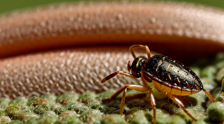
Immediate Reactions to a Tick Bite
The Bite Mark Itself
Initial Appearance
A tick attachment typically leaves a small, erythematous papule at the bite site. The lesion measures 2–5 mm in diameter, appears as a flat or slightly raised red spot, and may contain a central punctum where the tick’s mouthparts remain embedded. The surrounding skin is often free of swelling or pain in the first 24 hours.
Common early visual features include:
- Uniform redness without a surrounding halo
- A pinpoint central dot or tiny scar indicating the attachment point
- Absence of vesicles or necrosis during the initial phase
The appearance may persist for several days before evolving into a larger erythema or a target‑shaped rash if infection develops. Immediate inspection of the bite area should focus on these characteristics to differentiate a simple tick bite from early signs of disease.
Size and Shape Variations
After a tick attachment, the cutaneous manifestation exhibits a spectrum of dimensions and configurations. The initial mark often appears as a minute puncture, typically 1–3 mm in diameter, corresponding to the mouthparts that penetrated the epidermis. In many cases, an expanding erythematous halo follows, reaching 5–10 mm within a few days and sometimes extending beyond 20 mm when an allergic or infectious response intensifies.
Common size categories include:
- Micro‑lesion: ≤ 3 mm, limited to the bite point.
- Small erythema: 3–10 mm, circular or slightly oval.
- Medium rash: 10–25 mm, often uniform in coloration.
- Large area: > 25 mm, may involve surrounding tissue and indicate systemic involvement.
Shape variations reflect the tick’s feeding behavior, host skin tension, and immune reaction. Typical forms are:
- Round: symmetric, centered on the bite site.
- Oval: elongated along the direction of skin stretch.
- Target (bull’s‑eye): concentric rings of erythema and pallor, characteristic of certain infections.
- Linear or serpentine: follows a skin crease or vessel, suggesting migration of inflammatory mediators.
- Irregular: uneven borders, often associated with secondary irritation or secondary infection.
The specific pattern observed provides clues about the tick species, duration of attachment, and the host’s immunological response, aiding clinicians in diagnosis and management.
Common Skin Reactions
Redness and Swelling
Redness and swelling are the most common cutaneous manifestations after a tick attaches to the skin. The bite site typically develops a localized erythema that may expand outward from the point of attachment. Edema accompanies the erythema, producing a raised, often tender area.
The reaction results from the host’s inflammatory response to tick saliva, which contains anticoagulants, anesthetics, and immunomodulatory proteins. Histamine release and vasodilation cause the characteristic redness, while increased vascular permeability leads to fluid accumulation and swelling.
Typical presentation includes:
- A circular or oval area of redness, usually 0.5–2 cm in diameter.
- A raised, firm or soft swelling surrounding the erythema.
- Mild to moderate pain or itching at the site.
- Persistence for several days; resolution often occurs within 1–2 weeks if no infection develops.
Medical evaluation is advised when:
- Redness exceeds 5 cm or expands rapidly.
- Swelling becomes increasingly painful, warm, or accompanied by fever.
- An ulcerated or necrotic center appears, suggesting secondary infection or tick‑borne disease.
- Symptoms persist beyond two weeks without improvement.
Prompt removal of the tick, cleaning the area with antiseptic, and monitoring for changes reduce complications and support recovery.
Itching and Irritation
Itching and irritation are among the most immediate cutaneous responses following a tick attachment. The bite introduces saliva containing proteins that trigger a localized immune reaction, leading to histamine release and nerve stimulation. This results in a pruritic, often erythematous area that may expand over several hours.
Typical characteristics include:
- A small, red papule at the attachment site, sometimes surrounded by a halo.
- Intense itching that intensifies when the area is scratched.
- Mild swelling or warmth, indicating inflammation.
The severity of pruritus varies with tick species, duration of attachment, and individual sensitivity. In most cases, symptoms subside within a few days after removal of the arthropod. Persistent or worsening irritation may suggest secondary infection or an allergic response and warrants medical evaluation.
Management strategies:
- Remove the tick promptly with fine‑tipped tweezers, grasping close to the skin and pulling straight upward.
- Clean the bite site with antiseptic solution.
- Apply a topical corticosteroid to reduce inflammation and itching.
- Administer an oral antihistamine if pruritus interferes with daily activities.
- Monitor for signs of infection—pus, increasing redness, or fever—and seek professional care if they appear.
Early intervention limits tissue irritation and reduces the risk of complications such as tick‑borne illnesses.
Small Bump or Nodule
A small, raised area on the skin is the most common immediate response to a tick attachment. The lesion typically appears within hours to a few days after the bite and may be described as a papule, nodule, or erythematous bump. Its size ranges from a few millimeters to about one centimeter in diameter. The surface can be smooth or slightly rough, and the surrounding skin often shows mild redness.
Key characteristics of the bump include:
- Firm or slightly tender to palpation.
- Uniform color, usually pink to light red; occasional central punctum marks the tick’s mouthparts.
- Persistence for several days; may enlarge slightly before gradually fading.
The bump may represent a localized inflammatory reaction to tick saliva, which contains anticoagulants and immunomodulatory proteins. In most cases, the lesion resolves without intervention. However, certain pathogens transmitted by ticks can modify the presentation:
- Borrelia burgdorferi (Lyme disease) may produce a larger, expanding erythema migrans that starts as a small papule and spreads outward.
- Rickettsia species can cause a vesicular or pustular lesion, often accompanied by fever and systemic symptoms.
When the bump persists beyond two weeks, enlarges rapidly, or is accompanied by fever, headache, joint pain, or a characteristic “bull’s‑eye” pattern, medical evaluation is warranted. Diagnostic steps may include serologic testing for tick‑borne infections and, if necessary, antibiotic therapy tailored to the identified pathogen.
Preventive measures focus on prompt removal of the tick, thorough skin inspection after outdoor activities, and early documentation of any cutaneous changes. Early recognition of a simple nodule versus a sign of systemic infection guides appropriate treatment and reduces the risk of complications.
Delayed Skin Manifestations and Potential Infections
Localized Infections
Bacterial Contamination
Bacterial infection is a common consequence of a tick attachment that can produce visible skin changes. The bite site may develop redness, swelling, or a raised lesion within hours to days. In some cases, the area evolves into an ulcerating wound or a necrotic patch, reflecting deeper tissue involvement.
Typical cutaneous presentations of bacterial contamination include:
- Erythematous macule that expands rapidly
- Purulent pustule or abscess formation
- Tender, indurated nodule with central necrosis (eschar)
- Lymphangitic streaking extending from the bite
These signs often accompany systemic symptoms such as fever, chills, or malaise, indicating that the pathogen has breached the local barrier. The most frequently implicated organisms are Staphylococcus aureus and Streptococcus pyogenes, but tick-borne bacteria such as Borrelia burgdorferi and Rickettsia species can also provoke secondary bacterial overgrowth.
Prompt antimicrobial therapy, guided by culture when feasible, reduces the risk of complications. Empiric coverage typically involves a beta‑lactam agent effective against gram‑positive cocci; severe or polymicrobial infections may require broader-spectrum regimens. Wound care, including debridement of necrotic tissue and regular dressing changes, supports healing and prevents further bacterial proliferation.
Pus and Inflammation
A tick bite frequently triggers a localized inflammatory response. The area becomes red, swollen, and warm, reflecting increased blood flow and immune cell activity aimed at containing any introduced pathogens.
When bacterial invasion follows the bite, pus may accumulate. Pus appears as a thick, yellow‑white exudate, often accompanied by a palpable lump that can progress to an abscess. Its presence signals a secondary infection that exceeds the normal sterile inflammatory process.
Key indicators that pus formation is occurring include:
- Persistent or enlarging swelling beyond the initial bite site
- Fluctuant, tender nodule suggestive of an abscess cavity
- Yellow or greenish discharge from the skin surface
- Increased pain unrelieved by standard anti‑inflammatory measures
Management requires prompt cleansing of the wound, application of antiseptic agents, and, when pus is evident, medical evaluation for possible drainage and systemic antibiotic therapy. Early intervention reduces the risk of complications such as cellulitis or tick‑borne disease progression.
Tick-Borne Diseases
Lyme Disease («Erythema Migrans»)
Lyme disease frequently presents with a distinctive cutaneous lesion after a tick attachment. The rash, known as erythema migrans, is the earliest visible sign of infection with Borrelia burgdorferi.
Erythema migrans typically emerges 3–30 days post‑bite. It begins as a small, flat erythema at the bite site and expands outward, often reaching 5–30 cm in diameter. The lesion commonly exhibits a concentric pattern, creating a “bull’s‑eye” appearance when central clearing occurs, though uniform redness is also observed.
Key characteristics of the rash include:
- Expansion of at least 5 cm in diameter
- Warmth and mild tenderness
- Absence of vesicles or purpura
- Possible central clearing, producing a target‑like outline
- Occurrence on trunk, limbs, or less frequently on the face
The lesion persists for several weeks if untreated, during which systemic manifestations—fever, headache, arthralgia, and fatigue—may develop. Prompt antimicrobial therapy, typically doxycycline for adults or amoxicillin for children, halts progression and reduces the risk of disseminated disease.
Early recognition of erythema migrans after a tick exposure remains critical for effective management of Lyme disease.
Rocky Mountain Spotted Fever («Rash Characteristics»)
A tick bite can introduce Rickettsia rickettsii, the agent of Rocky Mountain spotted fever, which is commonly recognized by a distinctive cutaneous eruption.
- Appearance: maculopapular lesions that become petechial; may evolve into purpuric spots.
- Onset: typically 2–5 days after the bite; fever often precedes rash.
- Distribution: begins on wrists and ankles, spreads centrally to trunk, palms, and soles; may involve the face later.
- Progression: lesions enlarge, coalesce, and may develop a “rose‑petal” pattern on the palms and soles.
- Duration: persists for 3–7 days if untreated; resolves with appropriate antimicrobial therapy.
The rash serves as an early diagnostic clue, differentiating Rocky Mountain spotted fever from other tick‑borne illnesses. Prompt recognition and treatment reduce morbidity and mortality.
Other Less Common Rashes
Tick bites may produce a range of cutaneous reactions beyond the typical erythema migrans. Less frequent rashes often signal alternative pathogen exposure or atypical immune responses.
-
Urticarial wheals: Raised, pruritic plaques appear within hours to days. Lesions are transient, often migrating, and may accompany systemic symptoms such as fever or malaise. Their presence suggests a hypersensitivity reaction rather than direct infection.
-
Vesiculobullous eruptions: Small vesicles or larger bullae develop on the extremities or trunk, sometimes resembling herpes simplex lesions. Onset typically occurs 3–10 days after the bite. Histology frequently reveals intraepidermal clefting with necrotic keratinocytes, indicating viral co‑infection or severe inflammatory response.
-
Pustular lesions: Localized pustules emerge around the attachment site, occasionally spreading to adjacent skin. These lesions may be sterile or culture‑positive for Staphylococcus aureus, reflecting secondary bacterial colonization. Pustules resolve within 1–2 weeks with appropriate antimicrobial therapy.
-
Papular‑purpuric rash: Discrete papules with a purplish hue appear on the lower limbs, sometimes coalescing into larger patches. The rash may persist for several weeks and is associated with tick‑borne rickettsial diseases such as Rocky Mountain spotted fever. Laboratory testing for specific antibodies aids diagnosis.
-
Erythema multiforme–like lesions: Target‑shaped macules and plaques develop, often on the palms and soles. The pattern suggests an immune‑mediated reaction to tick salivary antigens or to co‑transmitted pathogens. Lesions typically resolve spontaneously within 2–3 weeks.
Recognition of these uncommon presentations assists clinicians in differentiating tick‑related infections from allergic or infectious mimics. Prompt laboratory evaluation and targeted treatment reduce the risk of complications.
Allergic Reactions
Hives and Urticaria
Hives, medically termed urticaria, are a frequent cutaneous response after a tick attaches to the skin. They appear as raised, erythematous wheals that vary in size from a few millimeters to several centimeters. Individual lesions typically develop within hours of the bite and may coalesce into larger patches. The underlying mechanism involves rapid release of histamine and other mediators from mast cells, triggered by tick saliva proteins that act as allergens.
Key clinical features of tick‑induced urticaria include:
- Pruritic, pink‑to‑red plaques with well‑defined borders
- Transient nature; each wheal usually fades within 24 hours, while new lesions may arise elsewhere
- Possible accompanying angio‑edema of the lips, eyelids, or genitalia
- Absence of a central punctum, distinguishing the reaction from the bite mark itself
Differential diagnosis should consider erythema migrans, papular lesions, and secondary infection. Laboratory testing is rarely required; diagnosis rests on visual assessment and patient history of recent tick exposure.
Management recommendations:
- Remove the tick promptly with fine‑tipped forceps, grasping close to the skin and pulling steadily.
- Apply a second‑generation antihistamine (e.g., cetirizine 10 mg once daily) to reduce itching and wheal formation.
- If symptoms persist beyond 48 hours or intensify, add a short course of oral corticosteroids (e.g., prednisone 0.5 mg/kg daily for 5 days).
- Seek medical evaluation for signs of anaphylaxis, extensive angio‑edema, or systemic involvement such as fever or joint pain.
Prompt recognition and treatment of urticaria after a tick bite limit discomfort and prevent progression to more severe allergic reactions.
Anaphylaxis (Rare)
Anaphylaxis is an uncommon but severe systemic reaction that can manifest on the skin after a tick attachment. Cutaneous signs typically include widespread urticaria, erythematous wheals, and intense itching. These lesions often appear rapidly, within minutes to an hour of the bite, and may be accompanied by flushing, swelling of the lips or eyelids, and a sensation of warmth.
Systemic involvement may follow the dermatologic presentation, with hypotension, bronchospasm, and gastrointestinal distress. Prompt recognition of the skin changes is essential because they frequently precede life‑threatening airway compromise. Administration of intramuscular epinephrine, followed by antihistamines and corticosteroids, constitutes the first‑line treatment. Observation for at least four hours after symptom resolution is recommended to monitor for biphasic reactions.
Key clinical features of tick‑related anaphylaxis:
- Sudden onset of hives or large, blanching wheals
- Marked pruritus and erythema extending beyond the bite site
- Angioedema of facial or oral tissues
- Rapid progression to cardiovascular or respiratory instability
Early intervention based on these cutaneous cues reduces morbidity and prevents fatal outcomes.
When to Seek Medical Attention
Persistent or Worsening Symptoms
After a tick attachment, the initial skin reaction often resolves within a few days. When redness, swelling, or a rash continues beyond the typical healing period, or when the lesion expands, the condition is no longer a simple local response. Persistent erythema, enlarging annular lesions, or the development of a central punctum suggest that the bite may have transmitted a pathogen or triggered an exaggerated immune reaction, requiring medical evaluation.
Key indicators of a worsening cutaneous condition include:
- Redness that spreads more than 5 cm from the bite site
- Development of a raised, target‑shaped, or bullseye lesion
- Increasing pain, warmth, or tenderness around the area
- Appearance of fever, chills, headache, or joint pain alongside the skin change
- Formation of ulceration, necrosis, or drainage from the bite site
These signs often precede systemic infections such as Lyme disease, Rocky Mountain spotted fever, or tularemia. Prompt consultation with a healthcare professional is essential to confirm diagnosis, initiate appropriate antimicrobial therapy, and prevent complications.
Signs of Infection
Skin changes following a tick bite may indicate an infection. Early warning signs include localized redness that spreads beyond the bite site, swelling, warmth, and tenderness. Pus or other fluid discharge signals a bacterial invasion. An expanding circular rash, often described as a “bull’s‑eye,” suggests Lyme disease. Fever, chills, or flu‑like symptoms accompany systemic involvement. Enlarged or tender lymph nodes near the bite area reflect regional immune response. Severe pain, ulceration, or necrotic tissue points to more aggressive pathogens such as Rickettsia or Francisella.
Typical infection indicators
- Redness extending >2 cm from the bite
- Progressive swelling and heat
- Purulent drainage or crust formation
- Expanding erythematous ring (≈5–10 cm diameter)
- Fever ≥ 38 °C, chills, malaise
- Swollen, tender lymph nodes
- Persistent or worsening pain, ulceration, necrosis
Presence of any of these manifestations warrants prompt medical evaluation and possibly antimicrobial therapy. Early treatment reduces the risk of complications and long‑term sequelae.
Systemic Symptoms Indicating Disease
After a tick attachment, systemic manifestations may develop even when the bite site shows only a small erythema. These signs signal that a pathogen has entered the bloodstream and requires prompt medical evaluation.
Common systemic symptoms include:
- Fever or chills
- Severe headache, often described as throbbing
- Muscle and joint aches, sometimes with swelling
- Fatigue or malaise that worsens over days
- Nausea, vomiting, or abdominal discomfort
- Neck stiffness or photophobia, suggesting meningitis
- Rapid heart rate (tachycardia) or low blood pressure (hypotension)
These clinical features are associated with specific tick‑borne infections:
- Lyme disease – flu‑like illness, migratory joint pain, facial palsy; may accompany an expanding erythema migrans.
- Rocky Mountain spotted fever – high fever, headache, and a maculopapular rash that often spreads from wrists and ankles to the trunk.
- Ehrlichiosis and anaplasmosis – fever, chills, myalgia, and leukopenia; laboratory tests typically reveal elevated liver enzymes.
- Babesiosis – hemolytic anemia, jaundice, and dark urine, frequently accompanied by fever and fatigue.
The onset of systemic signs varies by pathogen: some appear within 2–7 days (e.g., Ehrlichiosis), while others may emerge weeks after the bite (e.g., Lyme disease). Persistent or worsening symptoms, especially when combined with a rash, warrant immediate laboratory testing and empiric antimicrobial therapy according to current clinical guidelines. Early identification of systemic involvement reduces the risk of long‑term complications.
