Understanding the Risk of Tick-Borne Diseases
The Danger of Improper Tick Removal
Why Manual Removal is Not Recommended
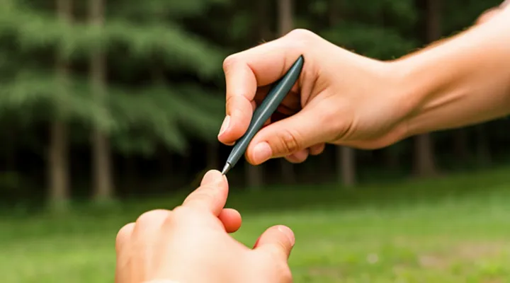
Manual extraction of a tick with fingers frequently leads to complications. Pressure applied by the hand can crush the arthropod, forcing saliva and pathogens deeper into the skin.
- Incomplete removal leaves mouthparts embedded, creating a persistent wound that can become infected.
- Crushing the body releases infectious fluids, increasing the chance of disease transmission such as Lyme disease or tick‑borne encephalitis.
- Improper grip may cause the tick to detach partially, making it difficult to retrieve the entire organism.
- Tissue trauma from pinching or pulling can result in bruising, swelling, or secondary bacterial infection.
Professional guidelines advise using fine‑pointed, non‑slipping tweezers to grasp the tick as close to the skin as possible and to pull upward with steady, even force. This method minimizes damage to the parasite’s body, reduces pathogen exposure, and ensures complete extraction, thereby lowering the risk of complications.
Potential Complications of Incomplete Removal
Improper extraction of a tick can leave mouthparts embedded in the skin, creating a gateway for pathogens and provoking local tissue damage. Retained fragments may dissolve slowly, causing prolonged inflammation and increasing the likelihood of secondary bacterial infection.
Key complications of incomplete removal include:
- Local infection: Bacterial colonization of residual parts leads to erythema, swelling, and possible abscess formation.
- Allergic reaction: Persistent foreign material can trigger hypersensitivity, resulting in intense itching, hives, or systemic rash.
- Transmission of tick‑borne diseases: Incomplete removal may allow pathogens such as Borrelia burgdorferi or Anaplasma to remain in contact with the host, raising infection risk despite prompt removal of the visible body.
- Granuloma formation: Chronic irritation from leftover mouthparts can provoke granulomatous nodules that may require surgical excision.
- Scarring: Prolonged inflammation or infection can lead to fibrotic tissue and visible scars, especially in cosmetically sensitive areas.
Ensuring complete extraction with fine tweezers, steady traction, and immediate disinfection minimizes these risks. When doubt remains about residual fragments, medical evaluation is advisable to prevent the outlined complications.
Recommended Tick Removal Methods
Proper Tools for Tick Removal
Fine-Tipped Tweezers: The Preferred Tool
Removing a tick without causing injury requires a tool that can grasp the parasite close to the skin while minimizing compression of its body. Fine‑tipped tweezers meet these criteria and are widely regarded as the optimal instrument for the task.
Key attributes of fine‑tipped tweezers:
- Needle‑thin, pointed tips that allow precise placement at the tick’s mouthparts.
- Non‑slipping surfaces that maintain a firm grip during extraction.
- Stainless‑steel construction, ensuring sterility and durability.
Standard procedure with fine‑tipped tweezers:
- Disinfect the tweezers with alcohol before use.
- Position the tips as close to the skin as possible, grasping the tick’s head or mouthparts.
- Apply steady, upward pressure; avoid twisting or squeezing the body.
- Release the tick once the mouthparts are detached, then place it in a sealed container for disposal.
- Clean the bite area with antiseptic and monitor for signs of infection.
Alternative implements—such as fingernails, blunt forceps, or petroleum‑based substances—often compress the tick’s abdomen, increasing the risk of releasing infectious fluids into the host. Fine‑tipped tweezers eliminate this hazard by enabling a clean, controlled pull.
Proper technique combined with the appropriate tool ensures that the tick is removed intact, reducing the likelihood of pathogen transmission and subsequent tissue damage.
Alternatives When Tweezers Are Unavailable
When standard tick‑removal tweezers are not at hand, several practical tools can achieve a safe extraction if used correctly.
- A fine‑pointed needle or pin can grasp the tick’s head. Position the tip as close to the skin as possible, press gently to secure the mouthparts, and pull upward with steady force.
- A clean pair of fingernails, preferably trimmed and disinfected, can pinch the tick’s body near the skin. Maintain a firm grip and lift straight out without twisting.
- The edge of a rigid credit‑card or similar plastic sheet can slide under the tick, lifting it away from the surface. Apply even pressure to avoid crushing the organism.
- A small piece of medical‑grade adhesive tape, pressed onto the tick and then pulled off, may detach the parasite when the adhesive adheres to the body.
Regardless of the instrument, follow these steps:
- Disinfect the area and the chosen tool with alcohol or iodine.
- Grasp the tick as close to the skin as possible to prevent the head from breaking off.
- Pull upward with constant, even pressure; avoid squeezing the abdomen to reduce the risk of pathogen transmission.
- Place the removed tick in a sealed container for identification if needed.
- Clean the bite site again and apply an antiseptic.
- Observe the area for several days; seek medical advice if redness, swelling, or flu‑like symptoms develop.
These alternatives allow effective manual removal while minimizing tissue damage and infection risk.
Step-by-Step Guide to Safe Tick Removal
Preparation Before Removal
Before attempting manual extraction, verify that the tick is still attached and not already detached, as incomplete removal increases infection risk. Clean the area with an antiseptic solution such as iodine or alcohol; allow it to dry to reduce skin irritation. Gather appropriate tools—fine‑point tweezers or forceps designed for tick removal—and ensure they are sterilized by boiling or using a disinfectant wipe.
Prepare the victim’s skin by applying a thin layer of petroleum jelly around the bite site; this creates a barrier that minimizes friction when the tweezers grip the tick. Position the victim comfortably, keeping the limb immobilized to prevent sudden movements that could cause the tick’s mouthparts to break off.
Key steps for safe removal:
- Inspect the tick’s orientation; the head should point toward the skin.
- Grasp the tick as close to the skin surface as possible, avoiding compression of the body.
- Apply steady, upward pressure; do not twist, jerk, or squeeze the tick’s abdomen.
- Release the tick once it separates cleanly; do not leave any part embedded.
- Immediately place the tick in a sealed container with alcohol for identification if needed.
- Re‑clean the bite area with antiseptic and monitor for signs of erythema, swelling, or fever over the next several days.
Document the removal time, location, and tick species (if known) for medical reference. If any part of the mouthparts remains, seek professional medical assistance promptly.
The Removal Technique
Removing a tick safely with fingers requires a precise grip, steady pressure, and immediate post‑removal care. The goal is to extract the parasite in one piece, preventing mouth‑part fragments from remaining embedded, which can trigger infection.
- Locate the tick’s head. The mouthparts point forward; the body is the larger, rounded section.
- Position fine‑point tweezers as close to the skin as possible, grasping the tick’s head rather than the abdomen.
- Apply steady, upward force. Pull straight out without twisting or jerking, which could separate the mouthparts.
- After removal, inspect the bite site. If any part of the tick remains, repeat the grip at the visible fragment and extract it using the same method.
- Disinfect the area with an antiseptic solution. Store the tick in a sealed container if disease testing is required.
- Monitor the site for signs of redness, swelling, or a rash over the next several weeks; seek medical evaluation if symptoms develop.
Key considerations: avoid squeezing the body, which may inject saliva or pathogens; do not use burnt matches, petroleum jelly, or folk remedies that attempt to force the tick out. A clean, controlled pull is the only technique proven to minimize tissue damage and infection risk.
Aftercare and Monitoring
After a tick is extracted manually, the wound requires prompt care and systematic observation.
First, cleanse the bite area with soap and water, then apply an antiseptic such as povidone‑iodine or alcohol. Avoid crushing the skin; use a gentle motion to remove any residual mouthparts. A sterile bandage can protect the site for the first 24 hours, after which the area should be left uncovered to air‑dry.
Monitoring should begin immediately and continue for at least four weeks. Record any of the following developments:
- Redness expanding beyond the immediate bite margin
- Swelling, warmth, or tenderness at the site
- Flu‑like symptoms (fever, chills, headache, muscle aches)
- A circular rash resembling a bull’s‑eye, typically appearing 3‑30 days post‑removal
If any of these signs emerge, seek medical evaluation without delay.
Document the date of removal, the tick’s size, and the location on the body. Retaining the specimen in a sealed container aids identification and may assist clinicians in selecting appropriate treatment.
Maintain the log for the entire observation period; absence of symptoms after 30 days generally indicates that infection has not developed.
When to Seek Medical Attention
Removing a tick with fingers can be safe if the parasite is grasped close to the skin and pulled straight out without squeezing its body. However, certain situations demand professional medical care.
- The tick’s mouthparts remain embedded after removal.
- The attachment time exceeds 24 hours.
- The bite is located on the face, scalp, or near the eyes.
- The person experiences fever, chills, headache, muscle aches, or joint pain within weeks of the bite.
- A rash resembling a bull’s‑eye (red center with a surrounding ring) appears.
- The individual has a weakened immune system, is pregnant, or has a history of allergic reactions to tick bites.
- The tick is unusually large, engorged, or belongs to a species known to transmit severe diseases (e.g., lone‑star, Asian long‑horned).
In these cases, seek prompt evaluation by a healthcare provider. Professional removal tools reduce the risk of breaking the tick’s body, and a clinician can assess the need for antibiotics, tetanus booster, or other preventive treatment. Immediate medical attention also allows for proper documentation of the bite, which is essential for any subsequent disease monitoring.
