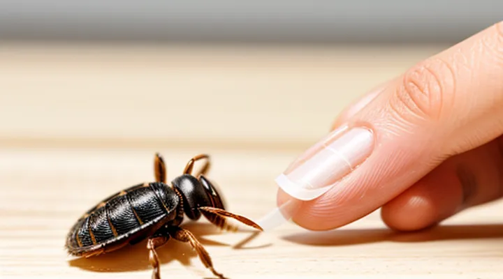
What to Prepare
Tools for Removal
Effective tick extraction relies on appropriate instruments that enable firm grip and precise control while minimizing skin trauma. The most reliable devices are:
- Fine‑point tweezers with slanted tips, preferably stainless steel, designed to grasp the tick’s head close to the skin.
- Specialized tick removal hooks, often shaped like a small, curved metal or plastic bar, allowing the mouthparts to be lifted without squeezing the body.
- Flat‑edge spatulas or blunt-edged tweezers, useful for pulling ticks that have embedded deeply but require careful positioning to avoid crushing.
- Disposable needle‑type extractors, which combine a narrow tip with a built‑in latch to secure the parasite before removal.
When selecting a tool, prioritize sterilizable materials, a narrow grasping surface, and a design that prevents the tick’s abdomen from rupturing. A clean work surface and disposable gloves further reduce contamination risk. After removal, the instrument should be disinfected with alcohol or an appropriate antiseptic to prepare it for subsequent use.
Disinfectants
When a tick is detached at home, disinfectants protect the wound from bacterial invasion and reduce the risk of secondary infection.
Before extraction, cleanse the skin surrounding the parasite with an antiseptic that rapidly kills microbes. Recommended agents include:
- 70 % isopropyl alcohol applied with a sterile swab.
- Povidone‑iodine solution (10 % w/v) applied for at least 30 seconds.
- Chlorhexidine gluconate (0.5 %–2 %) rubbed onto the area.
- 3 % hydrogen peroxide used sparingly to avoid tissue irritation.
After the tick is removed, treat the bite site with the same class of antiseptics to eliminate residual pathogens. Additionally, disinfect the tools used for removal—fine‑point tweezers or forceps—by immersing them in isopropyl alcohol for a minimum of one minute or by applying a chlorine‑based disinfectant followed by thorough rinsing.
Avoid applying disinfectants directly to the tick before removal, as this may cause the insect to release more saliva and increase pathogen transmission. Do not use harsh chemicals such as bleach or undiluted household cleaners on skin; they can cause chemical burns. Allow each antiseptic to dry completely before covering the wound with a sterile bandage.
Combining precise extraction with immediate antiseptic application ensures rapid, safe removal and minimizes complications.
Personal Protection
When a tick attaches to the skin, immediate action prevents disease transmission and reduces tissue damage. Personal protection begins with proper equipment: fine‑pointed tweezers, a pair of disposable gloves, antiseptic solution, and a clean cloth. Keep these items in a travel kit for outdoor activities.
- Wear gloves before touching the tick to avoid direct contact with saliva that may contain pathogens.
- Grip the tick as close to the skin as possible, using the tweezers’ tips to grasp the head or mouthparts.
- Apply steady, downward pressure; avoid twisting or jerking, which can leave mouthparts embedded.
- After removal, place the tick in a sealed container for identification or disposal; do not crush it.
- Clean the bite site with antiseptic, then wash hands thoroughly even though gloves were used.
Monitoring the bite area for several weeks is essential. A red ring or flu‑like symptoms may indicate infection; seek medical advice promptly if they appear.
Pre‑emptive personal protection reduces the likelihood of attachment. Use repellents containing DEET or picaridin on exposed skin, treat clothing with permethrin, and perform full‑body checks after outdoor exposure. Prompt removal, combined with these preventive steps, provides the most effective defense against tick‑borne illnesses.
Step-by-Step Tick Removal
Removing a tick promptly and properly reduces the risk of disease transmission. The following procedure provides a reliable method for safe extraction without specialized tools.
- Prepare equipment – Use fine‑pointed tweezers or a dedicated tick‑removal tool, a clean cloth, antiseptic solution, and a sealed container for disposal.
- Expose the tick – Gently part the skin around the attachment site with the cloth to see the entire body of the parasite.
- Grasp the tick – Position the tweezers as close to the skin as possible, holding the tick’s head or mouthparts, not the abdomen, to avoid crushing the body.
- Apply steady traction – Pull upward with constant, even force. Do not twist, jerk, or squeeze the tick, which could cause the mouthparts to remain embedded.
- Check for completeness – After removal, examine the bite area to confirm the entire tick, including the head, has been extracted. If any part remains, repeat the grasp‑and‑pull step.
- Disinfect the site – Clean the wound with antiseptic and allow it to air‑dry.
- Dispose of the tick – Place the specimen in the sealed container, then discard it in household trash or follow local public‑health guidelines for hazardous waste.
- Monitor the bite – Observe the area for several weeks. Note any redness, swelling, fever, or flu‑like symptoms and seek medical advice if they appear.
Following these steps ensures a swift, accurate removal while minimizing complications.
Immediate Actions After Tick Bite
After a tick attaches, act within minutes to reduce pathogen transmission risk.
- Remain calm; locate the tick’s head and body.
- Wash hands and the bite area with soap and water; avoid crushing the insect.
- Use fine‑point tweezers or a dedicated tick‑removal tool; grasp the tick as close to the skin as possible, parallel to the surface.
- Apply steady, even pressure to pull straight upward without twisting; detach the whole organism.
- Disinfect the wound with an antiseptic (e.g., iodine or alcohol).
- Preserve the tick in a sealed container with a damp cotton swab for possible laboratory identification; label with date and location of the bite.
- Monitor the bite site for redness, swelling, or a rash over the next 30 days; seek medical evaluation if symptoms develop.
- Record the incident in a personal health log, noting the removal time, tick stage (larva, nymph, adult), and any subsequent symptoms.
Prompt, precise removal and proper wound care are the essential immediate measures following a tick bite.
Correct Tick Removal Procedure
Using Tweezers
Tweezers are the preferred tool for extracting a feeding tick from human skin because they allow firm grip on the parasite’s head without crushing the body.
Procedure
- Choose fine‑point, non‑slip tweezers; disinfect the tips with alcohol.
- Grasp the tick as close to the skin as possible, holding the mouthparts, not the abdomen.
- Pull upward with steady, even pressure; avoid twisting or jerking motions.
- Continue until the tick releases completely; the entire mouthpart should be removed.
- Place the tick in a sealed container for identification if needed; discard safely.
Aftercare
- Clean the bite area with antiseptic solution.
- Apply a mild antiseptic ointment if irritation appears.
- Observe the site for several days; seek medical advice if rash, fever, or flu‑like symptoms develop.
Using a Tick Removal Tool
A tick removal device provides a secure grip on the parasite’s head, preventing the mouthparts from breaking off during extraction. The instrument typically consists of two fine, angled tips that converge on the tick’s mouthparts while a sliding mechanism holds the body steady.
- Disinfect the tool with alcohol or an antiseptic wipe.
- Grasp the tick as close to the skin as possible using the tips.
- Apply steady, downward pressure to pull the tick straight out; avoid twisting or jerking motions.
- Release the tick into a sealed container for proper disposal.
After removal, cleanse the bite area with soap and antiseptic. Observe the site for signs of redness, swelling, or fever over the next 48 hours; any abnormal symptoms require medical evaluation.
Clean the tool after each use, store it in a dry, protected case, and replace it if the tips become bent or dull. If the tick’s mouthparts remain embedded or the bite area shows signs of infection, seek professional care promptly.
What NOT to Do When Removing a Tick
Do not crush the tick’s body with fingers or tweezers. Pressing the abdomen may force saliva and infectious fluids into the bite site, increasing the risk of disease transmission.
Do not pull the tick with a rope, thread, or cotton swab. These materials lack the grip needed for a steady, controlled extraction and can cause the mouthparts to break off and remain embedded in the skin.
Do not apply heat, open flame, or a burning cigarette to the tick. Extreme temperature can cause the parasite to release more pathogens before it dies, and it does not facilitate removal.
Do not use petroleum jelly, oil, or alcohol to suffocate the tick. Chemical agents do not dislodge the organism and may irritate the skin, making it harder to grasp the tick firmly.
Do not twist, jerk, or yank the tick during removal. Sudden movements increase the chance of tearing the hypostome, leaving fragments that can cause local inflammation or infection.
Do not delay the extraction. The longer a tick remains attached, the greater the chance of pathogen transfer. Prompt removal reduces exposure time.
Do not ignore the need to clean the bite area after the tick is out. Failure to disinfect the wound can lead to secondary bacterial infection.
Do not discard the tick without proper documentation when disease monitoring is required. In regions with Lyme disease or other tick-borne illnesses, retaining the specimen for identification may be necessary for medical assessment.
Post-Removal Care and Observation
Disinfection of the Bite Site
After the tick is extracted, the wound must be treated promptly to reduce the risk of infection. Begin by washing the area with warm water and mild soap for at least 20 seconds. Rinse thoroughly, then pat dry with a clean disposable towel.
Apply an antiseptic solution directly to the bite site. Preferred agents include:
- 70 % isopropyl alcohol – effective against a broad spectrum of bacteria and viruses.
- Povidone‑iodine (Betadine) – provides sustained antimicrobial activity.
- Chlorhexidine gluconate (Hibiclens) – useful for patients with iodine sensitivity.
Allow the antiseptic to remain on the skin for a minimum of one minute before covering the area. If the chosen disinfectant requires a drying period, wait until it is completely evaporated.
Cover the cleaned site with a sterile, non‑adhesive dressing to protect against external contaminants. Change the dressing daily or whenever it becomes wet or soiled.
Monitor the bite for signs of inflammation, such as redness extending beyond the immediate area, swelling, or increased pain. Should any of these symptoms appear, seek medical evaluation promptly.
Tick Preservation and Testing
Removing a tick is only the first step; preserving the specimen allows accurate identification of the species and detection of disease‑causing agents. Proper handling prevents degradation of DNA and minimizes contamination, which is essential for reliable laboratory results.
Preservation procedure
- Use fine‑point tweezers to grasp the tick close to the skin, avoiding compression of the body.
- Transfer the tick immediately into a sealable plastic tube or a small container.
- Add a moist cotton ball or a few drops of sterile saline to keep the tick alive if testing for live pathogens is required; otherwise, submerge the tick in 70 % ethanol for long‑term storage.
- Label the container with the date, time of removal, body site, and patient information.
- Store the specimen at 4 °C (refrigerated) if it will be examined within 24 hours; otherwise, keep it at –20 °C (freezer) for longer periods.
Testing options
- Polymerase chain reaction (PCR) to detect bacterial, viral, or protozoan DNA.
- Enzyme‑linked immunosorbent assay (ELISA) for specific antigens or antibodies.
- Microscopic examination after staining to identify morphological features and internal parasites.
- Culture methods for certain bacteria, such as Borrelia spp., when viable organisms are required.
Result handling
- Submit the specimen to a qualified laboratory within the recommended timeframe (ideally within 48 hours).
- Request a detailed report indicating species, pathogen presence, and recommended clinical actions.
- Communicate findings to the treating physician promptly to guide post‑removal care and possible prophylactic treatment.
Monitoring for Symptoms
After a tick has been detached, observe the bite site and the individual for at least two weeks. Early detection of adverse reactions reduces the risk of complications.
Key signs to monitor include:
- Redness or swelling that expands beyond the immediate area of the bite.
- A rash resembling a bull’s-eye, especially if it appears 3–14 days after removal.
- Fever, chills, headache, muscle aches, or joint pain that develop within a week.
- Nausea, vomiting, or abdominal pain.
- Neurological symptoms such as facial weakness, numbness, or difficulty concentrating.
If any of these manifestations arise, seek medical evaluation promptly. Documentation of the tick’s appearance, date of removal, and symptom onset assists healthcare providers in diagnosing tick‑borne illnesses. Continuous self‑assessment, even in the absence of immediate symptoms, remains essential for effective post‑removal care.
When to Seek Medical Attention
Incomplete Tick Removal
Incomplete removal of a tick leaves mouthparts embedded in the skin, creating a portal for pathogens and provoking local inflammation. The retained fragments can cause prolonged itching, redness, and, in some cases, secondary bacterial infection. Prompt identification and correction are essential for effective treatment.
Signs that removal was incomplete include a visible hollow head, a small dark spot at the bite site, or persistent irritation after the tick is gone. If any of these indicators appear, the following actions should be taken without delay:
- Disinfect the area with an antiseptic solution (e.g., iodine or chlorhexidine).
- Use fine‑point tweezers or a specialized tick‑removal tool to grasp the exposed part of the mouthparts as close to the skin as possible.
- Apply steady, gentle traction directly outward, avoiding twisting or squeezing the body.
- Continue pulling until the entire structure detaches; the removal should be smooth, without resistance.
- Once the fragment is out, clean the wound again and apply a sterile dressing.
If the mouthparts cannot be extracted with tweezers, professional medical assistance is required. A healthcare provider may use a small scalpel or forceps under sterile conditions to excise the remaining tissue, minimizing tissue damage and reducing infection risk.
After successful removal, monitor the site for several days. Persistent redness, swelling, or fever warrants immediate medical evaluation, as these symptoms may signal early Lyme disease or other tick‑borne illnesses. Documentation of the removal date, tick appearance, and any subsequent symptoms assists clinicians in diagnosis and treatment planning.
Appearance of Rash or Fever
After a tick is removed, the emergence of a skin eruption or an elevated body temperature often signals that the bite transmitted a pathogen. A macular or papular rash typically appears within 3‑7 days, though some infections, such as Lyme disease, may produce a characteristic expanding erythema after 5‑14 days. Fever commonly develops alongside the rash, but it can also arise as an isolated symptom, indicating systemic involvement.
When a rash or fever is observed, immediate medical evaluation is required. Physicians will assess the lesion’s size, shape, and distribution, and may order serologic tests to identify the causative agent. Empiric antibiotic therapy, most frequently doxycycline, is initiated in suspected early Lyme disease or other tick‑borne infections to prevent progression and complications.
Patients should monitor the progression of symptoms for at least two weeks after removal. Documentation of the rash’s dimensions and any changes in temperature helps clinicians determine treatment efficacy. If symptoms worsen—expanding rash, persistent fever, joint pain, or neurological signs—prompt re‑evaluation is essential to adjust therapy.
Key actions when rash or fever occurs:
- Record the date of tick removal and onset of symptoms.
- Measure temperature twice daily; note peaks and duration.
- Photograph the rash for serial comparison.
- Contact a healthcare provider without delay for diagnostic testing and possible prescription of antibiotics.
Timely recognition and professional management of rash or fever following a tick bite markedly reduce the risk of long‑term sequelae.
General Concerns
Removing a tick from a person at home raises several health‑related concerns that must be addressed before attempting the procedure.
First, the risk of pathogen transmission increases with the duration of attachment. Ticks can carry bacteria, viruses, and parasites that cause diseases such as Lyme disease, anaplasmosis, and babesiosis. Prompt removal reduces the chance of pathogen entry, but incomplete extraction may leave mouthparts embedded, creating a portal for infection.
Second, improper technique can cause tissue damage. Grasping the tick’s body with fingers or squeezing its abdomen may force infected fluids into the host’s bloodstream. Using fine‑pointed tweezers, positioning them as close to the skin as possible, and pulling steadily without twisting minimizes trauma.
Third, environmental factors affect the procedure’s success. A well‑lit, clean surface prevents loss of the tick and reduces contamination. Disinfecting the removal site before and after extraction lowers secondary infection risk.
Key concerns to monitor after removal:
- Persistent redness, swelling, or rash at the bite site
- Fever, headache, fatigue, or joint pain within weeks of the bite
- Presence of a small, hard nodule that may indicate retained mouthparts
If any of these symptoms appear, professional medical evaluation is required.
