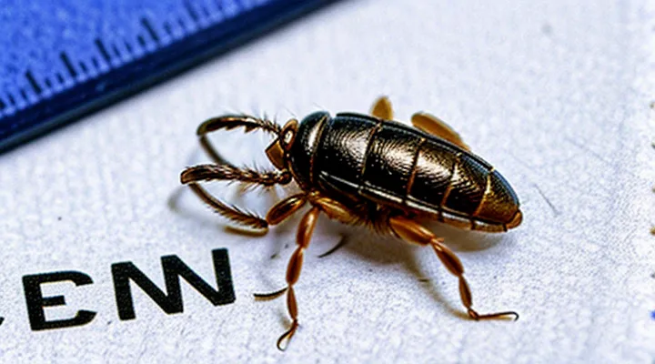
Understanding Tick-Borne Diseases
Common Tick-Borne Illnesses
Ticks transmit a limited set of pathogens that cause recognizable clinical syndromes. The interval between a bite and the appearance of symptoms varies by organism, influencing diagnosis and treatment decisions.
- Lyme disease (Borrelia burgdorferi) – erythema migrans may develop within 3–30 days; other manifestations (fever, headache, fatigue) often appear 1–2 weeks after infection.
- Rocky Mountain spotted fever (Rickettsia rickettsii) – fever, rash, and headache typically emerge 2–14 days post‑bite, most commonly around day 5.
- Anaplasmosis (Anaplasma phagocytophilum) – flu‑like symptoms and leukopenia usually begin 5–14 days after exposure.
- Babesiosis (Babesia microti) – low‑grade fever, hemolysis, and malaise often start 1–4 weeks after the bite.
- Ehrlichiosis (Ehrlichia chaffeensis) – fever, muscle aches, and leukopenia generally present 5–10 days post‑exposure.
- Tularemia (Francisella tularensis) – ulceroglandular form appears 3–5 days after inoculation; pneumonic form may develop 1–2 weeks later.
- Powassan virus disease – neurologic symptoms such as encephalitis can arise rapidly, within 1–5 days, but may also be delayed up to 2 weeks.
Understanding these typical latency periods helps clinicians recognize tick‑borne infections promptly and initiate appropriate therapy.
Factors Influencing Disease Transmission
Tick-borne disease transmission depends on several biological and environmental variables that directly affect the interval between exposure and clinical manifestation. Pathogen load within the tick, the duration of attachment, and feeding depth determine the quantity of infectious material transferred. Longer feeding periods increase inoculum size, often shortening the latency before symptoms emerge.
Host-related factors shape the incubation timeline. Age, immune competence, and pre‑existing conditions influence pathogen replication rates. Immunocompromised individuals may experience accelerated symptom onset, whereas robust immune defenses can delay or mitigate clinical signs. Genetic differences among host species also modulate susceptibility to specific tick‑borne agents.
Environmental conditions alter both vector behavior and pathogen viability. Temperature and humidity control tick activity, questing duration, and survival of the pathogen within the vector. Seasonal peaks in tick activity correspond with higher transmission risk and may compress the window between bite and symptom development.
Key influences on disease transmission timing:
- Pathogen species and strain virulence
- Tick attachment duration and feeding intensity
- Host immune status and comorbidities
- Ambient temperature, humidity, and seasonal patterns
- Geographic distribution of tick vectors and reservoir hosts
Factors Affecting Symptom Onset
Type of Tick-Borne Disease
Tick-borne infections differ markedly in the interval between attachment and the appearance of clinical signs. The latency period depends on the pathogen’s replication cycle, the site of inoculation, and host immune response.
- Lyme disease (Borrelia burgdorferi): erythema migrans may develop within 3–30 days; systemic symptoms such as fever or arthralgia often follow weeks later.
- Rocky Mountain spotted fever (Rickettsia rickettsii): fever, headache, and rash typically emerge 2–5 days after the bite; severe complications can arise by the end of the first week.
- Anaplasmosis (Anaplasma phagocytophilum): flu‑like illness appears 5–14 days post‑exposure; laboratory abnormalities may precede overt symptoms.
- Babesiosis (Babesia microti): nonspecific symptoms emerge 1–4 weeks after inoculation; severe hemolysis can be delayed further in immunocompromised patients.
- Tick‑borne encephalitis virus: initial febrile phase occurs 3–14 days; neurologic involvement may develop 1–2 weeks after the first signs.
- Ehrlichiosis (Ehrlichia chaffeensis): incubation averages 5–10 days, with fever and malaise often preceding laboratory changes.
Monitoring for at least four weeks after a known or suspected bite captures the majority of symptom onsets. Early laboratory testing should be considered when the incubation window of the suspected pathogen aligns with emerging signs. Prompt recognition of the specific latency pattern enables timely treatment and reduces the risk of complications.
Individual Immune Response
The speed at which clinical signs become evident after a tick attachment depends largely on how the host’s immune system reacts to the inoculated pathogens. Immediate innate mechanisms, such as skin‑resident macrophages and neutrophils, attempt to contain the foreign material within hours. If these cells recognize pathogen‑associated molecules, they release cytokines that generate localized redness, swelling, or itching, often visible within 1–3 days.
When the pathogen evades early detection, adaptive immunity must be engaged. Individuals with prior exposure to the same tick‑borne organism typically mount a faster antibody‑mediated response, shortening the latency to several days. Conversely, naïve hosts, the elderly, or immunocompromised patients may experience delayed symptom onset, extending the interval to 1–3 weeks.
Key determinants of the timing include:
- Previous sensitization – memory B‑cells accelerate antibody production.
- Genetic factors – variations in Toll‑like receptor expression modify innate signaling strength.
- Age – reduced immune vigor in older adults slows both innate and adaptive phases.
- Health status – conditions such as HIV, chemotherapy, or corticosteroid therapy impair response speed.
- Tick species and pathogen load – higher inoculum can trigger earlier inflammation, while low‑dose exposures may remain subclinical longer.
Thus, the individual immune profile shapes the interval between tick bite and observable symptoms, ranging from a few days in robust responders to several weeks in compromised or inexperienced hosts.
Duration of Tick Attachment
Ticks must remain attached long enough to transfer pathogens. The interval between attachment and symptom emergence depends on how quickly a specific organism is transmitted and the host’s response.
- Borrelia burgdorferi (Lyme disease): transmission usually requires ≥ 36 hours of attachment; symptoms may appear 3–30 days after the bite.
- Anaplasma phagocytophilum (Anaplasmosis): can be transmitted after 24 hours; fever and malaise often develop within 1–2 weeks.
- Rickettsia rickettsii (Rocky Mountain spotted fever): transmission may occur within 6–12 hours; rash and fever typically emerge 2–5 days post‑bite.
- Babesia microti (Babesiosis): needs at least 48 hours of feeding; clinical signs usually arise 1–4 weeks later.
- Powassan virus: can be transmitted in as little as 15 minutes; neurological symptoms may develop within 1 week.
Factors that modify these intervals include tick species, life stage, attachment site, host immune status, and ambient temperature. Prompt removal—grasping the tick close to the skin and pulling straight upward—reduces the chance of pathogen transfer. After extraction, monitor the bite site and overall health for at least several weeks; seek medical evaluation if fever, rash, joint pain, or neurologic signs appear.
Typical Incubation Periods for Major Tick-Borne Diseases
Lyme Disease
Lyme disease manifests after a tick bite according to a defined temporal pattern. The bacterium Borrelia burgdorferi requires a minimum of 36–48 hours of attachment before transmission becomes likely; bites removed earlier rarely result in infection.
Typical onset timeline
- Early localized stage – erythema migrans (expanding skin rash) appears in 3–30 days, most commonly 7–14 days post‑exposure. Flu‑like symptoms (fever, chills, headache, fatigue, muscle aches) may accompany the rash within the same window.
- Early disseminated stage – multiple erythema migrans lesions, cardiac involvement (e.g., atrioventricular block), neurologic signs (facial palsy, meningitis) emerge weeks to a few months after the bite, often 2–6 weeks.
- Late disseminated stage – chronic arthritis, particularly of large joints, and neurocognitive deficits develop months to years later, typically beyond six months if untreated.
The interval between tick attachment and symptom appearance varies with the stage of infection, the duration of attachment, and host immune response. Prompt removal of the tick before the 36‑hour threshold markedly reduces the risk of any clinical manifestation.
Rocky Mountain Spotted Fever
Rocky Mountain spotted fever (RMSF) is a bacterial infection transmitted primarily by the American dog tick (Dermacentor variabilis) and the Rocky Mountain wood tick (Dermacentor andersoni). The causative agent, Rickettsia rickettsii, replicates within endothelial cells, producing a systemic vasculitis.
The interval between tick attachment and the appearance of symptoms usually ranges from two to fourteen days, with most patients developing signs within five to seven days. Shorter incubation periods are reported for heavily infected ticks, while longer intervals may occur when the bacterial load is low.
Typical clinical evolution proceeds as follows:
- Days 1‑3: abrupt fever, chills, severe headache, and myalgia.
- Days 4‑6: appearance of a maculopapular rash that may become petechial, often starting on wrists and ankles before spreading centrally.
- Days 7‑10: possible progression to edema, hypotension, and organ dysfunction if untreated.
Prompt recognition relies on clinical suspicion, recent tick exposure, and laboratory evidence such as elevated hepatic transaminases, thrombocytopenia, or a positive PCR assay for R. rickettsii. Empiric doxycycline therapy should begin immediately after suspicion; delays increase mortality.
Early antimicrobial intervention, initiated within the first 48 hours of symptom onset, markedly reduces severe complications and fatality rates. The rapid onset of RMSF after a tick bite underscores the necessity for vigilance in endemic regions.
Anaplasmosis
Anaplasmosis is transmitted to humans by the bite of an infected Ixodes tick. After exposure, the pathogen begins to replicate in neutrophils, and clinical signs usually emerge within a defined window. The incubation period most commonly ranges from 5 to 21 days, with occasional reports of onset as early as 3 days or as late as 30 days following the bite.
Typical early manifestations that appear during this interval include:
- Fever (often ≥ 38 °C)
- Chills
- Headache
- Myalgia
- Malaise
- Nausea or vomiting
Laboratory findings frequently show leukopenia, thrombocytopenia, and elevated liver enzymes, supporting the clinical suspicion.
Factors that may shorten or prolong the latency period are:
- Age (children and elderly may present sooner)
- Immunocompromised status (accelerated progression)
- Co‑infection with other tick‑borne agents (may modify symptom onset)
Prompt recognition of the time frame between tick exposure and symptom development is essential for early diagnostic testing and initiation of doxycycline therapy, which reduces disease severity and prevents complications.
Ehrlichiosis
Ehrlichiosis is a bacterial infection transmitted by the lone‑star tick (Amblyomma americanum) and, less frequently, by other ixodid species. The pathogen, primarily Ehrlichia chaffeensis, invades monocytes and neutrophils, producing a systemic febrile illness.
The interval between a tick attachment and the appearance of clinical signs usually spans 5–14 days. Cases have been documented with onset as early as 2 days and as late as 21 days after the bite, reflecting variability in bacterial inoculum and host immune response.
Typical progression:
- Days 1‑3: Asymptomatic; tick may still be attached.
- Days 4‑7: Low‑grade fever, headache, malaise, myalgia; laboratory abnormalities (thrombocytopenia, leukopenia) may emerge.
- Days 8‑14: Intensified fever, rash (occasionally), elevated liver enzymes; severe forms can develop acute respiratory distress or organ failure.
- Beyond day 14: Persistent or relapsing symptoms if untreated; risk of complications increases.
Factors that shorten or extend the latency period include:
- Tick attachment duration (longer feeding delivers more organisms).
- Species of Ehrlichia (different strains have distinct virulence).
- Host age and immunocompetence (immunosuppressed individuals may manifest earlier or more severe disease).
- Prompt removal of the tick (reduces bacterial load).
Diagnostic accuracy depends on timing. Polymerase chain reaction (PCR) yields the highest sensitivity within the first two weeks, whereas serologic conversion is reliably detectable after 7–10 days. Blood smear examination may reveal morulae in leukocytes during the acute phase but is less sensitive.
Early recognition of the typical incubation window enables timely antimicrobial therapy, most commonly doxycycline, which reduces morbidity and prevents progression to severe disease.
Babesiosis
Babesiosis is a tick‑borne infection caused primarily by Babesia microti in the United States and by Babesia divergens in Europe. After an infected ixodid tick feeds, the parasite enters the bloodstream and multiplies within red blood cells. The period between the bite and the appearance of clinical signs varies, but most patients develop symptoms within 1 to 4 weeks. A minority may remain asymptomatic for up to two months, especially if the immune response limits parasite growth.
Typical timing of symptom onset:
- 1–7 days: Rare; may occur in heavily infested individuals or those with pre‑existing immunosuppression.
- 7–14 days: Most common window; fever, chills, sweats, and fatigue often emerge.
- 14–28 days: Progressive anemia, hemoglobinuria, and jaundice become more apparent.
- Beyond 28 days: Delayed presentation possible in immunocompromised hosts or when co‑infection with Lyme disease masks early signs.
Factors influencing the interval include the tick’s infection load, the host’s age, immune status, and whether prophylactic antibiotics were administered after the bite. Early laboratory confirmation—via blood smear, PCR, or serology—facilitates prompt treatment, reducing the risk of severe hemolytic complications.
Powassan Virus
Powassan virus is a tick‑borne flavivirus that can cause severe encephalitis and meningitis. The period between a bite from an infected tick and the appearance of clinical signs is short compared with many other tick‑borne infections.
The incubation interval typically ranges from five days to four weeks, with most cases presenting around ten to fourteen days after exposure. Rarely, symptoms may emerge as early as three days or as late as thirty days post‑bite.
Early manifestations often include:
- Fever
- Headache
- Nausea or vomiting
- Fatigue
- Confusion or altered mental status
Neurological complications such as seizures, focal deficits, or coma can develop within days of the initial fever, sometimes progressing rapidly despite prompt medical care.
Because the window for symptom onset is brief, clinicians should consider Powassan virus in patients who develop acute febrile illness or neurologic signs shortly after a tick bite, especially in endemic regions. Early recognition and supportive treatment improve the likelihood of a favorable outcome.
Recognizing Early Symptoms
General Symptoms to Watch For
After a tick attaches, the host may experience a range of early signs. Most reactions appear within hours to a few days, while others develop over several weeks.
- Local redness or swelling at the bite site, often accompanied by a small, raised bump.
- Itching or mild pain around the attachment point.
- A rash that expands outward, sometimes forming a target‑shaped lesion (often called a “bull’s‑eye” pattern).
- Fever, chills, or a general feeling of malaise that emerges days after the bite.
- Muscle or joint aches, frequently reported one to two weeks post‑exposure.
- Headache, nausea, or fatigue that may accompany systemic involvement.
- Neurological signs such as tingling, numbness, or facial weakness, typically appearing later in the incubation period.
The timing of each symptom varies with the pathogen transmitted and the individual’s immune response. Prompt recognition of these manifestations enables early medical evaluation and treatment.
Disease-Specific Early Indicators
Tick-borne infections display distinct early clinical cues that help estimate the interval between exposure and symptom manifestation. Recognizing these disease‑specific signals enables prompt diagnosis and treatment.
Early indicators and typical latency periods:
- Lyme disease (Borrelia burgdorferi): Expanding erythema migrans appears 3–30 days after the bite; accompanying fatigue, headache, or mild fever may precede the rash.
- Rocky Mountain spotted fever (Rickettsia rickettsii): Sudden high fever, severe headache, and myalgia emerge within 2–14 days; a maculopapular rash often follows on day 3–5, beginning on wrists and ankles.
- Anaplasmosis (Anaplasma phagocytophilum): Fever, chills, and malaise develop 5–14 days post‑exposure; laboratory tests frequently reveal leukopenia and thrombocytopenia early in the course.
- Ehrlichiosis (Ehrlichia chaffeensis): Similar to anaplasmosis, fever and myalgia arise 5–10 days after the bite, with possible rash on the trunk after day 7.
- Babesiosis (Babesia microti): Nonspecific flu‑like symptoms, including fever and chills, present 1–4 weeks after infection; hemolytic anemia may become evident early in severe cases.
These concise timeframes align each early manifestation with the underlying pathogen, providing clinicians a practical reference for estimating the period between tick attachment and observable illness.
When to Seek Medical Attention
Immediate Concerns After a Bite
After discovering a tick attached to the skin, the first priority is to eliminate the parasite safely. Use fine‑point tweezers to grasp the tick as close to the surface as possible, pull upward with steady pressure, and avoid crushing the body. Immediately clean the bite site with soap and water or an antiseptic solution.
- Grasp tick with tweezers at the head‑base
- Pull upward with constant force, no twisting
- Disinfect area after removal
- Store the tick in a sealed container for identification, if needed
- Record the date, location, and estimated duration of attachment
Following removal, monitor the bite for early warning signs that may precede the typical latency of tick‑borne diseases. Prompt medical evaluation is warranted if any of the following occurs:
- Expanding redness or a bullseye‑shaped rash around the bite
- Fever, chills, or flu‑like symptoms within days of exposure
- Severe headache, neck stiffness, or neurological changes
- Joint pain, swelling, or unexplained fatigue
These immediate concerns demand attention regardless of the usual incubation window for infections transmitted by ticks. Early intervention can reduce the risk of complications and improve treatment outcomes.
Delayed Symptoms and Follow-Up
After a tick attachment, certain infections may not produce noticeable signs until several days or weeks have passed. Early recognition of these delayed manifestations is essential for timely treatment.
Typical late‑appearing signs include:
- Expanding red skin lesion with central clearing (often called a “bull’s‑eye” rash)
- Persistent fever or intermittent chills
- Severe headache, sometimes accompanied by neck stiffness
- Muscle or joint aches, especially in larger joints
- Nausea, vomiting, or abdominal pain
- Neurological disturbances such as facial palsy, tingling, or memory problems
Incubation periods vary by pathogen. Lyme disease commonly shows a rash within 3‑30 days; anaplasmosis and ehrlichiosis present fever and malaise after 5‑14 days; babesiosis symptoms may emerge 1‑4 weeks post‑bite; Rocky Mountain spotted fever typically manifests 2‑14 days after exposure. These intervals guide clinicians in correlating exposure history with clinical presentation.
Follow‑up actions should include:
- Immediate medical assessment if any delayed sign appears.
- Laboratory testing tailored to suspected agents (e.g., serology, PCR, blood smears).
- Consideration of a single dose of doxycycline for prophylaxis when the tick was attached ≥36 hours and the region has a high incidence of Lyme disease.
- Scheduled re‑evaluation, usually within 2‑4 weeks, to monitor symptom progression and test results.
- Documentation of tick removal date, attachment duration, and geographic location to aid diagnostic accuracy.
Adhering to this protocol reduces the risk of complications and ensures appropriate therapeutic intervention.
