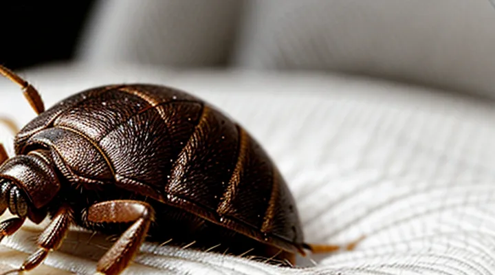
The Bed Bug's Mouthparts
The Proboscis: A Specialized Feeding Tube
The proboscis of a bedbug functions as a highly specialized feeding apparatus. It consists of a slender, elongated tube formed from the mandibles and maxillae, which together create a canal for saliva injection and blood extraction. The tip of the proboscis is equipped with a pair of serrated stylets that pierce the epidermis, allowing access to dermal capillaries.
During a feeding event, the insect first inserts the stylets into the skin, then releases saliva containing anticoagulants and anesthetic compounds. These substances prevent clot formation and mask the bite, enabling continuous blood flow. The proboscis then draws blood upward through the canal by muscular contractions of the cibarial pump located in the head.
Key features of the proboscis:
- Dual‑stylet configuration for precise penetration
- Salivary glands attached to the tube for immediate injection of bioactive fluids
- Muscular pump mechanism that generates suction pressure
- Flexible, sclerotized cuticle that resists deformation during repeated feeds
These adaptations allow bedbugs to obtain a blood meal with minimal detection by the host.
Maxillary and Mandibular Styles
Bedbugs attach to the skin using their specialized mouthparts, which consist of a maxilla and a mandible that function in distinct styles during the blood‑feeding process. The maxilla is elongated, serrated, and equipped with a series of fine, hook‑like teeth. These teeth anchor the insect to the epidermis, preventing displacement as the insect inserts its proboscis. The mandible, shorter and more robust, serves primarily to cut through the superficial layers of the stratum corneum, creating a narrow incision through which the maxilla can advance.
During a feeding episode, the sequence proceeds as follows:
- The mandible pierces the outer skin, producing a micro‑incision of approximately 0.2 mm.
- The maxilla slides into the incision, its serrated edges gripping the tissue and stabilizing the feeding site.
- Salivary enzymes are injected; they contain anticoagulants and anesthetics that facilitate uninterrupted blood intake.
- The cibarial pump, powered by thoracic muscles, draws blood through the maxillary canal into the insect’s gut.
The combination of cutting (mandibular) and anchoring (maxillary) actions enables bedbugs to feed efficiently while remaining undetected, accounting for the characteristic delayed itching and welts observed in human hosts.
The Biting Process
Locating a Host
Bedbugs locate a human host through a combination of sensory cues that indicate the presence of warm‑blooded organisms. The insects are equipped with thermoreceptors that detect temperature gradients, allowing them to move toward the heat emitted by skin. Simultaneously, they sense carbon dioxide released during exhalation; elevated CO₂ levels trigger an oriented response toward the source.
Chemical signals from the skin also guide the search. Volatile compounds such as fatty acids, lactic acid, and ammonia act as attractants, prompting the bugs to follow the concentration trail toward a potential meal. Mechanical stimuli, including subtle movements and vibrations, further refine the approach, helping the insects pinpoint a specific location on the host’s body.
Key cues used in host detection:
- Body heat gradients
- Carbon dioxide plumes
- Skin‑derived volatile compounds (fatty acids, lactic acid, ammonia)
- Low‑frequency vibrations and motion
These cues operate synergistically, enabling bedbugs to locate a suitable feeding site with precision even in low‑light environments. Once the host is identified, the insect positions itself to insert its proboscis and commence blood ingestion.
Anesthetic and Anticoagulant Saliva
Bedbugs inject a complex saliva mixture that serves two primary functions during a blood meal: sensory suppression and hemostasis inhibition.
The anesthetic fraction contains small neuroactive peptides that block voltage‑gated sodium channels in peripheral nerve endings. By preventing the initiation of action potentials, these compounds eliminate the immediate itch and pain that would otherwise alert the host. Research identifies the protein called “BED‑AN” as a potent blocker of Nav1.7 channels, producing a localized numbness lasting several minutes.
The anticoagulant fraction comprises enzymes and binding proteins that interfere with the host’s clotting cascade. Key components include:
- Apyrase, which hydrolyzes ATP and ADP, reducing platelet aggregation.
- A thrombin inhibitor that binds the active site of thrombin, preventing fibrin formation.
- A metalloprotease that degrades fibrinogen, further weakening clot stability.
Together, these agents maintain a fluid feeding site, allowing the insect to extract blood uninterrupted for up to ten minutes. The combined effect of numbing and anticoagulation explains why many bites remain unnoticed until after the insect has detached, often manifesting only as delayed erythema or swelling.
The Feeding Duration
Bedbugs attach to the host for a limited period, usually completing a blood meal within a few minutes. The feeding process consists of four phases: (1) locating a suitable site, (2) inserting the stylet, (3) ingesting blood, and (4) withdrawing and detaching.
- Initial probing: 10–30 seconds. The insect inserts its mouthparts and assesses skin temperature and carbon‑dioxide levels.
- Blood intake: 2–5 minutes for a hungry adult; 1–2 minutes for a nymph. During this stage the insect expands its abdomen up to three times its unfed size.
- Detachment: 15–30 seconds. The bug releases saliva containing anticoagulants and anesthetics, then slides away.
Feeding duration varies with environmental temperature (warmer conditions accelerate metabolism), host activity (movement can interrupt feeding), and the insect’s hunger level (starved individuals may feed longer). Life‑stage differences also influence time: first‑instar nymphs require less than a minute, while mature females may need up to seven minutes when heavily engorged.
Short feeding intervals reduce the chance of detection, yet the rapid engorgement allows the insect to acquire enough blood to sustain development and reproduction. Understanding the precise timing of each phase aids in designing monitoring tools and timing insecticide exposure for maximum efficacy.
The Aftermath of a Bed Bug Bite
Bite Characteristics
Bedbug feeding results in a recognizable set of skin reactions that differ from many other insect bites. The puncture marks are typically small, ranging from 1 to 3 mm in diameter, and appear as raised, red welts. Often a central dark spot marks the site where the insect’s mouthparts penetrated the epidermis.
- Onset: Redness and swelling develop within a few minutes to several hours after the bite. In some individuals, the reaction may be delayed up to 24 hours.
- Distribution: Bites commonly appear in linear or clustered patterns, reflecting the insect’s tendency to move along a host’s skin while feeding.
- Itching intensity: The lesions provoke moderate to severe pruritus, which can persist for several days. Scratching may lead to secondary infection.
- Variation: Allergic sensitivity influences the severity; highly sensitized persons may experience larger, more inflamed wheals, whereas others exhibit only faint erythema.
- Healing: Lesions typically resolve within 7–10 days without scarring, unless complicated by infection or excessive scratching.
Common Reactions and Symptoms
Bedbug bites typically produce small, raised welts that appear as red or pink spots on exposed skin. The lesions develop within a few minutes to several hours after the insect feeds and often cluster in linear or zig‑zag patterns, reflecting the insect’s movement across the host.
Common reactions include:
- Intense itching that may persist for several days
- Localized swelling or edema surrounding the bite site
- Redness that can spread outward from the central puncture point
- A central punctum or dark spot where the bug inserted its mouthparts
In some individuals, the immune response is more pronounced, leading to larger plaques, hives, or a generalized rash. Allergic sensitization may cause:
- Rapid onset of widespread urticaria
- Shortness of breath or wheezing in severe cases (anaphylaxis)
Secondary complications arise if the bite is scratched, allowing bacterial entry. Signs of infection comprise:
- Increasing pain, warmth, and purulent discharge
- Fever or chills accompanying the local reaction
Symptoms usually resolve within one to two weeks without medical intervention, although persistent itching may require topical corticosteroids or antihistamines. Prompt cleaning of the bite area and avoidance of excessive scratching reduce the risk of secondary infection.
Factors Influencing Bite Severity
Bedbug feeding results in a puncture wound that can vary widely in intensity. The degree of discomfort and skin reaction depends on several measurable factors.
- Host immune response – Individuals with heightened histamine release experience larger, more inflamed welts.
- Number of insects feeding simultaneously – Multiple bites in a confined area amplify tissue damage and swelling.
- Duration of blood extraction – Longer attachment times allow more saliva injection, increasing irritation.
- Anatomical location – Thin‑skinned regions such as the face or neck show sharper reactions than thicker areas like the thighs.
- Age and skin condition – Children and people with compromised or aged skin exhibit more pronounced lesions.
- Previous exposure – Repeated encounters can either sensitize the host, leading to stronger reactions, or induce tolerance, reducing severity.
- Blood volume and flow – Higher perfusion rates facilitate faster feeding, potentially limiting the amount of saliva deposited and moderating the bite’s impact.
These variables interact to shape the observable outcome of a bedbug bite, explaining why some victims develop merely a faint red spot while others suffer extensive swelling and itching. Understanding each factor aids in predicting bite severity and tailoring appropriate medical or preventive measures.
