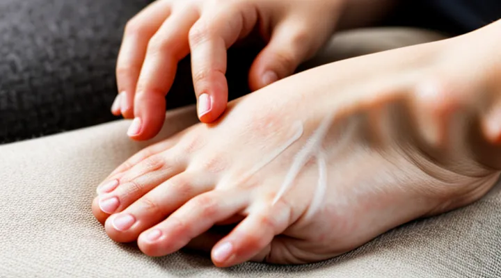
Understanding Scabies
What Scabies Is
The Scabies Mite «Sarcoptes Scabiei»
The mite responsible for scabies, «Sarcoptes scabiei», measures 0.2–0.4 mm and burrows within the stratum corneum. Female mites create serpentine tunnels where eggs are deposited; these tunnels become visible as thin, grayish lines on the skin.
Typical manifestations include intense nocturnal pruritus, erythematous papules, and linear burrows most often located on wrists, interdigital spaces, elbows, waistline, and genital region. The presence of a visible tunnel or a raised papule at the end of a tunnel strongly suggests active infestation.
Diagnostic methods applicable to self‑assessment:
- Visual inspection of characteristic burrows using a magnifying lens (10×–20×).
- Dermoscopic examination; a handheld dermatoscope reveals a triangular “delta” sign corresponding to the mite’s head and legs.
- Adhesive tape test; press clear medical tape onto a suspected burrow, lift, and examine the strip under a light microscope for mite bodies or eggs.
- Skin scraping; collect superficial material from the edge of a burrow, place on a slide, and observe under a microscope at 100×–400× for adult mites, eggs, or fecal pellets.
For practical self‑examination, wash hands thoroughly, dry skin, and systematically scan common sites with a magnifier. Mark any linear lesions, then apply adhesive tape to the most suspicious area and inspect the collected sample with a handheld microscope or send it to a laboratory for confirmation. Early identification enables prompt treatment and prevents secondary transmission.
How Scabies Spreads
Scabies mites transfer primarily through prolonged skin‑to‑skin contact. A single encounter of a few seconds rarely results in transmission; exposure lasting several minutes increases risk. Close relationships—family members, sexual partners, caregivers—represent the most common pathways because continuous contact allows mites to crawl from one host to another.
Secondary routes involve objects that retain live mites for up to 72 hours. Contaminated clothing, bedding, towels, or upholstered furniture can serve as vectors, especially in crowded living conditions where items are shared frequently. Direct transmission remains dominant, but indirect exposure contributes to outbreaks in institutions such as nursing homes, prisons, and shelters.
The life cycle influences spread. After a female mite burrows and lays eggs, the next generation emerges within 10 days, becoming capable of moving to a new host. During this period, any person who touches the infested skin or contaminated items may acquire mites before symptoms appear. The incubation interval ranges from 2 weeks in naïve individuals to 1 day in previously sensitized hosts, shortening the window for unnoticed transmission.
Key factors that amplify dissemination include:
- Frequent close contact with an infected individual.
- Sharing of personal items without washing at high temperatures.
- Living in densely populated environments.
- Delayed diagnosis or treatment, extending the period of contagiousness.
Understanding these mechanisms clarifies why early identification of the mite on one’s own skin is essential for interrupting the chain of infection.
Common Scabies Symptoms
Intense Itching «Pruritus»
Intense itching, medically termed «pruritus», serves as a primary indicator of a possible scabies infestation. The sensation typically peaks during nighttime, prompting frequent awakenings and uncontrollable scratching.
Key characteristics of scabies‑related «pruritus» include:
- Distribution on interdigital spaces, wrists, elbows, waistline, and genitals;
- Appearance of linear or serpentine burrows accompanying the itch;
- Rapid escalation of intensity over days, often after initial exposure;
- Absence of similar symptoms on the face or scalp in adults.
Self‑examination should focus on these patterns. Use a bright light source and, if available, a dermatoscope to locate raised, gray‑white tunnels within the epidermis. Gentle skin scraping of suspected burrows may reveal mites or eggs for microscopic review.
Persistent nocturnal itch, especially when combined with visible burrows, warrants prompt medical evaluation. Dermatological confirmation typically involves skin scraping, adhesive tape test, or dermoscopic identification of the mite’s characteristic “delta wing” sign. Early detection reduces transmission risk and facilitates timely treatment.
Rash Characteristics
The presence of a scabies infestation is most reliably indicated by a distinctive rash.
Typical lesions appear as tiny, erythematous papules, frequently accompanied by thin, gray‑white tunnels (burrows) that trace the mite’s path beneath the skin surface. The papules are often intensely itchy, especially during nighttime hours.
Distribution follows a predictable pattern: interdigital spaces of the hands, wrists, elbows, armpits, waistline, and genital region. In infants, the rash may extend to the face, scalp, and palms.
Key characteristics that differentiate scabies from other dermatological conditions include:
- Linear or serpentine burrows visible under magnification;
- Concentration in skin folds and areas where the skin contacts itself;
- Absence of primary lesions on the trunk in adults;
- Rapid spread to close contacts through direct skin‑to‑skin contact.
Recognition of these rash attributes enables self‑assessment of a possible scabies infestation and informs timely medical consultation.
Burrows on the Skin
Burrows appear as thin, gray‑white or skin‑colored lines on the surface of the skin. They are typically 2–10 mm long and follow the direction of hair growth. The most common locations include the wrists, between the fingers, the elbows, the armpits, the waistline, the buttocks and the genital area. Burrows often contain a small dot at one end, representing the female mite, and may be slightly raised or feel like a sandpaper texture when stroked gently.
Key visual cues for self‑examination:
- Linear or serpentine tracks aligned with hair follicles
- Presence of a tiny, dark speck at the terminus of the line
- Intense itching that worsens at night and after bathing
- Slight elevation or roughness of the track compared to surrounding skin
To confirm suspicion, examine the skin in a well‑lit area, using a magnifying lens if available. A close inspection may reveal the mite’s silhouette within the burrow. If burrows are identified, prompt medical consultation and treatment are advised.
Self-Detection of Scabies
Visual Examination of Skin
Identifying Scabies Burrows
Scabies burrows appear as thin, gray‑white or slightly reddish lines on the skin. They measure 2–10 mm in length and follow the natural direction of hair growth. Common sites include the web spaces of the fingers, wrists, elbows, axillae, waistline, buttocks, and genital area. The ends of the tunnels may terminate in a small vesicle or papule where the female mite lays eggs.
Visual inspection with adequate lighting is the first step. A magnifying glass or a handheld dermatoscope enhances the ability to distinguish the characteristic S‑shaped or linear pattern from ordinary scratches. When magnification is unavailable, a handheld LED lamp can improve contrast between the burrow and surrounding epidermis.
Key visual features of scabies burrows:
- Linear or serpentine track, often parallel to skin folds
- Color ranging from translucent to reddish‑brown
- Length of 2–10 mm, sometimes extending beyond visible borders
- Presence of a tiny raised papule at one terminus
- Localization to typical anatomical regions
If burrows are suspected, a skin scraping should be performed. A sterile blade collects material from the active edge of the tunnel. The sample is examined under a light microscope at 100–400× magnification. The presence of adult mites, eggs, or fecal pellets confirms infestation.
Prompt identification of burrows enables early treatment and reduces transmission risk. Regular self‑examination, especially after exposure to known cases, increases detection accuracy.
Recognizing Rash Patterns
Recognizing the specific characteristics of a scabies‑related eruption is essential for self‑assessment. The mite creates thin, gray‑white tracks known as burrows, typically visible as linear or serpentine lines under the skin. These lesions often appear on the wrists, interdigital spaces, elbows, waistline, and genital area. The surrounding skin may exhibit small papules, redness, and intense itching that intensifies at night.
Key visual cues distinguish scabies from other dermatologic conditions:
- Linear or curvilinear burrows measuring 2–10 mm, often containing a tiny dark dot at one end (the mite’s feces).
- Concentration of lesions in warm, moist skin folds rather than on exposed surfaces.
- Symmetrical distribution on both sides of the body, especially on hands and feet.
- Absence of vesicles or pustules, which are more typical of allergic reactions or bacterial infections.
When the described pattern is present, immediate consultation with a healthcare professional is advised. Laboratory confirmation, such as skin scraping examined under a microscope, provides definitive identification of the mite. Early treatment reduces transmission risk and alleviates symptoms.
Common Infestation Areas
Detecting a scabies infestation begins with recognizing the body regions most frequently colonized by the mite. The parasite prefers thin‑skinned areas where it can burrow and lay eggs, producing the characteristic rash and itching.
- wrists and the space between fingers
- elbows, particularly the inner surface
- armpits and under the breasts
- abdomen, especially around the waistline
- genital region, including the scrotum or labia
- buttocks and the perianal area
- feet, notably between the toes
In infants, the head, face, and neck may also be involved, whereas in adults the palms and soles are rarely affected. Lesions often appear as small, raised bumps or linear tracks of burrows. Early identification of these typical sites enables prompt self‑examination and timely treatment.
Understanding Itching Patterns
Nocturnal Itching
Nocturnal itching frequently signals a scabies infestation, because the mite becomes more active when the host is at rest. The sensation typically emerges after sunset, intensifies during the night, and may awaken the individual with persistent discomfort.
Key characteristics of night‑time pruritus include:
- Intense scratching episodes after 10 p.m.
- Small, raised papules concentrated on wrists, interdigital spaces, and the waistline.
- Presence of thin, gray‑white burrows visible under close inspection.
Detection steps without professional assistance:
- Examine skin in a well‑lit area using a magnifying lens or handheld dermatoscope.
- Identify linear or serpentine tracks (burrows) measuring 2–10 mm, often ending in a tiny vesicle.
- Capture high‑resolution images for later comparison with medical references.
- Perform a superficial skin scraping; place collected material on a microscope slide and observe for mite bodies, eggs, or fecal pellets.
- Correlate findings with the pattern of nocturnal itching to confirm suspicion.
Consistent observation of night‑time pruritus, combined with visual evidence of burrows or microscopic confirmation, provides reliable self‑assessment of a scabies mite presence.
Persistent Itching
Persistent itching often signals a mite infestation, especially when the sensation intensifies at night and spreads across wrists, elbows, waistline, and between fingers. The itch typically resists over‑the‑counter remedies and recurs despite temporary relief.
Self‑assessment can reveal the presence of the mite through several practical actions:
- Examine skin under bright light, focusing on common sites such as interdigital spaces, wrist creases, and the umbilical region. Look for tiny, raised tracks resembling thin, grayish lines.
- Employ a handheld magnifier (10×–20×) to inspect suspected tracks. The mite itself measures 0.3–0.4 mm and may appear as a faint, oval shape at the end of a tunnel.
- Perform a skin scraping: gently scrape the surface of a lesion with a sterile blade, collect the material on a glass slide, add a drop of mineral oil, and observe under a microscope. Presence of adult mites, eggs, or fecal pellets confirms infestation.
- Use a dermatoscope if available: the device enhances visualization of burrows and may display the characteristic “jet‑liner” pattern formed by the mite’s movement.
If microscopic examination yields no definitive findings but itching persists, consider seeking professional evaluation for laboratory analysis or dermatoscopic imaging. Early detection reduces transmission risk and facilitates prompt treatment.
Differentiating from Other Skin Conditions
Psoriasis
Psoriasis is a chronic inflammatory skin disorder characterized by well‑defined, erythematous plaques covered with silvery scales. The lesions typically appear on extensor surfaces such as elbows, knees, scalp, and lower back, and they persist for months or years without spontaneous resolution.
When attempting to identify a scabies mite on one’s own skin, psoriasis may be mistaken for scabies because both conditions can cause itching and skin changes. Distinguishing factors include:
- Distribution: scabies favors web spaces of fingers, wrists, and genital area; psoriasis shows a predilection for extensor surfaces and scalp.
- Scale appearance: psoriatic scales are thick, dry, and silvery; scabies burrows are thin, translucent, and linear.
- Lesion morphology: psoriasis presents as raised plaques; scabies produces papules and characteristic burrows.
- Response to treatment: topical corticosteroids improve psoriatic plaques; scabies requires acaricidal medication.
A definitive assessment involves visual inspection for the presence of mite tunnels (burrows) and, if necessary, microscopic examination of skin scrapings. Dermatological consultation can confirm the diagnosis and differentiate psoriasis from a mite infestation, ensuring appropriate therapeutic measures.
Eczema
Eczema manifests as red, inflamed patches that may ooze, crust, or thicken over time. Itching is common, often triggered by irritants, allergens, or stress. Lesions typically appear on flexural areas, hands, and face, and may display a chronic, relapsing pattern.
Scabies produces intense pruritus, especially at night, and generates thin, linear burrows caused by the mite’s tunneling activity. Burrows frequently locate in finger webs, wrists, elbows, waistline, and genital region—areas less typical for primary eczema involvement. The presence of visible mites or eggs within burrows distinguishes scabies from eczema.
Self‑examination for the mite includes:
- Using a magnifying lens or dermatoscope to inspect suspected sites.
- Identifying grey‑white, serpentine tracks (burrows) with a raised edge.
- Observing for tiny, moving specks at the burrow ends, indicating adult mites.
- Checking for small, white eggs adherent to skin surfaces.
When eczema coexists, secondary infection may obscure scabies signs. Persistent nocturnal itching, new burrow formation, or rapid spread despite eczema treatment warrants professional evaluation. Dermatological consultation provides definitive diagnosis through skin scraping and microscopic analysis.
Allergic Reactions
Allergic reactions to mite proteins often manifest as intense itching, erythema, and localized swelling. These symptoms can be mistaken for primary signs of a scabies infestation, complicating self‑assessment.
Key characteristics of a hypersensitivity response include:
- Rapid onset of pruritus within minutes to hours after exposure.
- Presence of wheals or hives (urticaria) that appear and fade quickly.
- Absence of the characteristic burrow tracks typical of mite activity.
In contrast, scabies lesions exhibit:
- Linear or serpentine tunnels (burrows) visible on the skin surface.
- Papules or nodules concentrated in interdigital spaces, wrists, and the abdomen.
- Persistent itching that worsens at night and persists for weeks without treatment.
Distinguishing between the two conditions requires careful visual inspection and consideration of temporal patterns. If itching appears suddenly and is accompanied by transient wheals, an allergic reaction is more likely. Persistent, night‑time pruritus with identifiable burrows points toward a mite infestation.
When uncertainty remains, a skin scraping examined under microscopy provides definitive evidence of mite presence, while a skin prick test or serum IgE measurement can confirm an allergic component. Prompt identification enables appropriate therapy: topical acaricides for infestation and antihistamines or corticosteroids for allergic inflammation.
When to Seek Medical Attention
Worsening Symptoms
Scabies infestations often begin with mild itching that intensifies at night. As the mite population expands, skin lesions become more numerous and severe, providing a reliable clue for self‑assessment.
- Increasing number of erythematous papules, especially on wrists, elbows, waistline, and interdigital spaces.
- Development of thin, gray‑white burrows visible under a magnifying lens.
- Emergence of vesicles or pustules around existing lesions.
- Persistent scratching leading to excoriations, crusting, or secondary bacterial infection.
- Extension of symptoms to previously unaffected areas such as the abdomen, buttocks, or genital region.
When any of these signs progress, immediate visual inspection with a handheld dermatoscope or magnifying glass is advisable. Identification of characteristic burrows confirms the presence of the mite. If burrows are absent but symptoms worsen, consider contacting a healthcare professional for microscopic skin scraping to verify infestation and initiate appropriate treatment.
No Improvement
Detecting a scabies mite on one’s own skin requires careful observation of characteristic signs.
Intense nocturnal itching, especially on wrists, fingers, elbows, waistline, and genital area, often precedes visible lesions. Thin, gray‑white serpentine tracks—burrows—appear where the mite has tunneled beneath the epidermis. A papular rash may develop at the ends of these tracks.
Effective self‑examination follows a three‑step protocol:
1. Expose suspected areas in bright, natural light; use a magnifying lens or handheld dermatoscope to enhance visibility of burrows and mite bodies.
2. Collect a superficial skin scraping from the edge of a burrow with a sterile blade; place the specimen on a glass slide.
3. Examine the slide under a microscope at 100–400× magnification; identify oval, translucent organisms measuring 0.2–0.4 mm, confirming the presence of Sarcoptes scabiei.
Recognition of these findings does not alter disease course. Without pharmacologic treatment, symptoms persist and skin lesions may spread, resulting in no improvement. Prompt initiation of topical scabicidal agents or oral ivermectin is required to halt infestation and achieve clinical resolution.
Continued reliance on visual detection alone provides diagnostic confirmation but yields no therapeutic benefit. Immediate medical intervention remains the decisive factor for recovery.
Suspicion of Complications
Suspicion of complications should arise when typical signs of a mite infestation are accompanied by additional symptoms that indicate a more severe condition.
Red, inflamed skin that develops crusts or thickened plaques suggests a hyperinfested form, often referred to as crusted scabies. Persistent sores, especially those that ooze pus or exhibit foul odor, point to secondary bacterial infection such as impetigo or cellulitis.
Systemic manifestations may include fever, malaise, or swollen lymph nodes, indicating that the infestation is affecting the body beyond the skin. Unexplained itching that spreads rapidly to areas not yet exposed to the mite can signal an allergic response or a hypersensitivity reaction.
Kidney involvement, although rare, may present as swelling of the face or ankles, dark urine, or hypertension, requiring immediate medical evaluation for possible post‑streptococcal glomerulonephritis.
When any of the following are observed, professional assessment is warranted:
- Thickened, scaly lesions covering large body areas
- Open, purulent wounds or crusted plaques
- Fever, chills, or generalized fatigue
- Rapidly spreading itch beyond typical infestation sites
- Signs of renal impairment (edema, dark urine, elevated blood pressure)
Prompt treatment of these complications reduces the risk of long‑term damage and prevents further spread. Early consultation with a dermatologist or primary‑care provider ensures appropriate therapy and monitoring.
