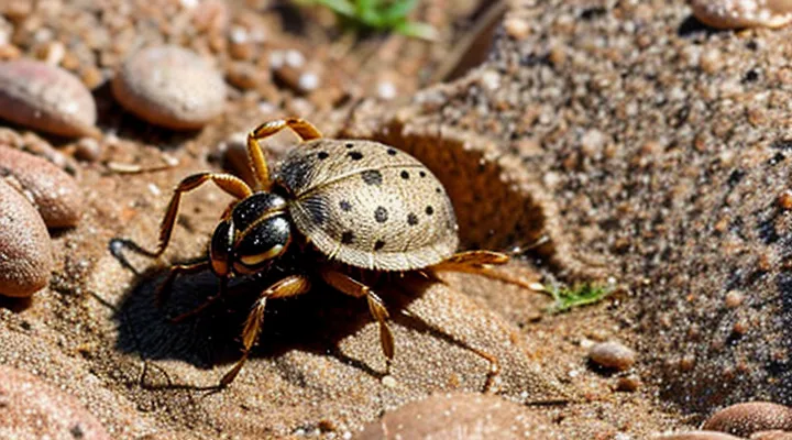
Understanding Tick Behavior
How Ticks Feed
Initial Attachment and Exploration
Ticks locate a host through heat, carbon dioxide, and movement. When a tick makes contact, the front legs grasp the skin and the tick tests the surface for suitability. If the site is acceptable, the tick secures itself by inserting its hypostome, a barbed feeding apparatus, into the epidermis. The hypostome does not penetrate beyond the outer skin layers; instead, it anchors within the superficial dermis while the tick’s salivary glands release anticoagulants and anesthetics.
During this initial phase the tick performs several actions:
- Sensory assessment: Tarsal organs on the front legs detect temperature and moisture gradients.
- Mouthpart deployment: The chelicerae cut a tiny opening, allowing the hypostome to slide into the tissue.
- Anchoring: Barbs on the hypostome lock the tick in place, preventing dislodgement.
- Saliva injection: Anticoagulant and immunomodulatory compounds facilitate blood flow and reduce host awareness.
The attachment is stable but superficial; the tick does not burrow completely beneath the skin. It remains attached to the outer dermal layer while it expands, feeds, and later detaches.
The Role of Barbs and Cement
Ticks attach to hosts by inserting their mouthparts into the epidermis and dermis. The attachment mechanism relies on two specialized structures: microscopic barbs on the hypostome and a proteinaceous cement that hardens after insertion.
Barbs interlock with collagen fibers, preventing the hypostome from being pulled out by the host’s movements. The cement, secreted from salivary glands, spreads around the barbed region and polymerizes within minutes, forming a firm seal that anchors the tick in place.
- Barbs: serrated, angled projections that grip tissue layers; resistance increases as the tick feeds.
- Cement: a mixture of glycine‑rich proteins and enzymes; it adheres to both the tick cuticle and host skin, creating a durable bond.
Because the cement solidifies only after the hypostome penetrates the skin, the tick does not burrow entirely beneath the surface. Instead, it remains partially exposed, with the mouthparts embedded while the body rests on the host’s exterior. This configuration allows continuous feeding while the tick can detach when required, facilitated by enzymatic breakdown of the cement.
Depth of Penetration
Ticks attach by inserting their specialized mouthparts, not by sinking their entire bodies into the host. The hypostome, a barbed structure on the feeding apparatus, penetrates the epidermis and reaches the dermal layer where it secures the tick and accesses blood vessels. The remainder of the tick’s body stays on the surface, supported by the surrounding legs.
Depth of penetration varies with species, developmental stage, and host tissue characteristics:
- Larvae and nymphs: typically 0.5–1 mm into the skin; the hypostome reaches the superficial dermis.
- Adult females: may extend 1–2 mm, occasionally reaching the deeper dermis but rarely the subcutaneous fat.
- Hard ticks (Ixodidae): exhibit the deepest insertion among common species; soft ticks (Argasidae) usually remain shallower.
Measurements from microscopic examinations of engorged ticks confirm that the hypostome does not traverse the full thickness of the skin. Even the deepest insertions stop before the subcutaneous layer, leaving the tick’s abdomen exposed on the host surface.
Consequences of this limited penetration include localized inflammation at the attachment site and the potential transmission of pathogens through the feeding canal. Because the tick’s body remains external, removal can be performed by grasping the mouthparts close to the skin without extracting deeper tissue.
Why Ticks Don't Fully Burrow
Anatomical Limitations
Size and Shape of the Tick Body
Ticks are arthropods with a compact, oval body that expands dramatically after a blood meal. Unfed adults range from 2 mm to 6 mm in length, while engorged specimens can reach 10 mm to 15 mm. Nymphs measure 1 mm to 2 mm, and larvae are typically 0.5 mm to 1 mm. The dorsal surface is covered by a hard scutum in males and partially in females; the ventral side is soft, allowing the abdomen to swell.
The shape of a tick’s mouthparts determines how far it can penetrate host tissue. The hypostome, a barbed structure, is 0.2 mm to 0.5 mm long in most species and anchors the parasite within the epidermis. The chelicerae, each about 0.1 mm, cut a small entry point but do not create a tunnel deep enough to place the entire body beneath the skin. Consequently, the majority of the tick remains external, with only the mouthparts embedded.
- Adult female (e.g., Ixodes scapularis): 3 mm unfed, up to 12 mm engorged; oval, dorsally flattened.
- Adult male: 2 mm–4 mm; similar shape, scutum covering entire dorsal surface.
- Nymph: 1 mm–1.5 mm; oval, less pronounced scutum.
- Larva: 0.5 mm–0.8 mm; spherical, soft cuticle.
Mouthpart Structure
Ticks attach to hosts using a specialized feeding apparatus located at the front of the mouth. The apparatus consists of four main components:
- Chelicerae – paired, blade‑like structures that cut the epidermis and create a small incision.
- Hypostome – a barbed, cone‑shaped organ that penetrates the tissue and anchors the tick by interlocking with host cells.
- Palps – sensory appendages that locate suitable feeding sites and guide the insertion of the chelicerae and hypostome.
- Salivary canal – a tube running through the hypostome that delivers anticoagulants and immunomodulatory substances during blood ingestion.
During attachment, the chelicerae open the skin surface, allowing the hypostome to be driven into the dermis. The barbs on the hypostome lock the tick in place, but the remainder of the body, including the legs and dorsal shield, stays above the skin. Consequently, the tick does not disappear entirely beneath the epidermis; only the mouthparts and a small portion of the hypostome are embedded in host tissue. This configuration enables prolonged feeding while maintaining a visible external presence.
Feeding Strategy and Efficiency
Maximizing Blood Meal Intake
Ticks attach to a host by inserting their hypostome into the epidermis and dermis, not by submerging their entire body beneath the skin surface. The feeding apparatus penetrates only a few millimeters, while the dorsal shield remains exposed to the environment. This partial embedding allows the parasite to remain anchored while maintaining a route for gas exchange and waste elimination.
Maximizing blood‑meal intake relies on several coordinated mechanisms:
- Hypostome anchorage – barbed, serrated structures create a secure lock in host tissue, preventing dislodgement during long feeding periods.
- Salivary cocktail – anticoagulants, vasodilators, and immunomodulators inhibit clot formation, maintain blood flow, and suppress host inflammatory responses.
- Extended feeding duration – ticks remain attached for days, gradually enlarging the feeding lesion to accommodate increasing blood volume.
- Cement secretion – proteinaceous glue solidifies around the mouthparts, reinforcing attachment and stabilizing the feeding site.
- Midgut expansion – elastic gut walls stretch to store large blood volumes, sometimes exceeding the tick’s unfed weight by several hundred percent.
The limited depth of insertion directly supports these strategies. By keeping the bulk of the body external, the tick reduces the risk of host detection and tissue damage, while the deep‑set mouthparts access a stable blood pool. This arrangement optimizes nutrient acquisition without the metabolic cost of full subcutaneous burrowing.
Understanding the distinction between superficial attachment and complete burial informs control measures. Interventions that disrupt cement formation, block salivary enzymes, or mechanically remove attached ticks before the feeding cavity expands can substantially lower the amount of blood ingested and limit pathogen transmission.
Minimizing Detection by Host
Ticks attach to a host by inserting their hypostome, a barbed feeding organ, into the epidermis. The mouthparts remain partially external, allowing the tick to remain visible only as a small, often unnoticed, protrusion. By limiting the depth of penetration, the parasite avoids triggering strong nociceptive responses that would alert the host.
During attachment, ticks secrete a complex mixture of pharmacologically active substances. These include:
- Anticoagulants that prevent clot formation and maintain blood flow.
- Immunomodulators that suppress local inflammatory signaling.
- Analgesic compounds that reduce pain perception at the bite site.
The secretion of a proteinaceous cement anchors the hypostome to surrounding tissue, stabilizing the feeding site without extensive tissue disruption. This cement forms a thin, flexible barrier that masks the presence of the feeding apparatus from the host’s immune surveillance.
Ticks also employ behavioral strategies to minimize detection. They select attachment sites with thin skin and low hair density, such as the scalp, armpits, or groin, where host grooming is less frequent. After feeding commences, the parasite reduces movement, limiting tactile cues that could be sensed by the host.
Collectively, these anatomical, chemical, and behavioral adaptations allow ticks to remain attached for days or weeks while evading host detection, without fully embedding beneath the skin surface.
Common Misconceptions About Tick Bites
The «Head Left Behind» Myth
The belief that a tick can detach leaving only its head embedded in the skin persists despite extensive research. Ticks attach by inserting a mouthpart called the hypostome, which contains barbed structures that lock into the host’s tissue. The hypostome, not the head, is the portion that remains anchored during feeding.
During a blood meal the tick’s body stays above the skin surface while the hypostome penetrates the epidermis and, in some species, reaches into the dermis. The barbs prevent the mouthpart from being easily pulled out, but the tick’s body remains attached until it voluntarily releases. When the tick is removed, the hypostome is withdrawn together with the rest of the organism; no separate fragment is left behind.
Scientific examinations of removed ticks and of skin lesions have never documented isolated head fragments. Studies involving microscopic analysis of attachment sites confirm that any remaining tissue consists of the hypostome and surrounding cement-like secretions, not a detached head.
Proper removal technique eliminates the risk of retained mouthparts:
- Grasp the tick as close to the skin as possible with fine‑point tweezers.
- Apply steady, upward traction without twisting.
- Clean the bite area with antiseptic after extraction.
If a small portion of the hypostome remains, it can be gently lifted with a sterile needle; the tissue will usually retract or be expelled naturally within days. Persistent irritation or infection warrants medical evaluation.
Full Burial vs. Partial Attachment
Ticks attach by inserting their mouthparts into the host’s skin. The hypostome, a barbed structure, anchors the parasite and creates a small cavity. In most species the cavity remains shallow, typically 0.5–2 mm deep, allowing the tick to feed while remaining partially exposed to the surface. This arrangement is referred to as partial attachment.
Full burial, defined as complete subcutaneous encasement of the body, occurs rarely. Certain larval or nymph stages of soft ticks (Argasidae) may embed deeper, but hard ticks (Ixodidae) generally do not surpass the epidermal‑dermal junction. The feeding tube stays within the epidermis and superficial dermis, preventing total concealment beneath the skin.
Key distinctions:
- Depth of insertion: Partial attachment – mouthparts only; Full burial – body fully surrounded by tissue.
- Visibility: Partial attachment – tick’s dorsal shield remains visible; Full burial – only a small puncture site observable.
- Risk of infection: Partial attachment – localized inflammation; Full burial – higher potential for secondary bacterial invasion due to deeper tissue disruption.
Risks Associated with Tick Bites
Disease Transmission
Ticks attach by inserting their hypostome, a barbed feeding organ, into the epidermis and dermis. The body remains on the surface; only the mouthparts penetrate. This partial embedment creates a channel through which saliva is delivered.
Pathogen transfer occurs when the tick’s saliva, containing bacteria, viruses, or protozoa, enters the host’s bloodstream. Transmission efficiency rises with feeding duration; many agents require at least 24 hours of attachment before being passed.
Common tick‑borne illnesses include:
- Lyme disease (Borrelia burgdorferi)
- Rocky Mountain spotted fever (Rickettsia rickettsii)
- Anaplasmosis (Anaplasma phagocytophilum)
- Babesiosis (Babesia microti)
- Powassan virus disease
Preventive actions focus on early removal and habitat management:
- Perform regular skin checks after outdoor exposure.
- Use fine‑tipped tweezers to grasp the tick close to the skin and pull upward with steady pressure.
- Apply EPA‑registered repellents to skin and clothing.
- Keep lawns trimmed and remove leaf litter to reduce tick habitats.
Localized Skin Reactions
Ticks attach by inserting their mouthparts into the epidermis and, in some species, into the dermis. The insertion creates a localized skin reaction that varies with the tick’s feeding duration, host immune response, and the presence of pathogen‑bearing saliva.
Typical manifestations include:
- Small, red papule at the attachment site, often surrounded by a faint halo.
- A central puncture wound that may be difficult to see because the tick’s feeding apparatus remains embedded.
- Mild swelling or edema that can extend a few millimeters beyond the puncture.
- Occasionally, a localized allergic wheal if the host reacts to tick saliva proteins.
These reactions are confined to the immediate area of attachment and do not indicate that the tick has tunneled completely beneath the skin surface. The tick’s hypostome anchors within the superficial layers, while the surrounding tissue responds with inflammation, vasodilation, and recruitment of immune cells. In most cases, removal of the tick eliminates the stimulus, and the skin lesion resolves within days to weeks, leaving only a faint scar if secondary infection occurs.
Safe Tick Removal Practices
Essential Tools and Techniques
Ticks can embed their mouthparts deep within the epidermis, sometimes leaving only the abdomen visible. Accurate assessment of the attachment depth requires precise visual aid and controlled extraction methods.
- Fine‑point tweezers with a non‑slipping grip
- 10‑40× magnifying lens or handheld dermatoscope
- Tick‑specific removal hooks or curved forceps
- Disposable nitrile gloves to prevent contamination
- Antiseptic solution (e.g., chlorhexidine) for post‑removal wound care
Effective practice begins with a thorough skin examination under magnification. The examiner should identify the tick’s capitulum and determine whether any portion protrudes from the skin surface. If the mouthparts are fully concealed, the removal instrument must grasp the tick as close to the skin as possible without crushing the body. A steady, upward pulling motion, maintained for 10–15 seconds, detaches the organism while minimizing tissue trauma. Immediate application of antiseptic reduces bacterial entry, and the bite site should be inspected after 24 hours for signs of infection or retained fragments. Regular use of the listed tools and adherence to the described technique ensure reliable evaluation of tick burial depth and safe extraction.
Post-Removal Care and Monitoring
After extracting a tick, clean the bite site with an antiseptic solution such as povidone‑iodine or chlorhexidine. Apply a sterile adhesive bandage if the wound is open; otherwise, leave it uncovered to air‑dry.
Monitor the area for at least four weeks. Record any of the following changes:
- Redness expanding beyond the immediate margin
- Swelling or a palpable lump
- Persistent itching or burning sensation
- Fever, headache, muscle aches, or rash elsewhere on the body
If any symptom appears, seek medical evaluation promptly. A healthcare professional may prescribe antibiotics or other treatment depending on the suspected pathogen.
During the observation period, avoid scratching the site and keep it dry. Replace the bandage daily if used, and wash hands before and after any contact with the wound. Document the date of removal, the tick’s developmental stage, and the location of the bite for reference during any future consultation.
