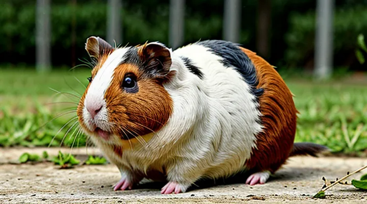
The Likelihood of Flea Infestation in Cavies
Understanding Host Specificity and Parasite Preferences
Guinea pigs are not primary hosts for common flea species such as Ctenocephalides felis or Ctenocephalides canis. These insects have evolved preferences for mammals with thicker fur, higher body temperatures, and frequent contact with other infested animals. Consequently, flea populations concentrate on cats, dogs, rodents, and wildlife that meet these criteria.
Host specificity influences flea survival in several ways:
- Temperature and humidity: Optimal ranges (22‑30 °C, 70‑80 % humidity) are more consistently maintained on larger mammals.
- Fur structure: Dense, oily coats facilitate attachment and movement; guinea pig hair is finer and less conducive to flea grip.
- Social behavior: Species that groom or nest together promote flea transmission; guinea pigs are solitary or kept in small groups, reducing opportunities for spread.
Parasite preferences can shift when environmental conditions force fleas onto suboptimal hosts. Overcrowded cages, poor hygiene, and the presence of other infested pets create opportunities for temporary colonization of guinea pigs. In such scenarios, flea eggs may be deposited on bedding, and emerging larvae can infest the animal despite its lower suitability.
Veterinary assessments confirm that genuine flea infestations on guinea pigs are rare but not impossible. Diagnosis relies on visual inspection of the skin and fur for adult fleas, eggs, or blood‑stained debris. Treatment protocols mirror those used for other small mammals, emphasizing environmental decontamination, regular cleaning, and, when necessary, topical or oral antiparasitic agents approved for guinea pig use.
In summary, flea presence on guinea pigs depends on the interplay between the parasite’s host specificity and the animal’s living conditions. Proper husbandry minimizes the risk, while atypical infestations signal lapses in environmental control or cross‑species exposure.
Why «Ctenocephalides» Rarely Affect Guinea Pigs
Guinea pigs are infrequently parasitized by fleas of the genus Ctenocephalides. The rarity stems from several biological and ecological factors.
- Host specificity: Ctenocephalides species have evolved to recognize chemical cues and body temperatures typical of cats, dogs, and rodents such as rats. Guinea pigs lack the pheromonal profile that triggers flea attachment and feeding behavior.
- Fur structure: The dense, coarse coat of a guinea pig creates a physical barrier that hinders flea movement and reduces the likelihood of successful penetration to the skin surface.
- Grooming habits: Guinea pigs engage in regular self‑grooming and mutual grooming within groups, removing adult fleas and larvae before they can establish a feeding site.
- Skin environment: The skin of a guinea pig maintains a slightly higher humidity and a different pH compared to typical flea hosts, making it less conducive to flea development and egg laying.
- Habitat conditions: Guinea pigs are commonly housed in indoor cages with controlled temperature and regular bedding changes, limiting the outdoor exposure where flea populations thrive.
Other ectoparasites, such as Trixacarus caviae (guinea pig mites) and Trichodectes spp. (lice), are more prevalent because they have adapted specifically to the guinea pig’s physiology and environment. Consequently, while fleas can occasionally be found on guinea pigs under atypical circumstances—such as cohabitation with heavily infested cats—their overall incidence remains low.
Identifying the True External Threats
Mites: The Most Common Cause of Dermatitis
Recognizing Symptoms of «Trixacarus caviae»
Guinea pigs can be infested by the mite Trichodectes caviae (commonly referred to as “cavy mites”), which is often mistaken for a flea problem. Accurate identification relies on observing specific clinical signs rather than assuming typical flea behavior.
Visible signs include:
- Intense itching that leads to frequent scratching or rubbing against cage bars.
- Hair loss in localized patches, especially around the neck, back, and hindquarters.
- Red, inflamed skin with possible crust formation or scabs.
- Small, moving specks on the fur that resemble tiny grains of sand; these are the adult mites.
- Weight loss or reduced appetite secondary to chronic irritation.
Additional indicators are the presence of dark debris (mite feces) on bedding or in the animal’s fur and thickened skin in areas of prolonged infestation. Early detection through these symptoms enables prompt treatment, preventing secondary infections and improving the animal’s welfare.
Hair Loss Patterns Associated with Mite Activity
Hair loss in guinea pigs most often results from mite infestations rather than flea bites. The two primary mite species—Trixacarus caviae (the fur mite) and Demodex caviae (the skin mite)—produce distinct alopecia patterns that help differentiate them from other dermatological problems.
The fur mite typically creates circular or irregular patches of hair loss on the back, flanks, and hindquarters. Affected areas appear smooth, with a fine, dry crust that may be slightly scaly. The edges of the patches are well defined, and the underlying skin often shows mild erythema. In severe cases, the mite spreads to the neck and shoulders, forming larger, confluent zones of alopecia.
The skin mite generates more diffuse thinning, especially around the face, ears, and forelimbs. Hair loss is accompanied by reddened, inflamed skin that may exude a thin, watery discharge. The fur in the affected regions becomes brittle and breaks easily, giving the impression of “patchy” baldness that expands gradually outward from the initial site.
Key observations for diagnosing mite‑related hair loss:
- Circular or irregular bald patches on the dorsal and lateral body (fur mite)
- Smooth, dry crusts with mild redness at patch margins
- Diffuse thinning on the face, ears, and forelimbs (skin mite)
- Red, inflamed skin with occasional watery exudate
- Brittle, easily broken hair in affected zones
Effective treatment requires topical acaricides applied according to veterinary guidelines, combined with environmental decontamination to prevent reinfestation. Regular grooming and inspection of the coat can detect early signs of mite activity before extensive hair loss occurs.
Lice: The Presence of Biting and Sucking Species
Visual Confirmation of Nits and Adult Lice
Visual assessment of a guinea pig’s coat is essential when evaluating ectoparasite concerns. Nits appear as oval, whitish‑tan shells firmly attached to hair shafts, typically 0.5–1 mm in length. They are immobile, positioned close to the skin, and often cluster near the neck, back, and hindquarters. Adult lice are tan to light brown, wingless insects measuring 2–3 mm, with a flattened body that clings tightly to hair. They move slowly, rarely leaving the host, and are most frequently observed on the ventral surface, around the ears, and under the tail.
Key visual differences from flea infestations include:
- Size: fleas are 1.5–3.5 mm, larger than nits and comparable to adult lice but possess a laterally compressed body.
- Mobility: fleas jump when disturbed; lice crawl and remain on the host.
- Body shape: fleas have a distinct “waist” and hind legs adapted for jumping; lice have a uniformly broad abdomen.
Effective inspection requires a bright, focused light source and a fine‑toothed grooming comb. Separate each hair strand and examine the base for attached nits; slide the comb slowly to expose any crawling lice. A magnifying lens (10×) enhances detection of the small, translucent nits and the subtle movements of adult lice. Consistent visual checks allow rapid identification and appropriate treatment of lice infestations in guinea pigs.
The Impact of «Gliricola porcelli»
Guinea pigs can host the flea Gliricola porcelli, a parasite that feeds on blood and occasionally transmits bacterial agents. Infestation manifests as hair loss, skin irritation, and anemia in severe cases. The flea’s life cycle—egg, larva, pupa, adult—occurs primarily in the animal’s bedding, allowing rapid population growth when hygiene is inadequate.
Health consequences extend beyond dermatological symptoms. Blood loss may reduce hematocrit levels, especially in young or underweight individuals. Secondary infections arise from scratching wounds, and the flea can serve as a mechanical vector for Bartonella spp., which cause febrile illness in rodents. Behavioral effects include increased restlessness and reduced food intake due to discomfort.
Control measures focus on environmental sanitation and targeted ectoparasiticide application. Recommended actions:
- Remove and replace all bedding weekly; wash cages with hot water and a mild disinfectant.
- Apply a veterinary‑approved topical flea treatment to each animal, following dosage guidelines.
- Conduct a thorough visual inspection of fur and skin at least twice a month, concentrating on the neck, back, and tail base.
- Treat all cohabiting animals simultaneously to prevent cross‑infestation.
- Monitor hematological parameters during treatment to detect residual anemia.
Clinical Signs and Behavioral Changes
Visible Indicators of Skin Irritation
Guinea pigs may exhibit skin irritation that mimics flea infestation. Recognizing the visual cues helps owners determine whether parasites are present or if another condition is causing discomfort.
- Red or pink patches on the skin, especially around the neck, back, and hindquarters.
- Small, circular areas of hair loss that may appear as bald spots.
- Dark, crusted scabs or scaly lesions that develop where the animal frequently scratches.
- Presence of tiny, dark specks that move rapidly across the fur, indicating live parasites.
- Excessive grooming or scratching behavior, often accompanied by audible squeaks of distress.
- Swollen or inflamed skin around the ears, paws, or tail base.
These signs should be evaluated alongside a physical examination of the coat. Live fleas are typically visible as moving specks and may leave tiny black droppings (flea dirt) on the fur. In contrast, allergic reactions, mites, or bacterial infections produce static lesions without the characteristic movement. Prompt veterinary assessment is essential to confirm the cause and initiate appropriate treatment.
Excessive Scratching and Self-Mutilation
Excessive scratching and self‑mutilation in guinea pigs often prompt owners to question the presence of ectoparasites. Flea infestations can cause intense pruritus, leading the animal to bite, chew, or rub affected areas until skin lesions develop. However, similar behaviors arise from mange, allergic dermatitis, fungal infections, or environmental irritants, so scratching alone does not confirm a flea problem.
Veterinarians assess the condition by:
- Inspecting the coat and skin for live fleas, flea dirt, or crusted lesions.
- Performing a skin scrape or tape test to identify mites or fungal elements.
- Reviewing diet, bedding, and enclosure hygiene for potential allergens.
- Conducting a complete physical exam to rule out systemic illness that may provoke discomfort.
Treatment focuses on eliminating the underlying cause and protecting the skin. When fleas are identified, a veterinarian‑approved topical or oral insecticide is applied, and the habitat is thoroughly cleaned, including bedding replacement and regular vacuuming. If other parasites or dermatological conditions are responsible, appropriate acaricides, antifungals, or anti‑inflammatory medications are prescribed. Preventive measures—routine health checks, clean bedding, and proper quarantine of new animals—reduce the risk of recurrence and help maintain skin integrity.
Secondary Infections Caused by Persistent Trauma
Guinea pigs rarely host flea infestations; however, any external parasite that irritates the skin can trigger repetitive scratching or biting. When such behavior continues, the tissue experiences persistent trauma, compromising the epidermal barrier and inviting opportunistic microorganisms.
Repeated mechanical damage creates micro‑abrasions that serve as entry points for bacteria and fungi. The loss of protective keratin layers allows colonization by organisms that normally reside on the skin surface or in the environment. In guinea pigs, secondary infections commonly arise from:
- Staphylococcus aureus – causes acute dermatitis with purulent exudate.
- Pseudomonas aeruginosa – produces moist, malodorous lesions that may spread rapidly.
- Dermatophyte species (e.g., Trichophyton mentagrophytes) – generate circular, alopecic patches with crusting.
- Mixed anaerobic flora – lead to ulcerative wounds that deteriorate without prompt intervention.
Clinical signs include localized swelling, heat, discharge, and progressive tissue necrosis. Diagnosis relies on cytology, culture, and, when necessary, histopathology to identify the causative agent.
Effective management combines:
- Immediate removal of the irritant source (e.g., parasite control, environmental enrichment).
- Thorough cleaning of the affected area with isotonic saline or mild antiseptic solutions.
- Targeted antimicrobial therapy based on culture sensitivity; empirical broad‑spectrum agents may be used initially.
- Topical antifungal preparations for dermatophyte involvement.
- Regular monitoring of wound healing and adjustment of treatment protocols as needed.
Preventive measures focus on maintaining clean bedding, providing adequate chew objects to reduce oral‑induced facial trauma, and conducting routine health checks to detect early skin irritation before secondary infection develops.
Diagnostic Procedures and Veterinary Consultation
Differentiating Parasitic Infections from Fungal Issues
Guinea pigs can host external parasites, but the presence of fleas is uncommon. When a pet shows skin irritation, it is essential to determine whether the cause is an arthropod infestation or a fungal condition, because treatment protocols differ markedly.
Parasites such as fleas, mites, or lice produce distinct clinical signs:
- Sudden itching, especially after handling
- Visible tiny insects or moving specks on the coat
- Dark specks resembling pepper (flea feces) on bedding
- Localized hair loss around the tail or hindquarters
Fungal infections, primarily caused by dermatophytes, present a different pattern:
- Gradual hair thinning forming circular patches
- Red, scaly skin that may crack or bleed
- Absence of live insects upon close inspection
- Positive result on a Wood’s lamp or fungal culture
Diagnostic steps to separate the two include:
- Conduct a thorough visual examination under bright light.
- Perform a skin scraping for microscopic analysis to detect mites or fungal hyphae.
- Use adhesive tape impressions to capture flea debris.
- Submit samples to a veterinary laboratory for definitive identification.
Effective management depends on accurate diagnosis. Parasite infestations require topical or systemic insecticides approved for rodents, while fungal cases respond to antifungal shampoos, oral medication, and environmental decontamination. Regular grooming, clean housing, and routine health checks reduce the risk of both problems.
Performing Skin Scrapings for Accurate Identification
Guinea pigs are occasionally suspected of harboring fleas, yet ectoparasitic infestations more frequently involve mites, lice, or ear ticks. Accurate determination requires direct examination of skin material rather than reliance on visual cues alone.
Skin scraping delivers a sample from the epidermis that can be examined microscopically to confirm the presence or absence of flea larvae, adult fleas, or other arthropods. The procedure consists of the following steps:
- Restrain the animal securely to prevent movement.
- Moisten a sterile scalpel blade with saline solution.
- Apply firm, controlled pressure to the target area (commonly the dorsal neck or flank) and scrape a thin layer of skin.
- Collect the dislodged material onto a glass slide, add a drop of mineral oil or lactophenol, and cover with a cover slip.
- Examine the slide under 10–40× magnification, identifying characteristic flea morphology (e.g., laterally compressed body, comb-like genal and pronotal structures) or distinguishing features of mites and lice.
Positive identification of flea elements confirms infestation; a negative result, combined with the absence of flea‑specific lesions, suggests alternative ectoparasites. Microscopic confirmation guides appropriate treatment, such as topical insecticides for fleas or acaricides for mites, and informs preventive measures to maintain a parasite‑free environment.
Effective Management and Safe Treatment Options
Approved Medications for Small Exotic Pets
Importance of Avoiding Flea Treatments Designed for Dogs and Cats
Guinea pigs rarely host fleas, yet owners often consider flea products meant for dogs or cats. Those medications are formulated for species with different skin absorption rates, metabolic pathways, and body weights. Applying them to rodents can cause severe toxicity.
Risks of using canine or feline flea treatments on guinea pigs include:
- Dermal irritation or chemical burns from strong insecticides.
- Systemic poisoning leading to liver or kidney failure.
- Neurological disturbances such as tremors or seizures.
- Fatal outcomes even at low doses.
Safe management relies on veterinary guidance. Veterinarians can prescribe products specifically labeled for guinea pigs or recommend non‑chemical methods, such as regular cage cleaning, environmental flea control, and manual inspection. Selecting only species‑approved treatments eliminates the danger posed by inappropriate flea medications.
Protocols for Thorough Environmental Decontamination
Guinea pigs can occasionally harbor fleas, making rigorous environmental decontamination essential to prevent reinfestation and protect other animals.
Before cleaning, isolate the affected enclosure, remove all animals, and don disposable gloves, gowns, and masks. Dispose of contaminated bedding, toys, and food dishes in sealed bags; wash reusable items in hot water (≥ 60 °C) with detergent.
- Use an EPA‑registered insecticide‑approved disinfectant (e.g., a quaternary ammonium compound or hydrogen peroxide‑based solution).
- Apply a detergent‑based cleaner to all surfaces, allowing at least 10 minutes of contact.
- Rinse with potable water, ensuring no residue remains.
- Apply the disinfectant, maintaining the manufacturer‑specified dwell time (typically 5–10 minutes).
- Fog or mist the enclosure with a residual‑acting insect growth regulator if the facility permits.
After treatment, verify efficacy by swabbing high‑touch areas and testing for flea DNA or eggs. Record results, and repeat the decontamination cycle if any trace remains.
Finally, introduce a preventive schedule: weekly cleaning, regular inspection of animals, and immediate isolation of any new infestations. This systematic approach eliminates fleas from the environment and reduces the likelihood of future outbreaks.
Preventive Measures to Ensure a Clean Habitat
Guinea pigs are susceptible to flea infestations; maintaining a pristine enclosure is essential for prevention.
- Clean the cage daily, removing droppings, food remnants, and loose bedding.
- Replace the entire bedding weekly with a dust‑free, absorbent substrate.
- Wash water bottles and food dishes with hot, soapy water at least once a week; rinse thoroughly before refilling.
- Vacuum the area surrounding the cage weekly to capture stray eggs or larvae.
- Use a flea‑free environment: keep the habitat away from other pets that may carry fleas and avoid outdoor exposure without proper protection.
- Apply a veterinarian‑approved, pet‑safe flea preventive on the animal according to the prescribed schedule.
Regular health checks complement environmental hygiene. Inspect the fur and skin for signs of irritation or movement, and consult a veterinarian promptly if any abnormalities appear. Consistent adherence to these measures minimizes the risk of flea presence and supports overall well‑being.
