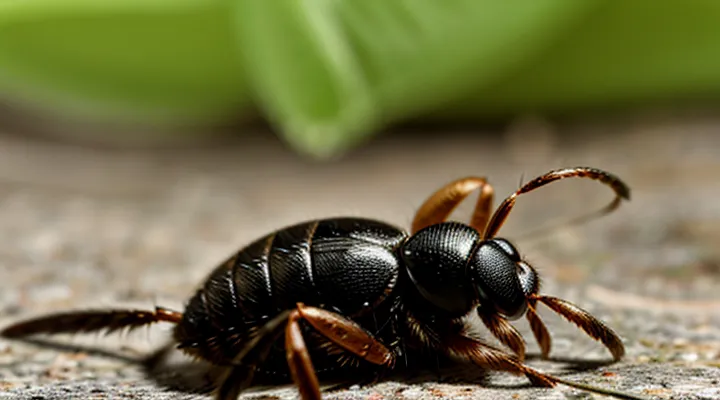
The Anatomy of a Tick Bite
The Tick's Mouthparts
The Hypostome: A Barbed Weapon
The hypostome is a hardened, barbed projection located on the ventral side of a feeding tick. Its microscopic teeth penetrate the host’s skin, anchoring the parasite firmly while it draws blood. This mechanical insertion disrupts epidermal and dermal layers, producing immediate nociceptive signals that the nervous system registers as pain.
During attachment, the hypostome releases anticoagulant and immunomodulatory proteins. These secretions prevent clot formation and dampen the host’s early immune response, allowing the tick to remain attached for days. The combination of tissue tearing and biochemical interference provokes inflammation, swelling, and heightened sensitivity around the bite site.
Key characteristics of the hypostome that contribute to painful bites:
- Barbed architecture: multiple microspines lock into collagen fibers, resisting removal.
- Hardened cuticle: provides rigidity, ensuring deep penetration without bending.
- Continuous feeding: the structure maintains a stable channel for blood flow, reducing the need for repeated probing.
- Associated salivary compounds: anticoagulants and anesthetic-like proteins intensify local irritation once the barrier is breached.
Chelicerae: Cutting and Lacerating
Ticks attach to the host using specialized mouthparts called chelicerae. The chelicerae consist of two sharp, blade‑like structures that act as cutting tools. During attachment, the chelicerae slice through the epidermis and dermis, creating a narrow, irregular wound. This mechanical disruption severs nerve endings and ruptures small blood vessels, producing immediate sharp pain.
The cutting action also produces micro‑lacerations that expose deeper tissue layers. These lacerations trigger the release of nociceptive chemicals such as substance P and prostaglandins, amplifying the pain signal. Additionally, the chelicerae’s movement generates friction, which further irritates surrounding sensory fibers.
Key effects of cheliceral cutting and lacerating:
- Direct transection of cutaneous nerves → rapid pain perception.
- Rupture of capillaries → localized pressure and inflammation.
- Exposure of dermal tissue → activation of inflammatory mediators.
- Creation of a feeding canal → sustained mechanical stimulation during blood extraction.
Together, these processes explain the acute discomfort experienced when a tick bites.
Salivary Glands: A Cocktail of Chemicals
Ticks attach by inserting a hypostome equipped with salivary glands that release a complex mixture of bioactive molecules. This secretion enables prolonged feeding while simultaneously provoking the sharp, localized sensation experienced at the bite site.
The cocktail includes several classes of compounds:
- Anticoagulants (e.g., apyrase, tick anticoagulant peptide) prevent clot formation, maintaining blood flow.
- Anesthetics (e.g., tick salivary protein 1) suppress immediate nerve signaling, delaying detection.
- Anti‑inflammatory agents (e.g., prostaglandin‑degrading enzymes) modulate host immune responses.
- Immunomodulators (e.g., Salp15, Iricin) interfere with cytokine release and complement activation.
- Proteolytic enzymes (e.g., metalloproteases) degrade extracellular matrix, facilitating mouthpart penetration.
These substances act together to create a paradoxical effect. Anticoagulants and proteases damage tissue and expose nociceptors, while anesthetic components temporarily mute the signal. As the anesthetic effect wanes, the accumulated irritation and inflammatory mediators trigger a rapid pain response. The brief, intense sting results from sudden activation of peripheral nerve endings once the suppressive chemicals diminish.
Thus, the pain associated with a tick bite derives directly from the biochemical strategy of the tick’s salivary glands, which balances feeding efficiency against host detection by delivering a precisely timed mixture of pain‑inducing and pain‑masking agents.
The Mechanisms Behind the Pain
Mechanical Injury
Tissue Damage Upon Penetration
The tick’s mandibles and hypostome pierce the epidermis, creating a narrow wound that severs epidermal cells and disrupts the dermal matrix. This mechanical breach directly activates cutaneous nociceptors, producing the immediate sharp sensation felt at the bite site.
During penetration the tick injects saliva that contains proteolytic enzymes, anticoagulants, and immunomodulatory proteins. These substances degrade extracellular proteins, dissolve fibrin clots, and suppress local hemostasis, which amplifies tissue injury and prolongs nociceptor stimulation.
Key aspects of tissue damage include:
- Rupture of epidermal cells, exposing nerve endings.
- Laceration of dermal collagen fibers, destabilizing the structural framework.
- Injury to small blood vessels, leading to micro‑bleeding and edema.
- Chemical irritation from salivary components, enhancing inflammatory mediator release.
The combination of physical disruption and biochemical irritation generates the pain associated with a tick bite.
Inflammation and Swelling
A tick’s mouthparts penetrate the skin and inject saliva that contains anticoagulants, proteases, and immunomodulatory proteins. These substances disrupt normal hemostasis and expose underlying tissue to foreign antigens, initiating an acute inflammatory response.
Inflammation is characterized by:
- Release of histamine and prostaglandins, causing vasodilation and increased vascular permeability.
- Recruitment of neutrophils and macrophages that secrete cytokines (IL‑1, TNF‑α) to amplify the response.
- Activation of nociceptors by inflammatory mediators, producing the sharp sensation felt at the bite site.
Swelling follows the same cascade. Fluid exudes from capillaries into interstitial spaces, producing edema that stretches surrounding skin and nerve endings. The mechanical pressure of the edema intensifies nociceptor activation, contributing further to pain.
The combined effect of chemical irritation and physical expansion explains why the bite is painful shortly after attachment and may persist for hours or days as the inflammatory process resolves. Persistent or excessive swelling can indicate an allergic reaction or secondary infection, warranting medical evaluation.
Chemical Reactions
Anesthetics: Masking the Initial Attack
Ticks inject saliva the moment their mouthparts penetrate the skin. The saliva contains a cocktail of bioactive molecules that interfere with the host’s pain pathways. Anesthetic peptides bind to voltage‑gated sodium channels on peripheral nociceptors, preventing the depolarization required for action‑potential generation. Simultaneously, anti‑inflammatory proteins inhibit prostaglandin synthesis, further dampening the sensory response.
Key components of the anesthetic mixture include:
- Ixolaris‑like peptides: block Na⁺ channel activation, silencing the initial nociceptive impulse.
- Sialostatin L: suppresses protease activity that would otherwise amplify pain signals.
- Histamine‑binding proteins: reduce vasodilation and edema, limiting secondary irritation.
The immediate suppression of pain allows the tick to remain attached for days, extending the window for pathogen transmission. Without the anesthetic effect, host detection would trigger rapid grooming or removal, curtailing feeding and reducing the likelihood of disease spread.
Anticoagulants: Preventing Clotting
Ticks inject saliva containing anticoagulant compounds that interfere with the host’s hemostatic cascade. These molecules bind to clotting factors, inhibit platelet aggregation, and block fibrin formation, allowing blood to remain fluid at the feeding site. Continuous blood flow reduces the pressure buildup that would otherwise trigger a rapid nociceptive response, extending the duration of attachment and increasing the likelihood of tissue irritation.
The prolonged exposure of nerve endings to saliva components contributes to the sensation of pain. Salivary proteins such as apyrase, thrombin inhibitors, and metalloproteases not only prevent clot formation but also provoke inflammatory mediators. The resulting vasodilation and edema amplify mechanical stimulation of cutaneous receptors, producing the characteristic ache after a bite.
Key anticoagulants present in tick saliva:
- Apyrase – hydrolyzes ADP, limiting platelet activation.
- Salp14 – inhibits the intrinsic pathway by targeting factor XIIa.
- Ixolaris – blocks tissue factor–factor VIIa complex, suppressing extrinsic clotting.
- Metalloproteases – degrade extracellular matrix, facilitating feeding and enhancing inflammation.
By maintaining a fluid feeding pool, these agents delay the host’s wound‑closure mechanisms. The delayed clotting, combined with inflammatory signaling, explains why the bite site often feels sore for hours or days after the tick detaches.
Vasodilators: Increasing Blood Flow
Tick saliva contains potent vasodilators that act on the host’s microvasculature. These compounds bind to endothelial receptors, trigger nitric‑oxide release, and relax smooth‑muscle cells in arterioles. The resulting dilation enlarges capillary networks around the attachment site, enhancing perfusion.
Increased blood flow delivers two effects that contribute to the bite’s discomfort. First, the expanded vascular pool lowers local tissue pressure, allowing more saliva to spread deeper into the skin. Second, the heightened circulation carries immune mediators—histamine, prostaglandins, and cytokines—rapidly to the area, amplifying nociceptor activation.
Key mechanisms of vasodilator‑induced pain:
- Nitric‑oxide–mediated smooth‑muscle relaxation → vessel diameter ↑
- Elevated perfusion → faster distribution of tick anticoagulants and anesthetics
- Accelerated recruitment of inflammatory cells → release of algogenic substances
- Swelling of interstitial space → mechanical stimulation of sensory endings
The combined action of these processes creates a localized, sharp sensation that signals the host to the presence of the parasite. Understanding the role of vasodilators clarifies why a tick bite is not merely a puncture but a biologically active event that provokes pain through vascular and inflammatory pathways.
Immune Response
Histamine Release
When a tick inserts its mouthparts into the skin, it injects saliva that contains anticoagulants, enzymes, and proteins designed to suppress the host’s immediate defensive reactions. Among these components, substances that trigger mast cells to release histamine are particularly relevant to the sensation of pain.
Histamine acts on nearby sensory nerve endings, increasing their excitability. The molecule binds to H1 receptors on peripheral nerves, causing depolarization and the transmission of pain signals to the spinal cord. Simultaneously, histamine dilates local blood vessels, leading to swelling that further stretches the tissue and amplifies discomfort.
The sequence of events can be summarized as follows:
- Tick saliva introduces immunogenic proteins.
- Mast cells in the dermis detect these proteins and degranulate.
- Histamine is released into the extracellular space.
- Histamine engages H1 receptors on nociceptors, lowering their activation threshold.
- Resulting nerve impulses are perceived as sharp or burning pain at the bite site.
The rapid onset of pain after a tick attachment is therefore a direct consequence of histamine‑mediated sensitization of cutaneous nerves, compounded by the inflammatory swelling that accompanies the reaction.
Allergic Reactions
Tick bites can provoke pain through immediate hypersensitivity. When a tick inserts its mouthparts, it introduces saliva containing anticoagulants and proteins that the immune system may recognize as foreign. Mast cells in the skin release histamine and other mediators, causing vasodilation, edema, and stimulation of nociceptors. The resulting sharp or burning sensation often appears within minutes of the attachment.
Allergic mechanisms that amplify discomfort include:
- IgE‑mediated reactions: Prior sensitization to tick salivary antigens leads to rapid IgE binding on mast cells, triggering degranulation and intense pruritus or pain.
- Late‑phase inflammation: Cytokines such as IL‑4 and IL‑5 attract eosinophils, prolonging tissue swelling and tenderness for hours to days.
- Cross‑reactivity: Structural similarity between tick proteins and allergens from other arthropods can provoke a broader immune response, increasing symptom severity.
Understanding these pathways clarifies why some individuals experience pronounced pain after a tick bite, while others report only mild irritation.
Potential Secondary Issues
Bacterial Infections
Ticks inject saliva containing anticoagulants and enzymes that irritate skin and nerve endings. Bacterial pathogens introduced with the saliva amplify this irritation, producing pronounced pain at the bite site.
Common tick‑borne bacteria responsible for painful reactions include:
- Borrelia burgdorferi – triggers inflammatory lesions that can become tender as spirochetes spread through dermal tissue.
- Rickettsia rickettsii – induces vasculitis; swelling and necrosis generate sharp discomfort.
- Anaplasma phagocytophilum – stimulates immune cell infiltration, leading to localized throbbing.
- Ehrlichia chaffeensis – provokes cytokine release, heightening sensitivity of peripheral nerves.
The pain mechanism follows a sequence: bacterial entry → activation of pattern‑recognition receptors → release of pro‑inflammatory mediators (e.g., interleukin‑1, tumor‑necrosis factor‑α) → sensitization of nociceptors → perception of sharp or burning pain. Rapid bacterial replication intensifies the inflammatory cascade, extending the duration and severity of the sensation.
Effective management requires prompt antimicrobial therapy to halt bacterial proliferation and reduce inflammatory mediator production. Early doxycycline administration typically diminishes pain within 24–48 hours, preventing chronic tissue damage and secondary complications.
Lyme Disease and Other Pathogens
Tick bites introduce saliva that contains anticoagulants, anesthetics, and inflammatory mediators. The mechanical puncture and rapid immune activation generate localized soreness, swelling, and sometimes a burning sensation.
Lyme disease, caused by Borrelia burgdorferi, often follows a painful bite. Early infection produces erythema migrans, joint discomfort, and muscle aches as spirochetes disseminate and provoke inflammatory pathways. Neurological involvement may add sharp or radiating pain.
Other tick‑borne agents contribute to bite‑related discomfort:
- Anaplasma phagocytophilum: induces fever, headache, and muscle pain through leukocyte infection.
- Babesia microti: triggers hemolytic anemia, leading to generalized aches and fatigue.
- Powassan virus: produces encephalitis, meningitis, and severe head or neck pain.
- Rickettsia spp.: cause spotted fevers with intense scalp tenderness and arthralgia.
- Ehrlichia chaffeensis: results in myalgia and joint soreness via macrophage infection.
Prompt removal reduces saliva exposure, yet pathogens may have already entered the bloodstream. Monitoring for expanding rash, joint swelling, fever, or neurological signs enables early diagnosis. Serologic testing, PCR, or culture confirms infection; doxycycline remains first‑line therapy for most bacterial agents, while antiviral or antiparasitic regimens address specific viruses and protozoa. Timely treatment mitigates pain progression and prevents chronic sequelae.
