Understanding Bed Bug Bites
The Culprit: Bed Bugs
What are Bed Bugs?
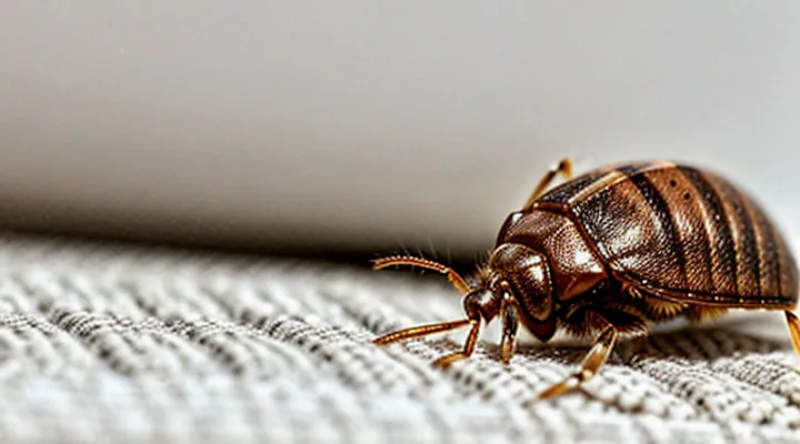
Bed bugs (Cimex lectularius) are small, wingless insects that feed exclusively on the blood of warm‑blooded hosts. Adults measure 4–5 mm in length, have a flattened oval shape, and are reddish‑brown after feeding. They hide in cracks, seams of furniture, mattresses, and wall voids, emerging at night to locate a host by detecting carbon dioxide and body heat.
Key biological traits:
- Hemimetabolous development: egg → five nymphal stages → adult, each molt requiring a blood meal.
- Rapid reproduction: a single female can lay 200–500 eggs over a lifetime.
- Survival without feeding: nymphs and adults can endure several months without a blood source, enabling persistence in vacant dwellings.
- Resistance to many insecticides: genetic mutations and behavioral avoidance reduce chemical efficacy.
Understanding these characteristics clarifies why bed‑bug bites commonly provoke itching, as the insects inject saliva containing anticoagulants and anesthetics that trigger immune responses in the skin.
How Bed Bugs Feed
Bed bugs locate a sleeping host by sensing body heat, carbon‑dioxide, and movement. Once a suitable spot is identified, the insect climbs onto the skin, anchors its mouthparts, and begins to feed.
- The elongated proboscis pierces the epidermis.
- Two slender stylets separate; one injects saliva, the other draws blood.
- Saliva is released continuously while the insect sucks blood for several minutes.
- After engorgement, the bug withdraws its mouthparts and drops off.
The saliva contains a mixture of biologically active substances. Anticoagulants prevent clotting, allowing a steady flow of blood. Vasodilators widen capillaries, increasing blood availability. Anesthetic compounds reduce the host’s immediate sensation of the bite. Proteins and enzymes act as allergens, triggering the immune system.
When the host’s skin encounters these foreign proteins, mast cells release histamine and other inflammatory mediators. Histamine dilates blood vessels, creates swelling, and stimulates nerve endings, producing the characteristic itch. The intensity of the reaction varies with the amount of saliva deposited, the individual’s sensitivity, and the frequency of exposure.
Thus, the feeding behavior of bed bugs directly introduces irritant substances into the skin, which provokes an immune response that manifests as itching.
The Science of the Itch
Allergic Reaction to Saliva
Components of Bed Bug Saliva
Bed bug saliva contains a complex mixture of biologically active substances that trigger the host’s immediate skin response.
- Anticoagulants such as apyrase and nitrophorin inhibit platelet aggregation, allowing continuous blood flow while exposing vascular endothelium to foreign proteins.
- Enzymes including proteases and hyaluronidases degrade extracellular matrix components, facilitating deeper needle penetration and dispersing other salivary compounds.
- Anesthetic peptides temporarily suppress pain receptors, delaying the perception of the bite and prolonging feeding.
- Immunomodulatory proteins like histamine‑binding proteins and allergen‑like molecules interfere with the host’s immune signaling, yet paradoxically provoke a rapid release of histamine from mast cells.
The sudden histamine surge dilates capillaries, increases vascular permeability, and stimulates nerve endings, producing the characteristic itch. Proteolytic activity further amplifies inflammation by generating peptide fragments that act as additional chemoattractants for immune cells. Collectively, these salivary components create a localized hypersensitivity reaction that manifests as itching, redness, and swelling around the feeding site.
Histamine Release and Immune Response
Bedbug saliva contains anticoagulant proteins and enzymes that breach the skin barrier and immediately interact with resident mast cells. These cells detect foreign proteins through surface receptors and degranulate, releasing histamine, prostaglandins, and leukotrienes into the surrounding tissue. Histamine binds to H1 receptors on sensory nerve endings, lowering the activation threshold and producing the characteristic pruritus.
The released mediators also increase vascular permeability, allowing plasma proteins and immune cells to infiltrate the bite site. Neutrophils and eosinophils arrive within hours, amplifying inflammation through additional cytokine secretion. This cascade sustains the itch and may prolong discomfort for several days.
- Mast cell activation → histamine release
- Histamine → H1 receptor stimulation → itch sensation
- Vascular leakage → edema and redness
- Leukocyte recruitment → prolonged inflammatory response
The combined effect of immediate histamine release and subsequent immune cell activity explains why bedbug bites are frequently accompanied by intense itching.
Delayed Reaction
Why Itching Doesn't Happen Immediately
Bedbug saliva contains proteins and anticoagulants that prevent blood clotting while the insect feeds. Those substances are foreign to the human immune system, but they are initially masked by the insect’s injection of a small amount of anesthetic. The anesthetic suppresses the immediate sensory response, so the bite often feels painless at the moment of contact.
Within minutes to several hours after feeding, the anesthetic dissipates and the immune system begins to recognize the foreign proteins. Mast cells in the skin release histamine and other inflammatory mediators as part of the delayed‑type hypersensitivity reaction. Histamine increases vascular permeability and stimulates nerve endings, producing the characteristic itching sensation.
The latency of the itch can be summarized as follows:
- Anesthetic injection → no immediate pain or itch.
- Clearance of anesthetic → exposure of salivary proteins.
- Activation of immune cells → histamine release.
- Stimulation of peripheral nerves → perception of itch.
The delay varies with individual sensitivity, the amount of saliva introduced, and the site of the bite. People with prior exposure may react faster because their immune system has already been sensitized, while naïve individuals often experience a longer interval before the itch emerges.
Variability in Individual Responses
Bedbug bites trigger itching through a cascade of immune reactions, but the intensity and duration of the sensation differ markedly among individuals. This variability stems from several physiological and environmental factors.
- Genetic differences influence the sensitivity of mast cells and the amount of histamine released upon exposure to bedbug saliva proteins. Some people possess alleles that amplify mediator release, leading to pronounced pruritus, while others exhibit muted responses.
- Prior sensitization alters reaction severity. Repeated encounters with bedbug antigens can prime the immune system, resulting in a heightened secondary response that intensifies itching. Conversely, individuals with no previous exposure may experience only mild irritation.
- Age and skin condition affect symptom expression. Elderly skin, with reduced barrier function, often shows delayed or diminished itching, whereas youthful, highly innervated epidermis tends to generate stronger itch signals.
- Co‑existing dermatological conditions, such as eczema or psoriasis, can exacerbate the inflammatory response to bedbug saliva, producing larger, more itchy lesions.
- Medications that modulate immune activity, including antihistamines, corticosteroids, or biologic agents, can suppress or modify the itch response, accounting for inter‑patient differences.
Understanding these determinants clarifies why some victims report severe, persistent itching while others notice only faint, transient discomfort after the same type of bite.
Factors Influencing Itch Severity
Individual Sensitivity
Allergic Predisposition
Allergic predisposition determines the intensity of the pruritic reaction to bedbug saliva. Individuals with heightened IgE‑mediated sensitivity experience rapid mast‑cell degranulation, releasing histamine and other mediators that stimulate nerve endings. The resulting neurogenic inflammation produces the characteristic itch.
Genetic and environmental factors contribute to this susceptibility:
- Atopic background (eczema, allergic rhinitis, asthma) correlates with stronger cutaneous responses.
- Elevated serum IgE levels indicate a primed immune system ready to react to foreign proteins.
- Prior exposure to arthropod saliva can sensitize the host, leading to amplified reactions on subsequent bites.
- Skin barrier defects allow easier penetration of salivary antigens, increasing immune activation.
Consequently, people with an allergic predisposition react more vigorously to the proteins injected by bedbugs, resulting in pronounced itching compared with non‑sensitized individuals.
Skin Type and Condition
Bedbug bites trigger an inflammatory response that varies with the characteristics of the host’s skin. The intensity of the itch depends on how the epidermal barrier and immune cells react to the insect’s saliva.
- Dry or compromised skin allows saliva proteins to penetrate more easily, exposing nerve endings to histamine and other mediators.
- Oily skin, with a thicker sebum layer, can slow the diffusion of irritants, often resulting in milder sensations.
- Sensitive skin, defined by a low threshold for irritation, amplifies the release of cytokines, producing pronounced pruritus.
Pre‑existing dermatological conditions further modulate the reaction. Individuals with eczema, psoriasis, or chronic dermatitis already exhibit heightened immune activity; a bite can exacerbate these pathways, leading to rapid swelling and intense itching. Age‑related changes, such as reduced barrier lipid content in the elderly, similarly increase susceptibility. Immunosuppressed patients may experience delayed or atypical responses, but when inflammation occurs, it can be disproportionately severe due to dysregulated cytokine release.
Understanding the interaction between skin type, underlying conditions, and bedbug saliva informs treatment choices. Topical barrier restorers, antihistamines, and corticosteroids are more effective when tailored to the specific cutaneous profile, reducing discomfort and preventing secondary infection.
Number and Location of Bites
Multiple Bites, Increased Irritation
Bedbug bites trigger a localized immune reaction. Each puncture introduces saliva containing anticoagulants and anesthetics, which the body recognises as foreign. Mast cells release histamine, producing the characteristic redness, swelling, and itch.
When several bites occur close together, the following mechanisms amplify discomfort:
- Cumulative histamine release – multiple injection sites increase the total amount of histamine in the affected skin area, intensifying the sensory signal that the brain interprets as itch.
- Overlapping inflammation – swelling from adjacent bites merges, creating a larger edematous zone that stretches nerve endings and heightens sensitivity.
- Increased nerve activation – a greater number of damaged cutaneous nerve fibers fire simultaneously, producing a stronger pruritic response.
- Enhanced scratching – stronger itch leads to more vigorous scratching, which can damage the epidermis, introduce bacteria, and prolong inflammation.
The combined effect of these factors explains why clusters of bites generate a more pronounced and persistent itching sensation than isolated lesions.
Sensitive Areas of the Body
Bedbug bites trigger an immediate immune response when the insect injects saliva containing anticoagulants and anesthetics. The body recognizes these foreign proteins, activating mast cells that release histamine and other mediators. Histamine binds to receptors on cutaneous nerves, producing the characteristic pruritus.
Histamine‑induced itching intensifies where the skin is thin, richly innervated, or contains a high density of blood vessels. In such locations, the inflammatory signal reaches nerve endings more efficiently, amplifying the sensation.
Sensitive regions that commonly exhibit pronounced itching include:
- Face, especially around the eyes and cheeks
- Neck and décolletage
- Upper arms and forearms
- Inner elbows and knees
- Genital area and perianal region
The heightened reaction in these areas results from a combination of reduced epidermal thickness, increased nerve fiber concentration, and greater vascular permeability. Consequently, even a small amount of saliva can produce a visible wheal and a strong urge to scratch.
Effective management involves promptly cleaning the bite with mild soap, applying a topical antihistamine or corticosteroid to block mediator activity, and avoiding scratching to prevent secondary infection. Persistent or severe reactions warrant medical evaluation.
Differentiating Bed Bug Bites from Other Bites
Visual Characteristics of Bites
Appearance and Pattern
Bedbug bites typically present as small, red, raised papules ranging from 2 to 5 mm in diameter. The lesions often develop a few hours after the feeding event and may become more pronounced over 24–48 hours as inflammation increases.
Common visual features include:
- Linear or “breakfast‑cereal” arrangement – several bites aligned in a row or clustered closely together, reflecting the insect’s movement across the skin while feeding.
- Grouped clusters – three to five bites grouped in a tight area, often on the forearms, shoulders, neck, or face, where the host’s skin is exposed.
- Central punctum – a faint, pinpoint indentation at the center of each papule, marking the entry point of the bug’s proboscis.
- Erythema and edema – surrounding redness and slight swelling that intensify as the immune response progresses.
The pattern of lesions assists clinicians in distinguishing bedbug reactions from those caused by mosquitoes, fleas, or allergic dermatitis. The characteristic linear or clustered distribution, combined with the delayed onset of itching, is a reliable indicator of bedbug exposure.
Common Misidentifications
Bedbug bite reactions are frequently mistaken for other arthropod injuries, which can delay proper treatment and control measures. Misidentifying the source of itching often leads to inappropriate interventions and persistent infestations.
Common errors include:
- Confusing bites with mosquito or sandfly lesions, which also produce a pruritic papule but differ in distribution and blood‑feeding behavior.
- Attributing the rash to flea bites, which typically appear on the lower legs and are accompanied by a distinct “sandpaper” sensation.
- Mistaking the marks for spider or tick bites, which may present with a central puncture or necrotic center, unlike the linear or clustered pattern of bedbug bites.
- Labeling the eruption as an allergic dermatitis or urticaria, despite the absence of widespread wheals and the presence of localized, erythematous welts.
- Assuming the irritation results from contact with household chemicals, when the timing of appearance aligns with nocturnal feeding.
Accurate identification relies on recognizing the characteristic grouping of three to five bites in a line or zigzag, often found on exposed skin such as the arms, shoulders, and neck. When the pattern deviates from these hallmarks, clinicians should consider alternative etiologies and adjust management accordingly.
Associated Symptoms
Beyond the Itch
Bedbug bites trigger a localized immune reaction that produces the characteristic itch, but the response extends far beyond simple skin irritation. The insect injects saliva containing anticoagulants, anesthetics, and proteolytic enzymes. These substances breach the epidermal barrier, exposing immune cells to foreign proteins. Mast cells release histamine and other mediators, generating the pruritic sensation. Simultaneously, cytokines such as interleukin‑4 and interleukin‑13 promote inflammation, leading to swelling and redness.
Beyond the itch, the bite’s effects include:
- Edematous lesions that persist for several days
- Entry points for skin‑resident bacteria, raising infection risk
- Development of sensitization, whereby repeated exposures amplify allergic reactions
- Disruption of sleep patterns due to nocturnal feeding behavior
- Heightened anxiety or stress, especially in infestations with extensive exposure
Repeated bites can shift the immune response from a mild, transient reaction to a chronic hypersensitivity state. In rare cases, systemic allergic manifestations, including urticaria or anaphylaxis, may occur. Moreover, persistent skin damage can exacerbate existing dermatological conditions such as eczema or psoriasis.
Effective management therefore requires more than topical antihistamines. Strategies should address inflammation, prevent secondary infection, and mitigate psychological distress. Early identification of these ancillary effects improves outcomes and reduces the overall burden of bedbug encounters.
Secondary Skin Reactions
Bedbug bites trigger an immediate immune response that releases histamine and other mediators, producing the characteristic pruritus. The primary irritation often leads to secondary skin reactions when the affected area is scratched or exposed to additional irritants.
Common secondary reactions include:
- Excoriation – skin breakdown caused by repeated scratching, which can deepen the wound and prolong itching.
- Eczematous dermatitis – localized eczema that develops around the bite site, marked by erythema, scaling, and persistent itch.
- Secondary bacterial infection – entry of skin flora such as Staphylococcus aureus into damaged tissue, resulting in pus formation, increased redness, and heightened discomfort.
- Post‑inflammatory hyperpigmentation – darkening of the skin after inflammation subsides, especially in individuals with higher melanin levels.
- Papular or wheal‑type lesions – raised, itchy bumps that may persist for days or weeks, reflecting ongoing inflammatory activity.
The progression from primary bite to these secondary conditions amplifies the perception of itch. Histamine release initiates vasodilation and nerve activation; scratching intensifies the response by mechanically stimulating nerve endings and disrupting the epidermal barrier. When the barrier is compromised, allergens and microbes gain access, extending the inflammatory cascade and sustaining the pruritic cycle.
Effective management requires interrupting this loop. Prompt cleansing of bite sites, application of topical corticosteroids or antihistamines, and avoidance of excessive scratching reduce the likelihood of secondary complications. In cases of infection, appropriate antimicrobial therapy is essential to prevent further tissue damage and persistent itching.
