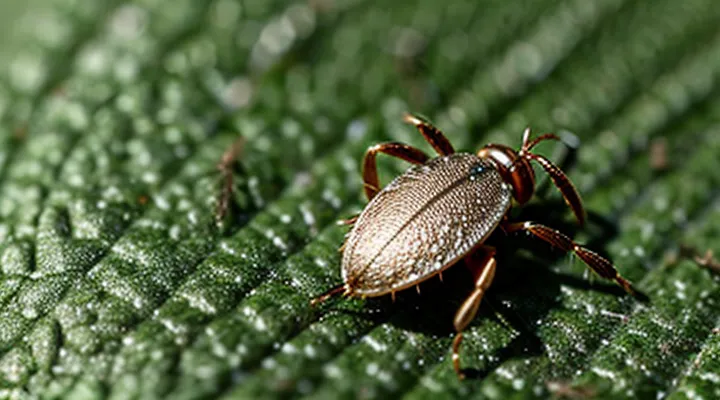
The Immediate Aftermath: What to Expect
Why Does the Head Detach?
Ticks attach to a host using a barbed feeding apparatus called the hypostome, which penetrates the skin and locks into tissue. The mouthparts are not fused to the tick’s body; they are connected by flexible muscles and a thin cuticular sheath. When a grip is placed on the tick’s abdomen and a rapid, forceful pull is applied, the body can separate from the hypostome before the latter disengages from the host. The resulting detachment leaves the head, containing the hypostome and surrounding salivary glands, embedded in the skin.
The separation occurs because:
- The hypostome’s barbs resist outward movement, while the body’s cuticle offers little resistance to traction.
- Muscular contraction during feeding tightens the attachment, making the mouthparts more likely to stay lodged when the body is jerked away.
- The tick’s exoskeleton lacks a rigid joint between the abdomen and mouthparts, allowing a clean break under sufficient force.
If the head remains, it can continue to release saliva that contains pathogens and inflammatory compounds, increasing the risk of local infection and disease transmission. Proper removal—grasping the tick as close to the skin as possible with fine‑point tweezers and pulling upward with steady, even pressure—minimizes the chance of head retention and reduces associated health hazards.
What Happens to the Embedded Head?
Localized Inflammation and Irritation
Removing a tick without extracting the mouthparts leaves foreign tissue embedded in the skin. The body recognizes the retained parts as a wound, triggering a localized inflammatory response. Blood vessels dilate, increasing blood flow to the area; leukocytes migrate to the site, releasing cytokines that cause redness, swelling, heat, and pain. Irritation arises from mechanical trauma and from tick saliva proteins that remain active in the tissue.
Typical signs of the reaction include:
- Erythema surrounding the bite site
- Edematous swelling that may extend a few centimeters from the attachment point
- Tenderness or throbbing pain on palpation
- Mild pruritus that can develop within hours
- Possible formation of a small ulcer or crust as the tissue attempts to expel the foreign material
If the inflammatory process persists beyond several days, secondary infection is likely. Common bacterial agents include Staphylococcus aureus and Streptococcus pyogenes. Indicators of infection are increasing warmth, purulent discharge, and expanding erythema.
Management focuses on eliminating the remaining mouthparts and controlling inflammation:
- Clean the area with antiseptic solution.
- Apply fine‑point tweezers to grasp the exposed portion of the mouthparts and pull straight outward with steady pressure.
- Disinfect again after removal.
- Administer a topical corticosteroid to reduce edema and itching, or an over‑the‑counter antihistamine for symptomatic relief.
- If signs of infection appear, initiate a short course of oral antibiotics targeting skin flora.
Monitoring the site for resolution is essential. Persistent or worsening symptoms warrant medical evaluation to assess for deeper tissue involvement or tick‑borne disease transmission.
Risk of Secondary Infection
Removing a tick without extracting its mouthparts leaves a foreign body in the skin that can serve as a conduit for microorganisms. The exposed cuticle and damaged tissue create an environment conducive to bacterial colonisation and subsequent infection.
Pathogens may enter through the puncture wound directly from the tick’s salivary secretions or from normal skin flora that colonise the residual fragment. Common agents include Staphylococcus aureus, Streptococcus pyogenes, and, less frequently, Borrelia burgdorferi if the tick was infected. The retained mouthparts can also provoke a local inflammatory response that compromises the skin’s barrier function, increasing susceptibility to opportunistic fungi and parasites.
Typical secondary infections present as erythema, swelling, warmth, and pain at the site, often progressing to cellulitis or abscess formation within 24–72 hours. Systemic signs such as fever, chills, or lymphadenopathy may indicate deeper involvement and require prompt medical attention.
Preventive and corrective actions
- Use fine‑point tweezers to grasp the tick as close to the skin as possible and pull upward with steady pressure.
- Inspect the bite area immediately after removal; if any part of the mouth remains, seek professional extraction.
- Clean the wound with an antiseptic (e.g., povidone‑iodine or chlorhexidine) and apply a sterile dressing.
- Monitor for signs of infection for at least a week; initiate antibiotic therapy if redness expands, pus appears, or systemic symptoms develop.
- Consider prophylactic antibiotics for high‑risk individuals (immunocompromised, diabetic, or with extensive bite sites) after consultation with a healthcare provider.
Distinguishing from an Embedded Mouthpart
Removing only the tick’s abdomen while leaving the capitulum embedded creates a foreign body that can continue to draw blood and provoke a localized inflammatory reaction. The retained mouthparts maintain a connection to the host’s dermal tissue, allowing limited feeding and providing a conduit for bacterial invasion. Pathogens transmitted by ticks, such as Borrelia burgdorferi or Anaplasma phagocytophilum, may still be introduced through the retained structures, although the probability diminishes compared to an intact feeding tick.
Clinical signs associated with residual mouthparts include erythema, pruritus, and a papular or nodular lesion at the attachment site. In some cases, a small, firm nodule persists for weeks, reflecting the body’s response to the foreign protein. If the lesion enlarges, becomes painful, or exudes pus, secondary bacterial infection is likely and warrants antimicrobial therapy.
Distinguishing a detached tick’s body from an embedded capitulum relies on visual and tactile cues:
- Size: the remaining part measures 1–3 mm, markedly smaller than the full tick.
- Morphology: only the ventral mouthparts (hypostome, chelicerae) are visible; legs, dorsal scutum, and eyes are absent.
- Color: the capitulum appears pale or translucent, contrasting with the darker, engorged abdomen of a whole tick.
- Texture: the embedded portion feels firm and anchored within the skin, whereas a detached body is smooth and separable.
Accurate identification ensures appropriate removal—typically by excising the mouthparts with sterile scissors or a fine‑point tweezer—thereby reducing the risk of prolonged tissue irritation and pathogen transmission.
Long-Term Concerns and Prevention
Potential Health Risks
Tick-Borne Diseases
Removing a tick without extracting the attached mouthparts leaves fragments embedded in the skin. Those fragments can continue to feed for several hours, providing a conduit for pathogens that the tick may have already transmitted.
The presence of retained mouthparts increases the likelihood of infection with common tick‑borne agents, including:
- Borrelia burgdorferi (Lyme disease)
- Anaplasma phagocytophilum (anaplasmosis)
- Ehrlichia chaffeensis (ehrlichiosis)
- Rickettsia spp. (spotted fever group rickettsioses)
- Babesia microti (babesiosis)
If the mouthparts remain, local inflammation may mask early systemic signs, delaying diagnosis. Serologic testing and polymerase chain reaction assays become less reliable when the infection source persists beneath the skin. Early antimicrobial therapy, typically doxycycline, is less effective if the pathogen continues to be introduced from the residual tissue.
Optimal removal requires a fine‑point tweezer or a specialized tick‑removal tool. Grasp the tick as close to the skin as possible, apply steady upward pressure, and avoid twisting. After extraction, disinfect the site, preserve the tick for identification, and monitor for fever, rash, or joint pain for at least four weeks. If any part of the tick is suspected to remain, seek medical evaluation for possible excision and prophylactic treatment.
Bacterial Infections
Removing a tick without extracting the mouthparts leaves a foreign object embedded in the skin. The retained hypostome can serve as a conduit for bacterial pathogens that the tick introduced during feeding. Immediate local inflammation often accompanies the retained fragments, creating an environment conducive to bacterial proliferation.
Common bacterial agents associated with improperly removed ticks include:
- Borrelia burgdorferi, the causative organism of Lyme disease
- Anaplasma phagocytophilum, responsible for human granulocytic anaplasmosis
- Rickettsia species, which cause spotted fever group rickettsioses
- Francisella tularensis, the agent of tularemia, though rare in tick bites
If the mouthparts remain, bacterial colonization may progress to cellulitis, abscess formation, or systemic infection. Early signs comprise redness, swelling, pain, and purulent discharge at the bite site. Systemic manifestations can include fever, chills, and malaise, reflecting dissemination of the pathogen.
Prompt medical evaluation is essential. Clinicians typically recommend surgical removal of the residual fragments, wound cleaning with antiseptic solutions, and empiric antibiotic therapy targeting the likely organisms. Empirical regimens frequently involve doxycycline, which covers most tick‑borne bacterial infections. Follow‑up monitoring ensures resolution of local inflammation and prevents chronic complications.
Viral Infections
Removing a tick while leaving the mouthparts embedded leaves a portal for pathogens that reside in the salivary glands or the attached fore‑leg. The incomplete extraction does not eliminate the risk of viral transmission; instead, it may increase the probability that viruses are introduced directly into the host’s dermal tissue.
Ticks are vectors for several neurotropic and hemorrhagic viruses. The most clinically relevant include:
- Powassan virus – causes encephalitis, can appear within days of a bite.
- Tick‑borne encephalitis virus (TBE) – produces fever, meningitis, or encephalitis.
- Crimean‑Congo hemorrhagic fever virus – leads to severe hemorrhagic disease.
- Omsk hemorrhagic fever virus – associated with fever and bleeding disorders.
When only the head remains, the detached body continues to secrete saliva containing viral particles. The residual mouthparts may act as a conduit, allowing the virus to migrate from the tick’s salivary glands into the surrounding tissue. This mechanism can shorten the incubation period compared with a fully removed tick, because the virus bypasses the natural barrier of the host’s skin.
Effective management requires immediate action. First, use fine‑pointed tweezers to grasp the mouthparts as close to the skin as possible and extract them with steady pressure, avoiding crushing. Second, disinfect the site with an antiseptic solution. Third, monitor for fever, headache, neck stiffness, or rash for at least four weeks, and seek medical evaluation if symptoms develop. Laboratory testing for viral RNA or serology may be indicated if exposure to a known endemic area occurred.
Proper Tick Removal Techniques
Tools and Materials
Removing a tick without extracting the mouthparts can leave the head embedded in the skin, increasing the risk of infection and disease transmission. Effective removal requires specific instruments and supplies designed to grasp the tick close to the skin surface and to manage the wound afterward.
- Fine‑point tweezers or straight‑edge forceps with serrated tips
- Tick removal device (e.g., a small plastic hook or a commercial tick key)
- Disposable nitrile gloves to prevent direct contact with the parasite
- Antiseptic solution (isopropyl alcohol, povidone‑iodine, or chlorhexidine) for skin preparation and post‑removal cleaning
- Sterile gauze pads or cotton swabs for pressure application and dressing
- Sealable plastic bag or specimen vial with 70 % ethanol for preserving the removed tick if laboratory testing is required
- Sharps container for safe disposal of used instruments
The tweezers or forceps should be positioned as close to the skin as possible, gripping the tick’s body without crushing it. A steady, upward motion removes the parasite while minimizing tissue damage. If the head remains, a sterile needle or the tick removal device can be employed to lift the embedded mouthparts gently. After extraction, the wound must be irrigated with antiseptic, then covered with gauze to control bleeding. The tick specimen, placed in the labeled bag or vial, should be stored at refrigeration temperature if analysis is anticipated. All tools are single‑use or must be autoclaved before reuse, and waste is disposed of in a designated sharps container.
Step-by-Step Guide
Removing a tick without extracting its mouthparts can leave the embedded parts in the skin, creating a portal for infection and increasing the risk of disease transmission. The following protocol minimizes complications and outlines the physiological response to incomplete removal.
- Assess the attachment – Examine the bite site for any visible portion of the tick’s head or mouthparts. If only the body is present, the mouthparts are likely still embedded.
- Disinfect the area – Apply an antiseptic (e.g., iodine or alcohol) to the surrounding skin to reduce bacterial load before manipulation.
- Use fine‑point tweezers – Grip the tick as close to the skin as possible, avoiding compression of the body, which could expel additional saliva.
- Pull upward with steady pressure – Maintain a straight, gentle traction until the entire tick, including the head, separates from the skin. Abrupt jerks increase the chance of breaking the mouthparts.
- Inspect the extraction site – Look for any residual fragments. If any part remains, do not attempt further removal with fingers; instead, seek professional medical care.
- Clean the wound again – Apply antiseptic once more and cover with a sterile dressing if bleeding occurs.
- Monitor for symptoms – Over the next 24‑48 hours, watch for redness, swelling, or a rash at the bite site. Persistent inflammation or flu‑like symptoms may indicate pathogen transmission and warrant immediate medical evaluation.
Leaving tick mouthparts embedded triggers a localized inflammatory response that can progress to secondary infection. Prompt, complete removal eliminates this risk and reduces the likelihood of disease vectors such as Borrelia or Rickettsia establishing infection. If any fragment is suspected, professional extraction is essential to prevent complications.
When to Seek Medical Attention
Persistent Symptoms
Removing a tick while the mouthparts remain in the skin can leave pathogens in contact with the host tissue. The retained hypostome may continue to deliver saliva containing bacteria, viruses, or protozoa for several hours after the initial bite, increasing the risk of infection. Early recognition of ongoing exposure is essential for timely treatment.
Persistent clinical manifestations often emerge weeks after the incomplete removal. Common long‑lasting symptoms include:
- Fever lasting more than 48 hours without an obvious source
- Severe headache or neck stiffness resistant to over‑the‑counter analgesics
- Muscular or joint pain that fluctuates or worsens with activity
- Skin lesions such as erythema migrans, papular rash, or ulcerated nodules
- Neurological signs, including tingling, numbness, or facial weakness
The duration and severity of these signs depend on the pathogen transmitted. For instance, Borrelia burgdorferi may cause chronic arthritis and neuropathy, while Anaplasma phagocytophilum frequently leads to prolonged fatigue and leukopenia. Persistent lymphadenopathy or unexplained weight loss may indicate a systemic response requiring laboratory confirmation.
Diagnostic work‑up should comprise serologic testing for tick‑borne diseases, complete blood count, inflammatory markers, and, when indicated, polymerase chain reaction assays on tissue samples. Empiric antimicrobial therapy may be justified if clinical suspicion is high, especially for Lyme disease or spotted‑fever rickettsiosis, because delayed treatment correlates with increased risk of chronic complications.
Follow‑up evaluations at intervals of two to four weeks allow clinicians to monitor symptom evolution, adjust therapy, and identify late‑onset sequelae. Documentation of the initial removal technique, including whether the head was left embedded, assists in risk stratification and patient counseling.
Signs of Infection
Removing a tick without extracting the embedded mouthparts creates an entry point for bacteria and other pathogens. The retained fragments can irritate tissue and trigger a localized infection.
Typical indicators of infection include:
- Redness spreading outward from the bite site
- Swelling that exceeds the immediate area of the tick attachment
- Warmth or heat sensation around the puncture
- Pain that intensifies rather than subsides
- Pus or clear fluid discharge
- Fever or chills accompanying the skin changes
If any of these signs develop, clean the area with antiseptic, apply a sterile dressing, and seek medical evaluation promptly. Healthcare providers may prescribe antibiotics, recommend tetanus vaccination if needed, and remove residual tick parts under sterile conditions. Early intervention reduces the risk of complications such as cellulitis, Lyme disease, or other tick‑borne infections.
Preventing Tick Bites
Personal Protection
Removing a tick without extracting its mouthparts leaves a fragment embedded in the skin. The retained piece can continue to feed, causing local irritation and increasing the chance of pathogen transmission. Pathogens such as Borrelia burgdorferi (Lyme disease) and Anaplasma can be introduced through the exposed tissue surrounding the fragment, potentially leading to infection even after the tick’s body is gone.
A fragment that remains attached can become a nidus for bacterial colonization. The body’s immune response may produce a chronic inflammatory nodule, sometimes requiring medical intervention. Early detection of residual parts reduces the risk of complications; visual inspection of the bite site after removal is essential.
Personal protection measures include:
- Use fine‑point tweezers or a specialized tick‑removal tool; grasp the tick as close to the skin as possible.
- Apply steady, upward pressure; avoid twisting or jerking motions that could detach the mouthparts.
- After extraction, disinfect the area with an antiseptic solution.
- Examine the bite site for any remaining fragments; if present, seek professional medical removal.
- Monitor the site for signs of redness, swelling, or a rash over the next several weeks; report any symptoms to a healthcare provider promptly.
Adopting these practices minimizes the likelihood of residual mouthparts and the associated health risks, reinforcing effective personal protection against tick‑borne diseases.
Yard Management
Proper yard management reduces the likelihood of encountering ticks that may be partially removed. Maintaining short grass, clearing leaf litter, and creating a barrier of wood chips or mulch around high‑traffic areas limit tick habitats. Regularly mowing and trimming vegetation eliminates the microclimate ticks need for survival.
When a tick is pulled without its head, the mouthparts remain embedded in the skin. The retained parts can continue to feed, increasing the chance of pathogen transmission. They may also cause localized irritation, swelling, or infection if bacteria enter the wound.
Effective yard practices to prevent such incidents include:
- Mow lawns weekly during peak tick season.
- Remove tall weeds, brush, and leaf piles.
- Install a 3‑foot strip of wood chips or gravel between lawns and wooded borders.
- Apply acaricide treatments to perimeter zones according to label directions.
- Inspect pets and family members after outdoor activities; promptly remove any attached ticks with fine‑point tweezers, ensuring the entire organism is extracted.
If a mouthpart remains after removal, clean the site with antiseptic, apply a sterile bandage, and monitor for signs of infection or rash. Seek medical advice if symptoms develop. Consistent yard maintenance and correct tick removal together minimize health risks associated with incomplete extraction.
