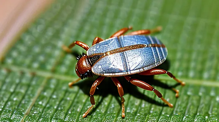
The Scale of Smallness
Comparing with Common Ticks
The tiniest tick, often a larval stage of Ixodes species, measures approximately 0.2 mm in length and appears as a translucent, rounded body lacking the hardened scutum that characterizes adult ticks. Its legs are proportionally long, giving the organism a spider‑like silhouette, and its mouthparts are slender and barely visible under low magnification.
Common ticks, such as adult Dermacentor or Rhipicephalus specimens, range from 2 mm to 5 mm when unfed and display a distinct, darkened scutum covering the dorsal surface. Their bodies are robust, with a clearly defined capitulum and shorter legs relative to body size. The coloration is typically brown to reddish‑brown, providing camouflage on host fur or vegetation.
Key morphological contrasts:
- Size: larval tick ≈ 0.2 mm; adult common tick ≥ 2 mm.
- Body covering: larva lacks scutum; adult possesses a hardened shield.
- Leg proportion: larva’s legs exceed body width; adult’s legs are shorter.
- Transparency: larva appears nearly invisible in water; adult is opaque and pigmented.
These differences enable precise identification when examining specimens under a microscope, facilitating accurate classification and appropriate control measures.
The Smallest Known Tick Species
The smallest tick species currently recognized is Ixodes microti, discovered in the Alpine region of Europe. Adult females measure approximately 1.2 mm in length when unfed, placing them at the lower extreme of tick dimensions. Males are slightly smaller, averaging 1.0 mm.
Morphologically, I. microti exhibits a compact, oval body with a smooth dorsal scutum that lacks the ornate patterns typical of larger ixodids. The mouthparts are proportionally short, reducing the overall profile. Legs are slender and bear fine, sensory setae that aid navigation through dense moss and leaf litter.
Ecologically, the species parasitizes small mammals such as voles and shrews, completing its life cycle within a confined microhabitat. Its diminutive size facilitates attachment to hosts with minimal detection.
- Length (unfed adult female): ~1.2 mm
- Length (unfed adult male): ~1.0 mm
- Dorsal scutum: smooth, unornamented
- Mouthparts: short, compact
- Preferred hosts: small rodents
- Habitat: alpine moss and leaf litter
These attributes define the visual and biological profile of the tiniest known tick.
Anatomy of a Tiny Tick
External Features
The tiniest tick species, such as Ixodes minutus, measures roughly 0.5 mm when unfed. Its external morphology is highly reduced, enabling it to remain unnoticed on host skin.
- Body shape: oval, dome‑shaped, lacking a pronounced scutum; dorsal surface appears smooth.
- Coloration: pale amber to translucent, facilitating camouflage against light‑colored fur or skin.
- Legs: eight slender, jointed legs, each less than 0.2 mm long; coxae are proportionally large, providing stability.
- Mouthparts: short, ventrally situated hypostome with a limited number of barbs, adapted for brief attachment.
- Sensory organs: simple eyes (ocelli) reduced to minute pigment spots; Haller’s organ on the first pair of legs remains functional for host detection.
- Cuticle: thin, flexible exoskeleton with minimal sclerotization, allowing expansion during blood intake.
Internal Structures
The tiniest member of the arachnid order Ixodida measures less than 0.2 mm when unfed. Its compact body consists of two primary regions: the gnathosoma, which houses the feeding apparatus, and the idiosoma, which contains the remaining internal systems.
The gnathosoma includes a pair of chelicerae for cutting skin and a hypostome equipped with backward‑pointing barbs that anchor the tick during blood extraction. Muscular fibers surrounding the chelicerae enable rapid opening and closing motions, while a small salivary gland duct delivers anticoagulant compounds directly into the host.
The idiosoma encloses the following structures:
- A dorsal shield (scutum) composed of hardened cuticle that protects internal organs.
- A ventral plate (ventrum) that supports the legs and houses the digestive tract.
- A simple tubular gut divided into fore‑, mid‑, and hind‑sections; the foregut leads to the hypostome, the midgut stores ingested blood, and the hindgut expels waste.
- Paired Malpighian tubules for excretion and osmoregulation.
- A rudimentary nervous system comprising a central ganglion in the opisthosomal region and segmental nerve cords extending to each leg.
- A pair of spiracles on the lateral margins that connect to a tracheal network for gas exchange.
- Small salivary and excretory glands embedded within the dorsal tissue, each lined with secretory cells.
The entire organ system is compacted within a soft hemocoel that circulates hemolymph, providing nutrients and immune factors throughout the body. The minute size of the tick imposes constraints on organ volume, resulting in highly specialized, densely packed structures that maintain functional efficiency during the brief feeding period.
Life Cycle and Habitat
Developmental Stages
The tick’s life cycle consists of four distinct stages, each with characteristic morphology and size.
- Egg: spherical, 0.5 mm in diameter, smooth surface, translucent to pale white.
- Larva: the smallest free‑living stage, typically 0.2–0.3 mm long, six‑legged, reddish‑brown, covered by a fine, translucent cuticle that reveals underlying internal structures.
- Nymph: 0.5–1.0 mm long, eight‑legged, darker brown to black, body more robust, scutum visible on dorsal surface.
- Adult: 2–5 mm (female up to 6 mm), fully developed scutum, deep brown to black coloration, pronounced mouthparts for blood feeding.
The larval tick, being the diminutive form, exhibits a compact, oval body with a soft, almost invisible outer layer. Its legs are proportionally longer than the body, allowing mobility on host surfaces. The coloration is uniformly light brown, lacking the hardened scutum seen in later stages. These traits define the appearance of the smallest tick in its developmental sequence.
Preferred Environments
The diminutive tick species thrives in microhabitats that provide stable moisture, moderate temperature, and frequent contact with suitable hosts. Ground litter composed of leaf fragments, moss, and fine detritus retains the humidity necessary for desiccation‑sensitive stages. Underneath the litter layer, temperatures usually remain within 10‑20 °C, a range that supports metabolic activity without triggering premature diapause.
Key environmental characteristics include:
- High relative humidity (≥ 80 %): prevents water loss during questing and molting.
- Shade or low‑light conditions: reduces temperature fluctuations and solar drying.
- Presence of small vertebrate hosts (e.g., rodents, shrews): ensures access to blood meals required for development.
- Stable substrate composition: fine, loosely packed organic matter allows easy movement and attachment to passing hosts.
- Protected microclimates: burrows, rodent nests, or rock crevices offer shelter from wind and predation.
In regions where these conditions co‑occur, the smallest tick exhibits its characteristic compact morphology, enabling it to navigate the narrow interstices of the substrate while remaining concealed from predators. Absence of any factor—particularly low humidity or extreme temperature—reduces survival rates and limits population density.
Microscopic Identification Challenges
Tools and Techniques
The investigation of the tiniest tick requires instrumentation capable of resolving sub‑nanometer dimensions or sub‑picosecond intervals, depending on whether the subject is a physical mark or a temporal increment.
High‑resolution imaging tools include:
- Scanning electron microscope (SEM) with field‑emission source for surface features down to a few nanometers.
- Transmission electron microscope (TEM) for internal structures at atomic resolution.
- Atomic force microscope (AFM) for topographical mapping with sub‑nanometer precision.
Temporal measurement techniques rely on:
- Picosecond‑resolution digital oscilloscopes employing interleaved sampling to capture rapid transitions.
- Time‑interval counters synchronized to a cesium or rubidium frequency standard, delivering uncertainties below 10 ps.
- Optical frequency combs referenced to an optical atomic clock, enabling direct measurement of intervals approaching the femtosecond regime.
Data acquisition systems must combine low‑noise front‑ends, high‑speed analog‑to‑digital converters, and precise triggering to avoid aliasing. Calibration against traceable standards guarantees that the observed tick corresponds to the true physical or temporal smallest unit under study.
Distinguishing from Mites
The tiniest tick species measure roughly 0.5 mm when unfed, a size that exceeds most free‑living mites, which commonly remain under 0.3 mm. This dimensional gap provides the first practical clue for separation in field samples.
Ticks possess a distinct body architecture: a ventrally located capitulum bearing a barbed hypostome, and an idiosoma that may bear a dorsal scutum in hard‑tick families. Mites lack a scutum and typically present a more uniform body without a specialized feeding apparatus.
Adult ticks bear eight legs; their larvae bear six before the second molt, after which eight legs appear. Mites retain eight legs throughout all active stages, and many species never develop a six‑legged larval form. The presence of a six‑legged larval stage followed by an eight‑legged nymph is therefore indicative of a tick.
Mouthparts differ sharply. Ticks insert a needle‑like hypostome equipped with backward‑pointing barbs, enabling prolonged attachment to hosts. Mites employ chelicerae that function as pincers or stylets, lacking barbs and generally unsuitable for deep tissue penetration.
Eyes are another discriminating feature. Many hard ticks exhibit a pair of dorsal eyes positioned near the scutum margin. In contrast, the majority of mites are eyeless or possess simple ocelli that do not resemble the tick’s well‑defined visual organs.
Key diagnostic points
- Size: ticks ≥ 0.5 mm; mites ≤ 0.3 mm.
- Body: tick scutum and capitulum; mite uniform body.
- Larval legs: six in ticks, eight in mites.
- Mouthparts: barbed hypostome (ticks) vs. chelicerae (mites).
- Eyes: distinct dorsal eyes (ticks) vs. absent or rudimentary (mites).
Applying these criteria enables reliable distinction between the smallest tick specimens and morphologically similar mites.
Ecological Role
Impact on Ecosystems
The diminutive ixodid, often measuring less than a millimeter, possesses a flattened, oval body, a smooth dorsal shield, and proportionally short legs. Its minute size enables penetration of host skin with minimal detection, facilitating blood extraction from a wide range of vertebrates.
Ecological consequences of this micro‑parasite include:
- Transmission of pathogenic microorganisms to mammals, birds, and reptiles, altering disease dynamics within populations.
- Regulation of host density through sublethal effects such as reduced reproductive output and increased susceptibility to predation.
- Contribution to nutrient cycling by returning blood‑derived organic material to the soil after detachment and death.
These interactions shape community structure, influence predator‑prey relationships, and affect overall biodiversity across habitats where the smallest tick species persists.
Disease Transmission Potential
The tiniest known tick species measures under one millimeter when unfed, possesses a flattened body, and exhibits limited mobility compared to larger relatives. Its diminutive size enables penetration of thin skin layers on small mammals, birds, and occasionally reptiles.
Disease transmission potential derives from three primary attributes: pathogen acquisition during blood meals, survival of the pathogen within the tick’s internal environment, and successful inoculation into a new host during subsequent feeding. Laboratory studies confirm that the smallest tick can harbor Borrelia burgdorferi sensu stricto, Anaplasma phagocytophilum, and tick‑borne encephalitis virus (TBEV). Field surveys have detected these agents in engorged specimens collected from grassland habitats.
Key factors influencing transmission efficiency include:
- Host range – preference for rodent and ground‑dwelling bird species creates a bridge between wildlife reservoirs and incidental human exposure.
- Feeding duration – attachment periods as short as 24 hours suffice for pathogen transfer in this species, compared with longer requirements for larger ticks.
- Environmental stability – tolerance of low humidity and temperature fluctuations extends questing activity throughout early spring and late autumn.
Public‑health implications demand targeted surveillance in regions where the smallest tick coexists with high‑density rodent populations. Control strategies focus on habitat modification to reduce leaf‑litter accumulation, use of acaricide‑treated bait stations, and education of at‑risk communities about early removal of attached ticks. Continuous monitoring of pathogen prevalence within these minute vectors informs risk assessments and guides resource allocation for disease prevention programs.
Research and Future Prospects
Current Studies
Recent investigations have focused on characterizing the morphology of the tiniest known tick species, often referred to as Ixodes sp. “micro‑tick.” Researchers have employed high‑resolution imaging techniques to capture detailed external features that were previously unobservable.
Advanced microscopy, including scanning electron microscopy (SEM) and confocal laser scanning microscopy, provides nanometer‑scale resolution of cuticular structures. Specimens are prepared in ethanol‑preserved or fresh‑frozen states to preserve delicate setae and sensilla. Molecular profiling accompanies morphological analysis, confirming species identity through mitochondrial 16S rRNA sequencing.
Key observations from current studies include:
- Body length ranging from 0.12 mm to 0.18 mm in unfed adult females.
- Dorsal shield (scutum) exhibiting a translucent, lightly sclerotized surface with faint reticulate patterning.
- Mouthparts reduced to a short, tapered hypostome lacking the typical barbs seen in larger relatives.
- Leg segments proportionally elongated, with tarsal claws barely discernible at 10 µm length.
- Absence of visible ocular structures; sensory pits dominate the anterior region.
These findings refine taxonomic placement, distinguishing micro‑ticks from juvenile stages of larger species. Morphological data support the hypothesis that extreme miniaturization influences host‑attachment strategies and may affect pathogen transmission dynamics. Ongoing research aims to link structural adaptations with ecological niches, enhancing predictive models of tick‑borne disease risk.
Unanswered Questions
The precise morphology of the tiniest tick remains uncertain. Existing microscopy images show only partial outlines, and the full structure of the smallest developmental stage has not been captured in a single, high‑resolution photograph.
Key unanswered questions include:
- What are the exact dimensions of the larval stage when it measures less than 0.1 mm?
- Which anatomical features become visible only under electron microscopy, and how do they differ from larger specimens?
- Does the cuticle of the smallest tick exhibit unique patterns or textures that distinguish it from other life stages?
- How does the coloration change during the transition from egg to the first active stage in environments with varying humidity?
Research efforts are hindered by the difficulty of isolating specimens without causing deformation. Current techniques rely on indirect measurement, leaving a gap between observed data and a definitive visual representation. Resolving these questions will require advances in imaging technology and refined collection methods.
