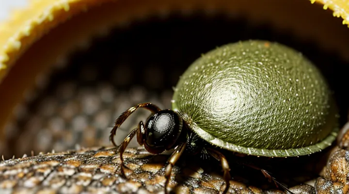
The Hunt: Before Engorgement
Questing Behavior
When a tick has filled its body with blood, the drive that previously directed it to climb vegetation and wait for a passing host—known as questing—diminishes sharply. The engorged specimen lowers itself from the host, often by crawling down the animal’s fur or by dropping directly onto the ground. This descent marks the transition from host‑seeking to a period of internal processing and reproduction.
After detachment, the tick undergoes several coordinated steps:
- Digestive expansion: Enzymes break down the ingested blood, providing nutrients for rapid tissue growth.
- Molting: In species that continue a life stage, the tick sheds its cuticle to accommodate the enlarged body.
- Egg production: Female ticks allocate the acquired resources to develop thousands of eggs within their enlarged abdomen.
- Nest construction: The tick seeks a protected microhabitat—leaf litter, soil, or crevices—where it can lay the egg mass safely.
During these phases, the tick no longer exhibits questing posture. Instead, it remains stationary, often concealed, until oviposition is complete. Once the eggs are deposited, the adult’s role ends, and the cycle restarts with newly hatched larvae that will resume questing to locate their first blood meal.
Host Detection Mechanisms
Ticks locate a suitable host through a combination of sensory cues that become critical once they have expanded with blood. Their detection system relies on:
- Carbon‑dioxide gradients: Receptors on the tick’s Haller’s organ sense rising CO₂ levels emitted by breathing vertebrates.
- Thermal contrast: Infrared‑sensitive sensilla detect temperature differences between the environment and the warm body of a host.
- Odorant molecules: Volatile compounds such as lactic acid, ammonia, and specific skin lipids trigger chemosensory neurons.
- Vibrational input: Mechanoreceptors respond to footfalls and movements, guiding the tick toward an approaching animal.
- Humidity cues: Hygroreceptors assess moisture levels, which increase near a host’s skin and help differentiate potential targets.
During the engorgement phase, the tick’s body swells, stretching cuticular structures that amplify sensory input. Enhanced receptor exposure improves the tick’s ability to pinpoint a host for the final blood meal. After reaching full engorgement, the tick disengages from the host, relying on the same sensory suite to locate a secure drop‑off site, typically a sheltered microhabitat where it can complete digestion and molt.
The Feeding Process: Engorgement Unveiled
Attachment and Saliva Secretion
During the final feeding phase a tick remains firmly anchored to the host’s skin. The mouthparts, especially the barbed hypostome, embed into the epidermis, and a proteinaceous cement secreted from the salivary glands hardens to prevent dislodgement. This attachment persists for several days while the tick expands its body size by ingesting blood.
Saliva production intensifies as the tick swells. The fluid contains a cocktail of bioactive molecules that facilitate rapid blood intake and suppress host defenses. Key components include:
- Anticoagulants that inhibit clot formation, allowing continuous flow.
- Vasodilators that widen capillaries and increase blood volume at the feeding site.
- Immunomodulators that dampen inflammatory responses and reduce itching.
- Enzymes that degrade host tissue, maintaining a clear feeding channel.
The combined action of cement and saliva ensures the tick can complete engorgement without interruption. Once the abdomen reaches maximal size, the cement weakens, the tick detaches, drops to the ground, and begins the reproductive cycle.
Blood Meal Acquisition
Ticks attach to a host using their hypostome, a barbed feeding organ that penetrates the skin and anchors the parasite. Salivary secretions contain anticoagulants, vasodilators, and immunomodulatory proteins, allowing uninterrupted blood flow. The tick’s foregut expands as it ingests plasma, red blood cells, and nutrients, increasing its body mass up to 100‑times its unfed size.
During engorgement, the following physiological events occur:
- Midgut distension: epithelial cells stretch, triggering rapid cell division to accommodate the volume of blood.
- Digestive enzyme activation: proteases and lipases break down proteins and lipids, providing amino acids and fatty acids for growth.
- Metabolic shift: aerobic respiration intensifies, and stored glycogen is mobilized to fuel muscular activity required for attachment maintenance.
- Pathogen acquisition: any microbes present in the host’s blood can be taken up and later transmitted to new hosts.
- Detachment preparation: the tick secretes enzymes that weaken the cement-like attachment, enabling it to drop off the host once the blood meal is complete.
After reaching full engorgement, the tick drops off the host, seeks a sheltered environment, and initiates the next developmental stage. The ingested blood is processed over several days; excess water is excreted as urine, while concentrated nutrients support molting or reproduction, depending on the species and life stage. This sequence ensures the tick converts a single blood meal into the resources necessary for survival and population continuity.
Physical Changes During Engorgement
During the feeding phase, a tick’s body undergoes rapid expansion as it absorbs the host’s blood. The cuticle, normally rigid, becomes pliable, allowing the abdomen to swell up to several times its unfed size. This enlargement is visible as a pronounced, rounded silhouette that replaces the earlier flattened profile.
Key physical transformations include:
- Weight increase: mass may rise from a few milligrams to over a gram, reflecting the volume of ingested blood.
- Color shift: the cuticle darkens, often turning from pale brown to a deep, almost black hue due to hemoglobin concentration.
- Cuticular stretching: micro‑fibers within the exoskeleton realign, preventing rupture while accommodating the expanding gut.
- Midgut dilation: the digestive chamber expands dramatically, creating space for blood storage and facilitating enzyme activity.
- Leg positioning: legs retract slightly toward the body, reducing leverage and stabilizing the engorged form.
These alterations prepare the tick for detachment and subsequent development, ensuring that the stored nutrients support molting, reproduction, or overwintering depending on the species’ life stage.
Duration of Feeding
Ticks attach to a host and remain attached for a species‑specific period while they ingest blood. The feeding interval determines when the tick reaches its engorged state and varies among life stages. Larvae typically feed for 2–5 days, nymphs for 3–7 days, and adult females for 5–10 days, depending on the species. In Ixodes scapularis, adult females require 7–10 days to fill, whereas Dermacentor variabilis females may complete engorgement in 5–6 days under optimal conditions.
Factors influencing the feeding duration include ambient temperature, host immune response, and the tick’s metabolic rate. Higher temperatures accelerate blood digestion, shortening the feeding period by up to 30 percent. Host grooming or immune defenses can interrupt feeding, causing premature detachment and incomplete engorgement.
Key points about feeding time:
- Species determines baseline duration (larva < nymph < adult female).
- Temperature modifies metabolic speed and thus feeding length.
- Host defenses can truncate the feeding cycle, affecting engorgement success.
Post-Engorgement: The Tick's Next Steps
Detachment
When a tick reaches full engorgement, the insect initiates separation from its host. The abdomen expands dramatically, stretching the cuticle and weakening the attachment organs. Salivary secretions that previously maintained a firm grip become reduced, allowing the hypostome to lose its anchoring grip.
Physiological changes drive this process. Hormonal spikes trigger muscle relaxation in the legs and mouthparts. The cement-like substance deposited during feeding begins to degrade, and the tick’s internal pressure pushes the body away from the skin surface. These mechanisms operate without external assistance from the host.
Environmental cues influence the timing of detachment. Ambient temperature, humidity, and daylight length affect the tick’s metabolic rate, accelerating or delaying the release. For instance, higher temperatures increase blood flow, shortening the feeding period and prompting earlier separation.
Key aspects of the detachment phase include:
- Abdominal swelling that stretches the attachment structures.
- Decrease in cement secretion and enzymatic breakdown of the bonding matrix.
- Hormone‑mediated muscle relaxation in the legs and hypostome.
- External conditions (temperature, humidity) that modify the feeding duration.
After detachment, the tick drops to the ground, seeks a protected microhabitat, and begins the post‑feeding molt. The engorged body, now detached, contains a fully developed batch of eggs ready for oviposition in the next reproductive cycle.
Digestion of the Blood Meal
When a tick swells with blood, the meal initiates a cascade of digestive processes that transform the ingested plasma and cells into usable nutrients. The midgut epithelium secretes a suite of proteases, lipases, and carbohydrases that break down hemoglobin, albumin, and lipids. Hemoglobin is cleaved into amino acids and heme, the latter bound by specialized proteins to prevent oxidative damage. Lipid droplets are emulsified and hydrolyzed, supplying fatty acids for energy storage.
Absorption occurs through microvilli lining the gut lumen. Amino acids and fatty acids enter the hemolymph, where they are distributed to the salivary glands, ovaries, and developing tissues. The tick synthesizes vitellogenin, a yolk precursor, using the surplus proteins; vitellogenin is transported to the ovaries for egg maturation. Concurrently, the tick up‑regulates genes involved in cuticle expansion, allowing the exoskeleton to accommodate the increased volume.
Excess water and nitrogenous waste are expelled via the Malpighian tubules and rectal glands. The rectal sac concentrates the waste, which is later eliminated during the subsequent molt or detachment from the host. The entire digestion phase spans 3‑7 days, depending on species and ambient temperature, after which the tick either drops off to lay eggs or seeks a new host for the next life stage.
Reproductive Cycle Initiation
When a female tick reaches full engorgement, the sudden increase in body mass and nutrient intake activates the reproductive cascade. Stretch receptors in the cuticle detect expansion, sending neural signals that stimulate the brain’s neuroendocrine centers. These centers release prohormones that are converted into ecdysteroids, the primary hormones driving vitellogenesis.
The hormonal surge initiates several coordinated events:
- Vitellogenin synthesis in the fat body, providing yolk precursors for developing oocytes.
- Oocyte maturation within the ovaries, progressing from previtellogenic to vitellogenic stages.
- Spermatophore storage in the spermatheca, which may have been acquired during the preceding blood meal or from a subsequent mating encounter.
- Egg shell formation through chitin deposition, preparing embryos for environmental exposure.
Once oocytes are fully stocked with yolk, the tick begins oviposition. Egg laying occurs in a protected microhabitat, often within the engorged female’s own exuviae or in the surrounding leaf litter. The number of eggs correlates with the volume of blood ingested; larger meals yield higher fecundity.
In summary, engorgement triggers a neurohormonal pathway that converts the acquired blood resources into reproductive output, completing the tick’s life cycle and ensuring the propagation of the next generation.
Health Implications of Engorged Ticks
Disease Transmission Risks
When a tick expands to its maximum size after ingesting blood, the physiological changes facilitate the movement of pathogens from the tick’s salivary glands into the host’s bloodstream. This stage coincides with the highest likelihood of disease transmission because the feeding apparatus remains attached for an extended period, allowing pathogens to be injected continuously.
The risk of infection rises markedly during the engorged phase. Pathogens that reside in the tick’s midgut must migrate to the salivary glands before they can be transmitted; this migration is accelerated as the tick swells and its metabolic activity increases. Consequently, the longer a fully fed tick remains attached, the greater the probability that viable organisms will be delivered to the host.
Key diseases associated with engorged ticks include:
- Lyme disease (caused by Borrelia burgdorferi)
- Rocky Mountain spotted fever (Rickettsia rickettsii)
- Anaplasmosis (Anaplasma phagocytophilum)
- Babesiosis (Babesia microti)
- Tick-borne encephalitis virus
- Ehrlichiosis (Ehrlichia chaffeensis)
Factors influencing transmission risk are:
- Duration of attachment – risk escalates after 24 hours for most bacterial pathogens and after 48 hours for some viruses.
- Tick species – certain species, such as Ixodes scapularis and Dermacentor variabilis, are more efficient vectors.
- Pathogen prevalence in the tick population – regional variations affect the probability of encountering an infected tick.
- Host immune status – immunocompromised individuals experience more severe outcomes.
Prompt removal of an attached tick, preferably within 24 hours, dramatically reduces the chance of pathogen transfer. After extraction, the bite site should be inspected for signs of infection, and individuals at high risk should seek medical evaluation for possible prophylactic treatment.
Localized Reactions and Symptoms
When a tick expands after a blood meal, the bite site often exhibits a confined inflammatory response. The skin around the attachment point may become erythematous, swelling slightly, and feel warm to the touch. These changes result from the tick’s salivary proteins that suppress local immunity and promote blood flow.
Typical localized manifestations include:
- Redness extending 2–5 mm from the puncture.
- Mild to moderate swelling that peaks within 24 hours.
- Pruritus that intensifies as the tick detaches.
- A small, painless papule or nodule that may persist for several days.
- Occasionally, a faint, linear track of erythema reflecting the tick’s mouthparts.
If the lesion enlarges rapidly, becomes increasingly painful, or develops pus, secondary bacterial infection should be considered. Persistent or atypical reactions warrant medical evaluation to rule out early signs of tick‑borne disease.
Prevention and Removal Strategies
Personal Protection Measures
Ticks enlarge dramatically after a blood meal, creating a higher risk of pathogen transmission. Reducing exposure to engorged ticks requires proactive personal protection.
- Wear long sleeves and trousers; tuck shirts into pants to block attachment sites.
- Apply EPA‑registered repellents containing DEET, picaridin, IR3535, or oil of lemon eucalyptus to exposed skin and clothing.
- Treat boots, gaiters, and pants with permethrin; reapply after washing.
- Perform thorough body checks within 30 minutes of leaving tick‑infested areas; focus on scalp, armpits, groin, and behind knees.
- Shower promptly after outdoor activity; water pressure helps dislodge unattached ticks.
- Remove any attached tick with fine‑point tweezers, grasping close to the skin and pulling straight upward without crushing the body; preserve the specimen for identification if needed.
- Store removed ticks in a sealed container for at least 24 hours to confirm complete removal before disposal.
Consistent use of these measures limits contact with feeding ticks, thereby decreasing the chance that an engorged tick will transmit disease.
Safe Tick Removal Techniques
An engorged tick expands dramatically as it fills with blood, increasing the risk of pathogen transmission. Prompt, correct removal reduces that risk and prevents the mouthparts from remaining embedded.
- Use fine‑pointed tweezers or a dedicated tick‑removal tool.
- Grasp the tick as close to the skin surface as possible, holding the head and body together.
- Apply steady, upward pressure without twisting or jerking.
- Continue pulling until the tick releases completely; avoid squeezing the abdomen to prevent rupture.
- Disinfect the bite area with alcohol, iodine, or soap and water.
- Place the detached tick in a sealed container with a label (date, location) for possible identification.
- Observe the site for several days; seek medical advice if redness, swelling, or flu‑like symptoms develop.
Improper techniques—such as burning, freezing, or using petroleum products—can increase infection risk and damage the tick, making pathogen identification more difficult. Following the outlined steps ensures safe extraction and minimizes health hazards associated with a swollen tick.
