Initial Appearance
Small, Dark Spot
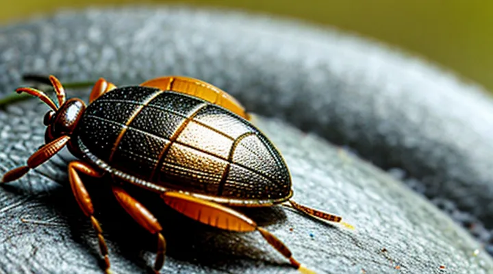
A tick that has begun feeding presents as a compact, dark spot on the skin. The lesion measures roughly 2‑5 mm in diameter and appears as a uniform, almost black discoloration. The surface may be slightly raised, reflecting the engorged body of the parasite beneath the epidermis.
Key visual indicators include:
- Dark, almost black coloration contrasting with surrounding skin
- Small, rounded shape with smooth edges
- Minimal elevation, often indistinguishable from a simple bruise without close inspection
- Absence of a distinct outer shell; the tick’s body is partially concealed within the skin
Recognition of these characteristics enables prompt identification and removal, reducing the risk of pathogen transmission.
Raised Bump or Nodule
A tick that has attached to the skin typically presents as a small, firm elevation. The lesion feels like a raised bump, often dome‑shaped, and may be slightly tender to pressure. The surface can appear smooth or slightly rough, depending on the species and duration of attachment. In many cases, the head and mouthparts are visible as a dark point at the apex of the nodule, while the body may be engorged and change color from pale to reddish‑brown as it feeds. The surrounding skin may show a faint halo of erythema, but inflammation is usually minimal. Observation of a discrete, raised nodule with a discernible central point strongly suggests a feeding tick rather than a simple skin papule.
Common Characteristics
Body Shape and Size
A tick that has attached to human skin presents a distinct morphology that changes as blood is ingested. In its early attachment stage the organism is flat, oval, and roughly 2–5 mm long, resembling a miniature seed. The dorsal surface is smooth and the legs are visible, extending outward from the corners of the body.
During feeding the tick’s abdomen expands dramatically. An engorged specimen can reach 5–10 mm in width and 8–12 mm in length, sometimes exceeding 15 mm when fully saturated. The shape transitions from a flattened oval to a rounded, balloon‑like profile, with the ventral side pressed against the skin and the dorsal side bulging outward. The leg positions become less conspicuous as the body swells.
Key dimensions:
- Unengorged adult: 2–5 mm length, 1–2 mm width.
- Partially engorged: 5–8 mm length, 3–5 mm width.
- Fully engorged: up to 12 mm length, 10–15 mm width.
The coloration shifts from brown or reddish‑brown in the unfed state to a grayish‑blue or tan hue when engorged, reflecting the blood content beneath the cuticle. The overall appearance—flattened oval to swollen sphere—provides a reliable visual cue for identification.
Leg Visibility
Ticks commonly attach to the lower extremities, especially the legs, because vegetation contacts the skin during outdoor activities. When a tick is attached, it appears as a small, rounded swelling that may be mistaken for a spider bite or a skin tag. The body of the tick is typically dark brown to black, with a smooth, glossy surface that contrasts with the surrounding skin tone. The head, or capitulum, protrudes slightly from the body and can be seen as a tiny, lighter‑colored point.
Visibility on the leg depends on several factors:
- Hair density: dense leg hair can conceal the tick, making visual detection more difficult.
- Skin tone: lighter skin provides higher contrast, enhancing the tick’s visibility.
- Clothing: socks, pants, or boots obscure the area, reducing the chance of noticing the parasite.
- Tick size: unfed nymphs measure 1–2 mm and may be nearly invisible, whereas engorged adults can reach up to 10 mm, becoming more apparent.
Effective inspection of the legs requires systematic examination from the ankle upward, using a mirror or a partner when necessary. Lightly parting hair and stretching the skin can reveal the tick’s characteristic shape. Prompt removal reduces the risk of disease transmission and prevents the tick from expanding further within the skin.
Coloration Variations
Ticks that have attached to the skin display a range of colors depending on species, life stage, and feeding status. The base coloration of an unfed tick typically reflects its taxonomic group. Common patterns include:
- Light brown or tan exoskeleton in many larval and nymph stages.
- Dark brown to black shields (scutum) in adult females of Ixodes species.
- Reddish‑orange hue in Dermacentor species before blood intake.
- Gray‑ish or mottled appearance in Amblyomma nymphs.
Feeding induces marked changes. As the tick expands, its cuticle stretches, revealing a pale, almost translucent abdomen that may appear whitish or pinkish. Engorged females often exhibit a dramatic color shift to a deep brown or black body with a lighter, balloon‑like abdomen. In contrast, engorged males retain a flatter shape and maintain the original dorsal coloration, though the abdomen may appear slightly swollen.
Some ticks possess distinctive markings that aid identification. Stripes, spots, or patterned scutums can be present on the dorsal surface, ranging from faint silvery lines to bold dark bands. These markings may fade as the tick fills with blood, making the overall color appear more uniform.
Environmental factors also influence coloration. Ticks that have spent time in leaf litter or grass may acquire a dusted, greenish tint from surrounding vegetation, while those exposed to sunlight can develop a slightly darker exoskeleton due to melanization.
Understanding these coloration variations assists in accurate recognition of attached ticks and informs appropriate removal and medical assessment.
Differentiating from Other Skin Conditions
Moles and Freckles
Moles are typically well‑defined pigmented lesions ranging from 1 mm to several centimeters. Their color may be uniform brown, black, or a mixture of hues; occasional pink or flesh tones occur. Borders are usually smooth, though some lesions display slight irregularity. Surface texture can be flat, slightly raised, or dome‑shaped, and the lesion is generally firm to the touch.
Freckles appear as small, flat macules, often 1–2 mm in diameter. They are uniformly tan to light brown and become more pronounced after exposure to ultraviolet light. Borders are indistinct, blending seamlessly with surrounding skin. The lesions do not elevate or change in texture over time.
Ticks attached to the skin differ markedly. An engorged tick presents as a rounded, raised structure, often dark brown or black, with a clear anterior attachment point where the mouthparts embed. Legs may be visible around the perimeter, and the body can swell to several millimeters in height, creating a palpable bulge.
Key distinguishing features:
- Size: moles can exceed several centimeters; freckles remain ≤ 2 mm; ticks enlarge primarily in height, not diameter.
- Elevation: moles may be flat or raised; freckles are flat; ticks are distinctly raised with a central attachment groove.
- Borders: moles possess defined edges; freckles have diffuse margins; ticks exhibit a sharp outline around the engorged body.
- Texture: moles feel firm; freckles feel smooth; ticks feel soft, sometimes gelatinous, and may shift slightly when pressed.
Recognition of these characteristics enables accurate differentiation between benign pigmented lesions and an attached arthropod, reducing unnecessary concern and guiding appropriate medical response.
Scabs or Ingrown Hairs
A tick attached to the skin typically presents as a small, rounded body, often darker than surrounding tissue, with a visible mouthpart embedded in the epidermis. The surrounding area may show a faint halo of redness, but the tick itself remains distinct from other skin lesions.
Scabs and ingrown hairs can be mistaken for a tick because they also appear as raised, pigmented formations. Differentiating features include:
- Shape: scabs are irregular, flaky, and lack a defined head; ingrown hairs form a narrow, cylindrical protrusion, sometimes surrounded by a halo of inflammation.
- Attachment: a tick’s mouthparts are firmly anchored, often visible as a tiny point at the center; scabs are loosely attached, and ingrown hairs are anchored by the hair shaft itself.
- Mobility: ticks remain stationary until removed; scabs may peel, and ingrown hairs can shift slightly with movement.
- Texture: ticks feel firm and may have a slightly raised, dome‑shaped surface; scabs are soft to crumbly, while ingrown hairs feel like a firm, hair‑filled nodule.
Accurate identification prevents unnecessary removal attempts and reduces risk of infection. If uncertainty persists, professional evaluation is recommended.
Other Insect Bites
Ticks attached to the skin appear as small, dome‑shaped lesions that may be dark brown or black. The body often enlarges after feeding, creating a noticeable bulge. A clear, disc‑shaped mouthpart, sometimes called a capitulum, may be visible at the center of the lesion.
Other insect bites exhibit distinct visual patterns:
- Mosquito bite: raised, red welts surrounded by a halo of mild swelling; typically itchy and transient.
- Flea bite: clusters of tiny puncture marks, each surrounded by a red halo; often located on the lower legs or ankles.
- Bed‑bug bite: linear or zig‑zag arrangement of three to five small, red papules; may develop a central punctum.
- Spider bite: localized erythema with a central necrotic spot in some species; surrounding area may be painful or swollen.
- Ant bite or sting: immediate swelling and erythema; may include a puncture wound with a visible stinger tip.
Distinguishing features such as lesion size, shape, presence of a central disc, and pattern of distribution aid in differentiating a tick attachment from other insect bites. Accurate identification supports appropriate medical response.
Signs of a Recent vs. Engorged Tick
Early Stage Attachment
During the initial hours after a tick latches onto human skin, the parasite appears as a small, oval body measuring roughly 2–5 mm in length. The dorsal surface is smooth and light brown to reddish‑brown, lacking the pronounced swelling seen in later stages.
Visible features include:
- A flat, unengorged abdomen that remains thin and elongated.
- Distinct, dark‑colored capitulum (mouthparts) protruding forward, often visible as a tiny “beak” at the attachment site.
- Legs positioned laterally, each bearing tiny claws that grip the skin.
The attachment point is usually in warm, protected areas such as the scalp, armpits, groin, or behind the knees. The tick’s legs may be partially hidden beneath the skin but can be detected by gentle palpation.
Early‑stage ticks differ from other arthropods by their lack of rapid movement, the presence of a hard dorsal shield (scutum), and the characteristic forward‑pointing mouthparts. Recognition of these traits enables prompt removal before significant blood intake and engorgement occur.
Partially Engorged Tick
A partially engorged tick presents a distinct morphology that differs from both unfed and fully engorged stages. The body elongates and swells, but the abdomen does not reach the maximal roundness seen in a fully fed specimen. The anterior portion, including the capitulum and legs, remains relatively narrow, while the posterior abdomen shows moderate expansion, often appearing oval or slightly oblong.
Key visual characteristics include:
- Dark brown to reddish‑brown coloration, sometimes with a lighter posterior margin.
- Abdomen length increased by 30‑60 % compared with an unfed tick.
- Visible segmentation of the scutum, which may appear slightly stretched.
- Legs still clearly articulated, allowing the tick to remain mobile on the host.
- Presence of a small, translucent window near the ventral surface, indicating partial blood intake.
These features enable identification of a tick that has begun feeding but has not yet reached full engorgement. Recognizing this stage assists in assessing the duration of attachment and potential pathogen transmission risk.
Fully Engorged Tick
A fully engorged tick attached to the skin presents as a markedly enlarged, oval or spherical body. The abdomen expands to several times its original size, often reaching 10–15 mm in length, while the head and legs remain relatively small and tucked against the body. The cuticle becomes thin and translucent, allowing the blood‑filled interior to appear pale yellow, pink, or reddish, depending on the host’s blood color.
The surface texture changes from the smooth, matte appearance of an unfed tick to a glossy, stretched skin that may appear slightly wrinkled where the abdomen expands. The ventral side shows the mouthparts firmly embedded in the epidermis, with the hypostome visible as a short, barbed projection. The legs, though still present, are less noticeable due to the bulk of the engorged abdomen.
Key visual indicators for identification:
- Size increase to several times normal dimensions
- Translucent, blood‑colored abdomen
- Glossy, thin cuticle with subtle wrinkles
- Visible mouthparts anchored in the skin
Recognition of these characteristics enables prompt removal and reduces the risk of pathogen transmission.
What to Do After Identification
Safe Removal Techniques
Ticks that are firmly attached to the skin appear as small, dark, oval bodies embedded in the epidermis. Their heads may be obscured, while the abdomen often swells as the parasite fills with blood. The attachment point can be slightly raised, sometimes surrounded by a faint halo of irritation.
Safe removal requires precision and avoidance of pressure on the tick’s body. The following procedure minimizes the risk of pathogen transmission:
- Grasp the tick as close to the skin as possible using fine‑point tweezers or a specialized tick‑removal tool.
- Apply steady, downward pressure to pull the tick straight out without twisting or squeezing the abdomen.
- Inspect the extracted specimen to ensure the mouthparts are completely removed; retained parts may cause local inflammation.
- Disinfect the bite site with an antiseptic solution such as povidone‑iodine or alcohol.
- Store the tick in a sealed container with a label indicating the date and location of removal for potential laboratory analysis.
- Dispose of the container by sealing it and discarding in regular waste, or by freezing the tick for later testing.
If the mouthparts remain embedded, consult a healthcare professional for removal. Monitoring the bite area for signs of rash, fever, or flu‑like symptoms over the next several weeks is essential, as these may indicate infection. Prompt medical evaluation should occur if any such symptoms develop.
When to Seek Medical Attention
A tick that has attached to the skin may appear as a flat, gray‑brown disc that enlarges into a rounded, darkened nodule as it feeds. The body can become noticeably swollen, especially near the head, and the surrounding skin may show slight redness.
Seek immediate medical evaluation if any of the following signs develop:
- Persistent fever, chills, or severe headache within weeks of the bite.
- A rash resembling a target, expanding rapidly, or any unusual skin lesions.
- Joint pain, swelling, or stiffness that does not resolve quickly.
- Neurological symptoms such as confusion, facial weakness, or difficulty concentrating.
- Prolonged attachment exceeding 24 hours, or difficulty removing the tick completely.
Professional assessment should include proper tick extraction, documentation of the attachment duration, and consideration of prophylactic antibiotics when indicated. Follow‑up appointments are advisable to monitor for delayed symptoms, especially in regions where tick‑borne diseases are prevalent.
