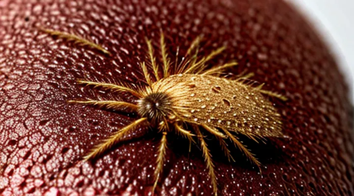
Understanding Scabies: The Invisible Invader
What is Scabies?
Scabies is a contagious skin infestation caused by the microscopic arthropod Sarcoptes scabiei var. hominis. The adult female mite burrows into the epidermis to lay eggs, triggering an immune response that produces intense itching and characteristic skin lesions.
Key clinical features include:
- Intense pruritus that worsens at night.
- Small, gray‑white or yellowish papules and vesicles.
- Linear or serpentine tracks (burrows) measuring 2–10 mm, often found between fingers, on wrists, elbows, waistline, and genital areas.
- Secondary bacterial infection from scratching.
Transmission occurs through prolonged skin‑to‑skin contact or sharing of clothing, bedding, or towels. The mite cannot survive more than 48–72 hours off the human host.
Diagnosis relies on clinical presentation and confirmation by microscopic examination of skin scrapings, which reveal mites, eggs, or fecal pellets. Effective treatment consists of topical scabicidal agents—such as 5 % permethrin cream applied overnight to the entire body—or oral ivermectin in resistant cases. All close contacts should receive simultaneous therapy to prevent reinfestation.
The Scabies Mite: Anatomy and Life Cycle
Size and Microscopic Appearance
The human scabies mite, Sarcoptes scabiei var. hominis, measures approximately 0.2–0.4 mm in length, roughly the size of a grain of sand. Its body is oval, covered by a hard dorsal shield (sclerotized cuticle) that appears translucent under magnification. The mite possesses eight short legs, each ending in claw‑like structures that facilitate attachment to the epidermis.
Microscopically, the mite displays a compact, flattened profile. The dorsal shield is smooth, with faint striations visible at high power (×1000). Ventral surfaces reveal a series of ventral plates and a distinct genital opening. Two simple eyes are positioned near the anterior margin. The legs are visible as slender appendages bearing setae and terminal claws, which can be seen clearly in phase‑contrast or differential interference contrast microscopy.
- Length: 0.2–0.4 mm (200–400 µm)
- Width: ≈ 0.15 mm (150 µm)
- Dorsal shield: translucent, smooth, slightly striated at ×1000
- Legs: eight, short, clawed, bearing setae
- Eyes: two simple, anteriorly placed
- Ventral plates: visible, genital opening distinct
These dimensions and structural details enable identification of the mite within skin burrows, where it creates a characteristic linear or serpiginous track visible to the naked eye as a thin, raised line. Under microscopic examination, the mite’s compact form, shielded dorsal surface, and clawed legs confirm its identity.
Lifecycle Stages and Skin Interaction
Scabies mites progress through four distinct stages, each influencing the skin’s appearance and symptoms.
- Egg: Laid within a short, tortuous tunnel in the stratum corneum; each egg measures about 0.2 mm and remains invisible to the naked eye until it hatches.
- Larva: Emerging as six-legged organisms, the larvae migrate a few millimeters from the original burrow, creating new superficial tracks that appear as fine, linear lesions.
- Nymph: After two molts, the mite gains eight legs and enlarges to approximately 0.3 mm. Nymphal burrowing deepens existing tunnels, intensifying the characteristic serpiginous lines.
- Adult: The mature female, about 0.4 mm long, resides permanently in a burrow, laying 10–30 eggs over several weeks. Males remain on the surface, seeking mates, and are rarely observed.
Interaction with the epidermis begins when the female inserts her mouthparts into the outer skin layer, forming a microscopic channel. The channel’s entrance appears as a pinhead-sized punctum; the tunnel itself manifests as a raised, gray‑white line that may be slightly elevated or flat, depending on the depth of infestation. The mite’s movement and the host’s immune response generate intense itching, erythema, and occasional vesicles surrounding the burrow. In advanced cases, secondary bacterial infection can produce crusted plaques, markedly altering the skin’s texture and coloration.
Understanding these stages clarifies why early lesions present as isolated puncta, while later infestations display extensive, serpentine tracks that trace the mite’s progression through the cutaneous environment.
Visualizing Scabies on Human Skin
Direct Signs of Mite Activity
Burrows: The Signature Lesion
Burrows represent the pathognomonic lesion of scabies infestation. They appear as thin, linear or serpentine tracks cut into the stratum corneum, typically measuring 2–10 mm in length. The tracks are pale or skin‑colored, often flanked by a subtle erythematous halo that may be indistinct on heavily pigmented skin. The distal end frequently terminates in a small vesicle or papule, which can become pruritic and may develop a crust if scratched.
Key clinical attributes of scabies burrows:
- Location: interdigital spaces of the hands, flexor surfaces of the wrists, elbows, axillae, waistline, and genital region.
- Orientation: usually aligned with skin tension lines, following the direction of finger flexion.
- Texture: superficial, easily visible under magnification; may be concealed beneath scales or hyperkeratotic plaques in chronic cases.
- Evolution: newly formed tracks are clear and translucent; older lesions become hyperpigmented or scarred.
Identification of these characteristic tunnels enables accurate diagnosis and guides targeted therapy.
Rash Characteristics: Papules and Vesicles
Scabies infestation produces a distinctive rash that typically begins as small, firm papules. These raised lesions are often clustered in line‑like patterns, reflecting the burrowing path of the mite. The papules may be erythematous or flesh‑colored, measuring 1–3 mm in diameter, and are most common on the wrists, interdigital spaces, elbows, and genital area.
In addition to papules, vesicles can develop, especially in hypersensitive individuals. Vesicles appear as clear or serous‑filled blisters, ranging from pinpoint to several millimeters across. They frequently accompany intense itching and may rupture, leaving superficial erosions that can become crusted.
Key features of the rash include:
- Linear arrangement of lesions following mite tunnels
- Presence of both solid papules and fluid‑filled vesicles
- Predominant distribution on thin‑skinned regions and skin folds
- Intense pruritus that intensifies at night
Recognition of these characteristics aids in differentiating scabies from other pruritic dermatoses and guides timely therapeutic intervention.
Indirect Indicators and Symptoms
Itching: The Primary Complaint
The most common reason patients seek medical attention for scabies is intense pruritus. The itch usually intensifies at night and may become relentless, disrupting sleep and daily activities. It originates from the immune response to mite saliva and feces deposited in the superficial epidermis.
Typical characteristics of the itch include:
- Localized to the web spaces of the fingers, wrists, elbows, waistline, and genital region.
- Accompanied by small, raised tracks (burrows) that appear as gray‑white or skin‑colored lines, 2–10 mm long.
- Often associated with secondary lesions such as vesicles, pustules, or excoriations caused by scratching.
The presence of these signs together with nocturnal itching strongly suggests infestation by the microscopic arthropod that burrows into the stratum corneum. Prompt identification of the primary complaint—persistent, night‑time itch—facilitates early diagnosis and treatment, preventing widespread skin damage and transmission.
Secondary Skin Changes: Scabs and Infections
The initial mite activity produces characteristic linear or serpentine tracks, but the skin often progresses to secondary lesions that dominate the clinical picture.
Scabs develop where intense scratching disrupts the epidermis. They appear as dark‑brown or black crusts, usually irregular in shape, adherent to the underlying erythema. Frequently they overlay the original burrows on the wrists, interdigital spaces, and elbows, giving a mottled, lichenified appearance.
Superimposed bacterial infection is common after prolonged excoriation. Typical signs include increased warmth, swelling, purulent discharge, and a foul odor. The most frequently isolated pathogens are:
- Staphylococcus aureus (including methicillin‑resistant strains)
- Streptococcus pyogenes
These organisms exploit the compromised barrier, producing impetiginized lesions that may coalesce into larger erosive areas.
Effective management requires simultaneous control of the mite infestation and the secondary changes. Primary therapy with scabicidal agents (e.g., permethrin 5 % cream) eliminates the source of irritation. Adjunctive measures include:
- Gentle cleansing with antiseptic solutions to remove crusts.
- Topical antibiotics (mupirocin or fusidic acid) applied to localized infection.
- Oral antibiotics (dicloxacillin, clindamycin, or appropriate agents for MRSA) for extensive or systemic involvement.
Prompt treatment reduces the risk of further bacterial spread and accelerates skin healing, restoring a smoother, less inflamed surface.
Common Locations for Scabies Manifestations
Areas of Predilection
Scabies mites preferentially inhabit skin folds and regions where warmth and humidity are greatest. Typical sites include:
- Interdigital spaces of the hands, especially between the fourth and fifth fingers
- Flexor surfaces of the wrists and elbows
- Axillary folds
- Inframammary and subareolar areas of the chest
- Nipple–areola complex, particularly in infants and nursing mothers
- Waistline, including the belt and underwear regions
- Genitalia and perianal skin
- Buttocks and the upper thighs
- Feet, particularly the web spaces between the toes
These locations provide the microenvironment required for mite survival and reproduction, explaining the characteristic distribution of lesions.
Atypical Presentations and Vulnerable Groups
Scabies may manifest beyond the classic linear burrows on the wrists and interdigital spaces. In atypical cases the mite’s activity produces small, raised papules that lack visible tracks, often clustered on the torso, face, or scalp of infants. Nodular scabies appears as firm, itchy nodules, frequently on the elbows, knees, or genital area, persisting despite standard treatment. Crusted (Norwegian) scabies presents with thick, hyperkeratotic plaques that cover extensive body surfaces; the density of mites in these lesions is markedly higher, facilitating rapid spread. Unusual locations such as the palms, soles, and perianal region may host subtle erythema or vesicles that mimic other dermatologic conditions.
- Neonates and young infants
- Elderly individuals, especially those in long‑term care facilities
- Patients with HIV/AIDS, organ transplants, or chemotherapy‑induced immunosuppression
- Individuals with neurological disorders that impair scratching reflexes (e.g., dementia, stroke)
- Persons receiving systemic corticosteroids or other immunomodulatory drugs
Recognizing these variants prevents misdiagnosis and limits outbreaks among populations where the disease course is more severe or treatment response is compromised. Early identification of non‑classical lesions, combined with awareness of high‑risk groups, supports prompt, effective management.
Differentiating Scabies from Other Skin Conditions
Similarities to Allergic Reactions
Scabies infestations produce thin, gray‑white tunnels (burrows) that trace the mite’s path beneath the epidermis. The tunnels often appear as slightly raised lines, most commonly on the wrists, interdigital spaces, elbows, and waistline. Surrounding the burrows, small, firm papules may develop, sometimes forming vesicles or pustules when the immune response intensifies. These lesions are intensely pruritic, especially at night.
Allergic skin reactions generate comparable signs:
- Redness (erythema) that can mimic the inflamed borders of scabies papules.
- Itching of similar severity, often worsening with heat or sweat.
- Swollen, raised bumps (hives or wheals) that resemble the papular component of a mite infestation.
Both conditions involve a host immune response to a foreign antigen—mite proteins in scabies, allergens in hypersensitivity. Histamine release contributes to vasodilation and edema in each case, producing overlapping visual cues. However, scabies uniquely features linear burrows, whereas allergic reactions lack such defined tracks. Recognizing the presence of burrows distinguishes scabies from pure allergic dermatitis.
Distinguishing Features from Fungal Infections
Scabies infestation presents as tiny, whitish burrows embedded in the epidermis. The burrows appear as linear or serpentine tracks, typically 2–10 mm long, most often located between fingers, on wrists, elbows, waistline, and genital area. The mite itself is invisible to the naked eye; only the tunnel it creates can be seen. Accompanying lesions include small, erythematous papules and intense pruritus that intensifies at night.
Fungal infections, such as tinea corporis or candidiasis, display distinct characteristics:
- Lesions are usually raised, scaly, and may form a well‑defined, circular border with central clearing.
- Coloration ranges from pink to brown; no linear burrows are present.
- Scale is often adherent and can be removed, revealing a moist or dry base.
- Distribution follows a pattern of spreading patches rather than confined tunnels.
- Itching may be present but is generally less severe and not specifically nocturnal.
Key differentiators:
- Morphology – Scabies produces linear burrows; fungi generate round or oval plaques with peripheral scaling.
- Location – Scabies favors interdigital spaces, wrists, and genital folds; fungal lesions commonly affect warm, moist areas such as the groin, feet, and trunk.
- Symptom timing – Scabies itching peaks during sleep; fungal itch is continuous or intermittent without a nocturnal surge.
- Diagnostic signs – Direct visualization of burrows or mite eggs confirms scabies; KOH preparation revealing hyphae confirms fungal infection.
Recognizing these features enables accurate identification and appropriate treatment.
The Importance of Professional Diagnosis
Professional diagnosis is indispensable when evaluating the visual signs of scabies on the skin. Dermatologists can differentiate the characteristic burrow patterns, papules, and vesicles caused by Sarcoptes scabiei from other dermatological conditions that present similar lesions. Accurate identification prevents misinterpretation of normal skin variations or unrelated infections.
A clinician’s assessment provides several concrete benefits:
- Confirmation of mite presence through dermatoscopy or skin scraping, eliminating guesswork.
- Determination of infestation severity, guiding appropriate treatment duration and medication choice.
- Detection of secondary bacterial infection, which may require additional therapy.
- Documentation of spread patterns, informing public‑health measures and contact tracing.
Relying on self‑diagnosis often leads to delayed treatment, inappropriate medication, and prolonged contagion. Expert evaluation ensures that the distinctive visual cues of scabies are correctly recognized and managed, reducing complications and transmission risk.
