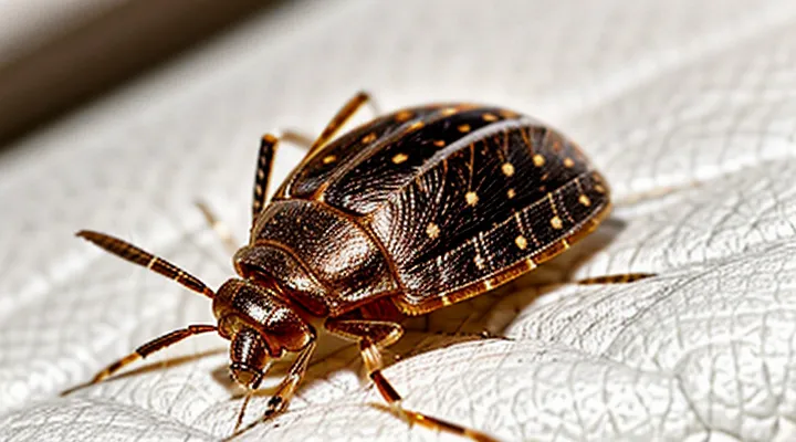
General Appearance
Size and Shape
Female bed bugs measure approximately 4 mm to 5 mm in length when unfed and can reach up to 7 mm after a blood meal. Their bodies are dorsoventrally flattened, allowing easy movement beneath mattress seams and furniture crevices. The overall silhouette is an elongated oval, broader at the abdomen and tapering toward the head.
Key dimensions:
- Length: 4–5 mm (unfed), up to 7 mm (fed)
- Width: 2.5 mm at the widest point of the abdomen
- Height: 1.5 mm, reflecting the flattened profile
The exoskeleton displays a smooth, glossy surface without prominent ridges. The thorax occupies roughly one‑third of the total body length, while the abdomen comprises the remaining two‑thirds, giving the insect a distinct, rounded rear. Antennae are slender, segmented, and extend no more than 0.5 mm, while the legs are short, each ending in a claw that facilitates gripping fabric fibers. The mouthparts form a concealed, needle‑like proboscis suited for piercing skin.
Coloration
Female bed bugs exhibit a characteristic reddish‑brown hue that darkens after a blood meal. The exoskeleton retains a matte finish, while the abdomen becomes more swollen and a deeper, almost mahogany shade when engorged. The thorax and legs remain lighter, typically a tan‑brown color, providing a contrast that aids identification.
Key coloration features:
- Base color: uniform reddish‑brown across the body.
- Abdomen after feeding: darker, mahogany to burgundy tone, reflecting the expanded gut.
- Legs and antennae: lighter tan‑brown, sometimes with a subtle yellowish tint.
- Nymphal stages: similar coloration but less intense, lacking the pronounced abdominal darkening seen in mature females.
These color patterns, combined with size and shape, allow reliable visual distinction of adult females from males and immature individuals.
Distinguishing Female from Male Bed Bugs
Abdominal Differences
Female bed bugs possess an abdomen that differs noticeably from that of males. The abdomen is broader, more rounded, and exhibits a distinct curvature toward the posterior end. This shape accommodates the reproductive organs and the expanding ovaries.
Key abdominal attributes include:
- Width: approximately 1.5 mm in unfed females, expanding to 2.5 mm after a blood meal.
- Length: proportionally longer than the male’s, reaching up to 5 mm when engorged.
- Segmentation: visible dorsal plates (tergites) appear smoother, with a subtle, pale median line that is less pronounced in males.
Coloration varies with feeding status. Unfed females display a uniform reddish‑brown hue across the abdomen. After ingesting blood, the abdomen turns a brighter, engorged red, often covering the entire dorsal surface. The ventral side may show a lighter, almost translucent appearance, revealing the underlying blood mass.
Post‑feeding changes are rapid. Within minutes, the abdomen expands, the cuticle stretches, and the dorsal surface becomes glossy. The expansion can increase abdominal volume by up to 200 %. This swelling is a reliable visual cue for distinguishing females from males in field observations.
Reproductive Structures
Female bed bugs possess a specialized reproductive system that enables egg production and storage of sperm after mating. The system consists of paired ovaries, each containing multiple ovarioles that generate mature oocytes. Oocytes travel through the lateral oviducts to the common median oviduct, which leads to the external genital opening.
Key components include:
- Ovaries – paired organs housing ovarioles where oocyte development occurs.
- Spermatheca – a sac‑like structure that retains sperm from the male, allowing fertilization of successive egg batches.
- Median oviduct – conduit that transports fertilized eggs toward the ovipositor.
- Ovipositor – elongated, segmented tube used to deposit eggs into crevices or fabric.
- Genital capsule – external sclerotized plate surrounding the genital opening, providing protection and structural support.
During oviposition, the ovipositor extends through the genital capsule, positioning each egg for deposition. The spermatheca releases stored sperm to fertilize oocytes as they pass through the median oviduct, ensuring continuous reproductive output throughout the female’s lifespan.
Female-Specific Characteristics
Post-Mating Indicators
After a successful insemination, the adult female of Cimex lectularius displays several physiological and morphological changes that differentiate her from an unmated counterpart. The abdomen expands noticeably to accommodate developing eggs, and the cuticle often acquires a slightly darker hue due to increased hemolymph circulation.
- Abdomen enlargement: lengthens by up to 30 % and becomes more convex.
- Color shift: cuticle darkens, especially on the dorsal surface.
- Ovipositor development: terminal segments become more pronounced.
- Behavioral alteration: increased tendency to seek sheltered sites for oviposition.
- Reduced locomotor activity: movement slows as energy is redirected toward reproduction.
These post‑mating markers provide reliable criteria for field identification of fertilized females, facilitating accurate population monitoring and targeted pest‑management interventions.
Ovipositor Presence (or lack thereof)
Female bed bugs (Cimex lectularius) can be distinguished by several anatomical features, among which the egg‑laying apparatus is frequently examined. The species does not possess a true, elongated ovipositor as seen in many other hemipterans. Instead, the female uses the terminal region of her abdomen, a shortened and sclerotized structure, to deposit eggs directly onto surfaces.
Key points regarding the ovipositor‑like structure:
- The organ is reduced to a blunt, ventral tip rather than a protruding needle.
- Eggs are released through a small opening called the gonopore.
- The lack of a specialized ovipositor does not hinder egg placement; females can embed eggs in crevices and fabric folds.
- The reduced structure is consistent across all developmental stages of the adult female.
Consequently, when assessing a female bed bug, the absence of a prominent ovipositor serves as a reliable diagnostic characteristic.
Developmental Stages of Female Bed Bugs
Nymph Stages
Female bed bugs develop through five distinct nymphal instars before attaining adult morphology. Each stage exhibits incremental growth and subtle changes in coloration, body shape, and the development of wing pads.
- First instar – Length 1.5 mm; translucent to pale amber; abdomen uniformly colored; no wing pads visible.
- Second instar – Length 2.2 mm; deeper amber hue; faint darkening of the dorsal surface; wing pads appear as minute, indistinct ridges.
- Third instar – Length 2.9 mm; coloration approaches the mature brown‑red tone; wing pads become more pronounced, extending halfway along the thorax.
- Fourth instar – Length 3.5 mm; dorsal coloration darkens further; wing pads cover roughly two‑thirds of the thorax; abdominal segments show slight segmentation.
- Fifth instar – Length 4.0 mm; coloration matches that of the adult female; wing pads nearly reach the posterior thoracic margin; body size approaches final adult dimensions.
Key visual cues for identifying a female nymph at any stage include the absence of fully developed genitalia, the progressive enlargement of wing pads, and the gradual deepening of the brown‑red exoskeleton. By the fifth instar, the nymph closely resembles an adult female, differing only in size and the incomplete development of the reproductive organs.
Adult Stage
Adult female bed bugs belong to the species Cimex lectularius and reach full development after several nymphal molts. At the adult stage the insect measures approximately 5–7 mm in length, with a flattened, oval‑shaped body. The dorsal surface exhibits a uniform reddish‑brown coloration that may appear darker after a recent blood meal.
Key morphological features include:
- A broad, convex abdomen that expands markedly when engorged, revealing a visible swelling of the ventral side.
- Six short, hair‑like sensory setae on the dorsal surface, aiding in host detection.
- A pair of short, non‑functional wings concealed beneath the tegmina; flight is absent.
- A slender, piercing rostrum extending forward from the head, used to penetrate skin and draw blood.
- Antennae composed of four segments, each bearing sensilla for chemical cues.
- Two large, compound eyes positioned laterally on the head, providing limited visual perception.
The exoskeleton remains relatively soft, permitting the abdomen to stretch during feeding. After oviposition, the female can lay up to 200 eggs over her lifespan, each deposited in concealed cracks and crevices near the host environment.
Where to Find Female Bed Bugs
Common Hiding Spots
Female bed bugs preferentially occupy locations that provide darkness, limited disturbance, and proximity to a blood source. Their choice of refuge influences detection and control efforts.
Typical concealment areas include:
- Mattress seams, folds, and tag strips
- Box‑spring voids and internal springs
- Bed‑frame joints and metal brackets
- Headboard or footboard cracks and decorative panels
- Upholstered‑furniture cushions, seams, and under‑cover folds
- Behind baseboards, wall trim, and inside wall cavities
- Behind picture frames, wall art, and decorative molding
- Inside luggage, backpacks, and travel bags
These sites share characteristics of tight spaces, low traffic, and easy access to a sleeping host. Regular inspection of each location improves early identification of infestations.
Signs of Infestation
The presence of a female bed bug in a dwelling manifests through distinct indicators that allow early detection and prompt intervention.
Visible evidence includes:
- Small, rust‑colored stains on bedding, mattresses, or furniture, representing digested blood excreted by the insect.
- Translucent exoskeletons left behind after molting, often found near seams, cracks, or folds.
- Live or dead specimens, typically 4–5 mm in length, with a flattened, oval shape and a reddish‑brown hue that darkens after feeding.
- Tiny, dark specks resembling pepper grains, indicating fecal deposits left on fabric or wall surfaces.
Additional clues encompass:
- Clusters of itchy, red welts on the skin, commonly appearing in linear or zigzag patterns.
- A faint, sweet, musty odor, especially in heavily infested areas, resulting from the insects’ pheromones and metabolic by‑products.
Prompt identification of these signs enables effective control measures before the population expands.
