The Anatomy of a Tick
External Features
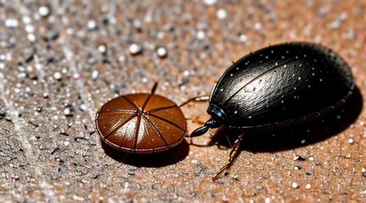
A flattened tick presents a distinct set of external characteristics that differ markedly from an intact specimen.
- Overall shape: elongated, oval silhouette with softened edges; the hard dorsal shield (scutum) appears compressed and may lose its defined outline.
- Size: length reduced to roughly one‑third of the original, while width expands slightly, giving a squat appearance.
- Color: dark brown to black, often mottled with reddish or amber patches where internal fluids have seeped to the surface.
- Surface texture: glossy or slightly wet sheen caused by released hemolymph; fine hairs (setae) become less visible, and the cuticle may appear torn or ragged.
- Visible internal contents: faint gray‑white or yellowish material may be seen through the translucent cuticle, indicating crushed internal organs.
These observable traits enable rapid identification of a tick that has been mechanically damaged.
Internal Structures
When a tick is flattened, the outer chitinous armor breaks apart, exposing the internal anatomy. The capitulum, located at the front, contains the hypostome—a barbed feeding tube—paired chelicerae used for cutting skin, and the palps that guide the mouthparts. The scutum, a hard dorsal plate, fragments, revealing the underlying soft cuticle that covers the body’s interior.
The abdomen, which expands during a blood meal, shows a translucent matrix filled with ingested blood. Within this matrix, the midgut epithelium lines a spacious chamber where digestion occurs. Salivary glands, positioned laterally, appear as pale, elongated sacs that release anticoagulant compounds during feeding. The nervous system consists of a ventral nerve cord with segmental ganglia, visible as faint, thread‑like structures beneath the cuticle.
Legs detach easily; each limb consists of segmented sclerites connected by flexible membranes. When crushed, the joint membranes rupture, leaving recognizable claw tips and pulvilli. The hemocoel, the primary body cavity, contains hemolymph that circulates nutrients and immune cells. In a flattened specimen, this fluid pools in irregular spaces, often mixed with residual blood.
Key internal components observable in a crushed tick:
- Capitulum (hypostome, chelicerae, palps)
- Salivary glands
- Midgut filled with blood
- Ventral nerve cord with ganglia
- Hemocoel with hemolymph
- Fragmented scutum and leg sclerites
These structures collectively define the morphology visible after the tick’s exoskeleton is compressed.
What Happens When a Tick is Crushed?
Physical Disruption of the Body
When a tick’s exoskeleton is subjected to crushing pressure, the rigid dorsal shield (scutum) fractures into irregular shards. The abdomen, composed of softer cuticle, collapses, forming a flattened, amorphous mass that often adheres to surrounding tissue. Internal organs—midgut, salivary glands, and reproductive structures—spill outward, appearing as translucent, gelatinous residues that may mix with hemolymph, creating a glossy sheen.
Observable characteristics include:
- Fragmented scutum edges, jagged and uneven.
- A compressed, dough‑like core with visible gut contents.
- Dispersed, milky fluid from salivary glands and hemolymph.
- Loss of the tick’s typical oval silhouette, replaced by a ragged, amorphous shape.
These changes result from mechanical disruption of the arthropod’s chitinous armor and soft internal tissues, producing a visual profile that contrasts sharply with an intact specimen.
Release of Internal Fluids
When a tick is flattened, its exoskeleton ruptures and internal fluids escape. The hemolymph, a pale yellow to amber liquid, spreads across the surrounding surface, often mixing with the tick’s dark body contents. Blood meals that the tick has ingested appear as a reddish‑brown mass, sometimes leaking from the abdomen and pooling around the crushed remains.
Key visual cues include:
- Color contrast: Transparent or lightly colored hemolymph against the tick’s deep brown or black cuticle.
- Viscosity: Fluid is thin and spreads rapidly, unlike the thicker, more gelatinous material of the engorged abdomen.
- Distribution: Fluid may form small droplets or a thin film, often seeping into cracks or fabric fibers.
The release of these fluids can contaminate surfaces, potentially leaving microscopic residues that are detectable under magnification. Proper disposal—placing the crushed tick in a sealed container and cleaning the area with disinfectant—prevents accidental exposure to pathogens that may be present in the expelled material.
Visual Characteristics of a Crushed Tick
Color Changes
A flattened tick reveals a distinct palette that changes from the intact exterior. The outer exoskeleton, normally dark brown or black, becomes translucent as pressure breaks the cuticle. Beneath the thin shell, the body releases a mixture of hemolymph and blood, creating the following observable colors:
- Pale yellow‑orange: hemolymph diluted with host blood, most common in adult females.
- Bright red: concentrated host blood when the tick has fed heavily.
- Dark brown‑black: residual gut contents and partially digested blood.
- Greenish‑gray: occasional pigment from the tick’s own digestive enzymes, especially in nymphs.
Color variations depend on the tick’s life stage and feeding status. Unfed larvae typically display a uniform light brown hue that turns pinkish after crushing. Nymphs may show a darker base with a reddish smear. Fully engorged adults produce the most vivid red or orange staining.
The presence of bright red fluid signals recent feeding and a higher likelihood of pathogen transmission. Conversely, a predominantly brown smear often indicates an older, partially digested meal. Recognizing these color cues aids in assessing exposure risk and determining appropriate decontamination measures.
Texture and Consistency
A flattened tick presents a compact mass approximately 2–4 mm in diameter. The exoskeleton becomes a smooth, matte surface, losing the distinct segmentation visible in an intact specimen. The outer shell appears semi‑translucent, allowing faint internal structures to be seen as faint, irregular shadows.
- Color shifts from reddish‑brown to a dull gray‑black tone after crushing.
- Surface feels slightly gritty due to the hardened cuticle, yet retains a soft, pliable core where internal tissues have been compressed.
- Consistency is uniform, lacking the raised legs, mouthparts, or body contours that characterize a live tick.
- Moisture content may leave a faint, sticky residue that dissipates quickly as the tissue dries.
Overall, the texture is a homogenous, flattened pellet with a muted hue, smooth outer layer, and a subtle, moist interior that hardens upon exposure to air.
Presence of Internal Contents
When a tick is flattened, its outer shell collapses, exposing the soft tissues that normally remain hidden. The cuticle becomes translucent, allowing the internal mass to be seen as a compact, reddish‑brown lump.
Visible internal structures include:
- Engorged blood meal, appearing as a dark, gelatinous core.
- Digestive tract contents, often a mixture of partially digested blood and residual proteins.
- Salivary glands, identifiable as pale, thread‑like strands radiating from the central mass.
- Midgut epithelium, which may show a faint, speckled pattern due to residual cellular debris.
The exposed contents provide a clear indication of recent feeding activity and can aid in determining the tick’s developmental stage. The presence of a dense blood core confirms that the arthropod has recently ingested a host’s blood, while the coloration of the surrounding tissues reflects the degree of engorgement.
Potential Risks of Crushing a Tick
Pathogen Release
A flattened, ruptured tick shows a collapsed exoskeleton, exposed internal organs, and a glossy, blood‑tinged smear where the abdomen has burst. The rupture releases the tick’s hemolymph, gut contents, and saliva onto surrounding surfaces.
When a tick is physically damaged, pathogens housed in its salivary glands, midgut, and hemolymph are expelled. The sudden exposure mixes these fluids with the environment, creating a direct route for infectious agents to contact human skin or mucous membranes. Immediate washing with soap and water reduces the likelihood of transmission, but the risk remains until the material is thoroughly decontaminated.
Key pathogens that may be released from a crushed tick include:
- Borrelia burgdorferi (Lyme disease)
- Anaplasma phagocytophilum (anaplasmosis)
- Babesia microti (babesiosis)
- Powassan virus (powassan encephalitis)
- Rickettsia spp. (spotted fever group rickettsioses)
Prompt removal of the tick before crushing eliminates the primary source of pathogen release. If crushing occurs, treat the area as a potential exposure site and seek medical advice if symptoms develop.
Skin Contact Concerns
A flattened tick on the skin appears as a small, flattened, brown‑to‑gray disc, often measuring 2–4 mm in diameter. The body loses its rounded shape, and the legs may be difficult to see. The surrounding skin may show a faint, reddish halo or a tiny puncture mark where the mouthparts entered.
Key concerns for skin contact include:
- Misidentification – a squashed specimen can be mistaken for a scab or debris, delaying removal and increasing infection risk.
- Residual mouthparts – fragments may remain embedded, potentially causing local irritation, inflammation, or transmission of pathogens.
- Secondary infection – broken skin can become colonized by bacteria if not cleaned promptly.
Recommended actions:
- Clean the area with antiseptic solution immediately after discovery.
- Inspect closely for any remaining tick parts; use fine tweezers to extract visible fragments.
- Apply a sterile dressing and monitor for redness, swelling, or fever over the next 48 hours.
- Seek medical evaluation if symptoms develop or if the tick was known to carry disease agents.
Prompt identification and proper skin care reduce complications associated with a crushed tick.
Proper Tick Removal Techniques
Tools and Methods
When a tick is flattened for examination, a controlled approach ensures that diagnostic features remain visible.
Fine-tipped forceps or micro‑dissection tweezers hold the specimen securely while a pair of clean glass slides compress it. A calibrated hand‑press or a spring‑loaded micro‑press provides consistent force, preventing accidental rupture of delicate structures. For precise sectioning, a disposable microtome blade or a sterile scalpel can make a single, shallow incision before compression, exposing internal anatomy without destroying the exoskeleton. A stereomicroscope or a low‑magnification dissecting microscope guides the process, allowing real‑time observation of the tick’s dorsal shield, capitulum, and leg bases as they flatten.
Typical workflow:
- Place the tick dorsal side up on a microscope slide.
- Secure with a second slide, aligning edges to avoid slippage.
- Apply uniform pressure using the press or calibrated forceps until the body collapses into a thin, translucent outline.
- Capture the result with a digital camera mounted on the microscope, or transfer the flattened specimen to a coverslip for further staining.
If chemical preservation is required, submerge the tick in 70 % ethanol for 10 minutes before crushing; the fixative maintains tissue integrity and reduces distortion. After compression, optional staining with a dilute iodine solution highlights mouthparts, while a brief rinse in distilled water removes excess reagent.
The resulting image displays a flattened, oval silhouette approximately 1–2 mm in length, with the scutum clearly outlined, the capitulum visible as a central raised area, and the legs reduced to faint, radiating streaks. These characteristics enable accurate species identification and assessment of engorgement level.
Aftercare and Monitoring
Crushing a tick can release saliva, gut contents, and pathogens onto the skin. Immediate cleaning reduces the risk of infection and minimizes irritation.
First, wash the area with soap and warm water for at least 30 seconds. Apply an antiseptic such as povidone‑iodine or chlorhexidine. Cover the site with a sterile adhesive bandage if the skin is broken.
Monitor the bite site for the following signs:
- Redness spreading beyond the immediate area
- Swelling or warmth
- Persistent itching or burning sensation
- Development of a rash, especially a bullseye pattern
- Fever, chills, headache, or muscle aches
Record any changes daily for at least two weeks. If any symptom persists beyond 48 hours, intensifies, or if a rash appears, seek medical evaluation promptly. Early treatment reduces the likelihood of tick‑borne disease complications.
