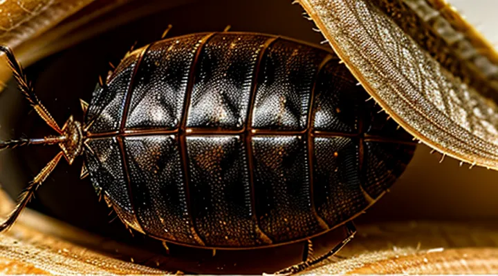
The Exoskeleton's Basic Anatomy
What is Chitin?
Chemical Composition of Chitin
The exoskeleton of a bed bug consists primarily of chitin, a linear polysaccharide formed by β‑(1→4)‑linked N‑acetylglucosamine residues. Each monomer contributes a carbonyl group and an acetyl moiety, creating a rigid backbone capable of extensive hydrogen bonding. This intermolecular network yields crystalline microfibrils that impart mechanical strength and resistance to deformation.
In addition to the polymeric carbohydrate, the cuticle incorporates:
- Structural proteins that interlock with chitin fibers, reinforcing the matrix.
- Cross‑linking agents such as catechol‑derived quinones, generated during sclerotization, which darken the shell and increase hardness.
- Minor lipid layers that provide waterproofing and contribute to the glossy surface appearance.
The combined effect of these components produces a translucent, slightly amber shell whose coloration and sheen result from the degree of protein‑catechol cross‑linking and surface lipid deposition.
Chitin's Role in Arthropods
Chitin, a β‑1,4‑linked N‑acetylglucosamine polymer, forms the backbone of the arthropod exoskeleton. Its crystalline fibers interlock with protein matrices, creating a composite material that resists deformation while remaining lightweight.
The cuticle results from successive layers: an outer epicuticle rich in waxes, a middle exocuticle where chitin‑protein fibers are densely packed, and an inner endocuticle with more loosely arranged fibers. Sclerotization, the cross‑linking of cuticular proteins, hardens the structure; mineral deposition, chiefly of calcium carbonate in some groups, adds rigidity.
In a bedbug, the chitinous shell presents as a series of overlapping sclerites that follow the insect’s segmented body plan. Visual characteristics include:
- Translucent to amber tones, darkening with age
- Smooth, glossy surface on the dorsal plates
- Fine, reticulate pattern visible under magnification
- Distinct lateral margins that hinge at intersegmental membranes
These features enable rapid identification in forensic and pest‑management contexts and illustrate how chitin, combined with protein cross‑links, yields a protective armor adapted to the bedbug’s hematophagous lifestyle.
Layers of the Bedbug Exoskeleton
The Epicuticle: Outer Protective Layer
The epicuticle forms the outermost layer of a bedbug’s chitinous exoskeleton, presenting a thin, waxy coating that masks the underlying rigid cuticle. Under microscopic observation it appears as a glossy, translucent film, often exhibiting a faint amber hue due to embedded pigments and surface lipids. Its smooth surface reduces friction and limits water loss, contributing to the insect’s desiccation resistance.
Key visual and structural attributes of the epicuticle include:
- Thickness of 0.5–2 µm, insufficient to be resolved without high‑resolution imaging.
- Uniform, featureless texture that lacks the ridges or ornamentation seen in deeper cuticular layers.
- Slight coloration ranging from pale yellow to light brown, influenced by cuticular sclerotization and environmental staining.
- Presence of a waxy layer composed of long‑chain hydrocarbons, visible as a thin sheen in scanning electron micrographs.
The Procuticle: Strength and Flexibility
The bedbug’s outer covering consists of a hardened exoskeleton in which the procuticle forms the bulk of the protective layer. This region lies beneath the thin, waxy epicuticle and is built from chitin fibers interlaced with a protein matrix.
The procuticle is organized into distinct sub‑layers:
- Exocuticle – densely cross‑linked, providing most of the rigidity.
- Endocuticle – loosely packed, allowing greater deformation.
- Phragmata – transverse ridges that reinforce structural integrity.
Strength derives from sclerotization, a biochemical process that creates covalent bonds between chitin and cuticular proteins. Additional reinforcement arises from:
- High degree of chitin crystallinity.
- Presence of catecholamine-derived pigments that harden the matrix.
- Limited incorporation of calcium salts in specific regions.
Flexibility is achieved through fiber orientation and lamination. In the endocuticle, chitin microfibrils are arranged at oblique angles, enabling the cuticle to bend without cracking. Variable thickness across the body permits articulation at joints while maintaining overall durability.
Visually, the procuticle appears as a semi‑transparent, brownish shell with a faint sheen. Light scattering by aligned chitin fibers can produce a subtle iridescent effect, especially on the dorsal surface where the exocuticle is thickest.
Exocuticle: Rigidity
The exocuticle forms the outermost layer of a bedbug’s chitinous armor. It is composed of highly cross‑linked sclerotized proteins and layered chitin fibers. This molecular architecture yields a hard, resistant surface that protects internal tissues from mechanical damage and desiccation.
Rigidity influences the visual texture of the shell. The hardened layer appears smooth and slightly glossy, reflecting light in a manner that can give the cuticle a faintly iridescent sheen. Because the exocuticle is thin yet stiff, the underlying pigmentation of the insect often remains visible, producing a semi‑transparent appearance in lighter‑colored individuals.
Key characteristics of the rigid exocuticle:
- High sclerotization level → increased hardness
- Dense chitin fiber arrangement → enhanced tensile strength
- Surface smoothness → reduced friction and wear
- Light‑reflective quality → subtle sheen observable under illumination
Endocuticle: Pliability
The bedbug’s exoskeleton consists of a multilayered cuticle in which the endocuticle forms the innermost structural component. This layer is composed of thin, overlapping chitin lamellae that are embedded in a matrix of protein cross‑links. The lamellae are oriented at slight angles to one another, creating a staggered arrangement that permits limited deformation without compromising integrity.
Pliability of the endocuticle derives from several microscopic features:
- Lamellar spacing: narrow gaps between sheets allow slight sliding under stress.
- Protein flexibility: resilin‑like proteins within the matrix absorb strain.
- Partial sclerotization: incomplete hardening preserves elasticity while maintaining protection.
These attributes give the bedbug’s shell a subtle give when the insect squeezes into cracks or expands after feeding. The outer epicuticle appears glossy and heavily sclerotized, while the underlying endocuticle remains comparatively softer, contributing to the overall texture that can be felt as a thin, slightly pliant film beneath a tougher surface.
Visual Characteristics of the Bedbug Shell
Coloration and Transparency
Factors Influencing Shell Color
The coloration of a bed bug’s sclerotized exoskeleton results from a combination of intrinsic and extrinsic variables. Genetic makeup determines the baseline pigment composition, primarily melanin and sclerotin, which sets the fundamental hue ranging from pale amber to deep brown. Variations in alleles encoding enzymes of the phenoloxidase pathway produce measurable differences among individuals and populations.
Environmental conditions modify the expressed coloration after each molt. Key influences include:
- Temperature: Higher rearing temperatures accelerate pigment synthesis, yielding darker shells; cooler environments slow the process, producing lighter tones.
- Relative humidity: Elevated moisture levels promote hydrolytic degradation of pigments, lightening the cuticle, while low humidity preserves pigment intensity.
- Light exposure: Prolonged ultraviolet radiation induces oxidative changes in melanin, deepening the shade.
- Dietary intake: Blood meals containing higher concentrations of heme and iron enhance melanin deposition, whereas nutrient‑deficient hosts result in paler exoskeletons.
- Chemical contact: Sublethal exposure to insecticides or detergents can disrupt enzymatic pathways, leading to atypical discoloration or mottling.
Age and molting frequency also affect coloration. Newly emerged nymphs exhibit a translucent, almost colorless cuticle that darkens progressively with each successive ecdysis as pigment layers accumulate. The rate of pigment accumulation slows in later instars, stabilizing the final adult hue.
Microbial symbionts residing in the gut may contribute auxiliary pigments or alter host metabolism, indirectly influencing shell coloration. Strains producing carotenoid‑like compounds can impart a subtle reddish tint, while others that metabolize phenolic precursors may reduce overall darkness.
Collectively, these factors interact to produce the observable spectrum of shell colors in Cimex lectularius, providing a reliable indicator of developmental stage, environmental history, and genetic lineage.
Texture and Surface Features
Bristles and Hairs
The chitinous exoskeleton of a bedbug is covered with a dense array of fine, hair‑like structures. These bristles are composed of hardened cuticular material that extends from the surface of each sclerite, giving the insect a slightly fuzzy texture when viewed under magnification.
- Length varies from a fraction of a millimeter to several millimeters, depending on body region.
- Diameter is uniformly thin, often less than 5 µm, creating a delicate appearance.
- Arrangement follows a patterned distribution: longer setae line the margins of the thorax, while shorter microtrichia populate the dorsal abdomen.
- Surface of each bristle is smooth, lacking pores, and tapers to a pointed tip that can interlock with neighboring hairs.
These hair‑like projections serve to interrupt light, producing a matte sheen on the shell and enhancing the insect’s ability to blend with fabric textures. Their rigidity also contributes to the protective function of the cuticle, resisting abrasion while retaining flexibility during locomotion.
Size and Shape Considerations
Variations Between Life Stages
The cuticle of a bedbug undergoes distinct transformations from egg to adult, reflecting changes in function and protection.
- Egg: The embryonic shell is a thin, translucent chorion that encloses the developing nymph. It lacks sclerotization, appears almost invisible against the substrate, and provides limited mechanical resistance.
- First‑through‑fifth instars: Each nymphal stage adds a new, partially hardened exoskeleton. The cuticle becomes progressively darker, shifting from pale amber to deep reddish‑brown. Sclerotization intensifies, thickening the dorsal plates while the ventral surface remains comparatively soft to allow growth. Segmentation becomes more pronounced, and sensory setae emerge on the thorax and abdomen. Molting occurs after each instar, shedding the previous cuticle and revealing a slightly larger, more robust one.
- Adult: The final exoskeleton is fully sclerotized, uniformly dark brown to black, and exhibits a glossy sheen. The dorsal shield (tegument) is thickest over the pronotum and abdomen, providing resistance to mechanical damage. The cuticle contains well‑developed sensory hairs and microstructures that aid in detecting host cues. Ventral plates remain thinner to facilitate locomotion and blood‑feeding.
These stage‑specific variations in coloration, hardness, and structural features define the visual and functional appearance of the bedbug’s chitinous armor throughout its life cycle.
Distinguishing Features of Adult Bedbugs
Adult bedbugs possess a hard, translucent exoskeleton that reveals a series of diagnostic characteristics. The cuticle is composed of layered chitin, giving the insect a glossy, amber‑brown hue. Surface texture is smooth, lacking setae or pronounced ridges, while the edges of the abdomen display a subtle, pale‑yellow band known as the “suture line.” The dorsal surface is covered by a thin, semi‑transparent membrane that allows internal organs to be faintly visible, especially the darkened gut filled with digested blood.
Key identifiers of mature specimens include:
- Length of 4.5–5.5 mm, width approximately 2 mm.
- Oval shape with a flattened dorsal surface.
- Two prominent, forward‑facing antennae, each bearing four segments.
- Six legs ending in tiny claws adapted for clinging to fabric.
- Pair of elongated, curved mouthparts (proboscis) concealed beneath the head when not feeding.
- Distinctive “c-shaped” wing pads that remain undeveloped, confirming the species’ wingless status.
These features, combined with the characteristic chitinous shell, enable reliable identification of adult bedbugs in field and laboratory settings.
