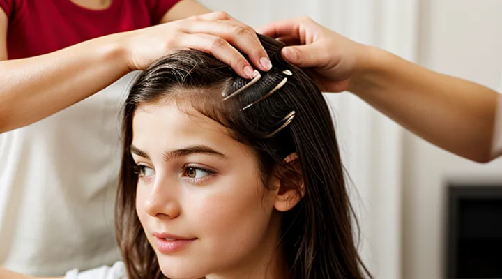
What are Head Lice?
Life Cycle of Head Lice
Head lice (Pediculus humanus capitis) complete a rapid development cycle that makes early detection essential. The cycle begins with the egg, commonly called a nit, which is firmly cemented to a hair shaft near the scalp. Nits measure 0.8 mm in length, appear oval, and range in color from ivory‑white when freshly laid to yellow‑brown as they mature. Their attachment point is a tiny, transparent shell that can be seen as a glossy, elongated speck positioned a few millimeters from the scalp surface.
- Egg (nit) – Deposited by the female, each egg is glued at an angle to the hair shaft. The shell is hard, translucent, and resistant to removal without specialized tools. The embryo inside remains invisible until the shell darkens.
- Nymph – Hatches after 7–10 days. The emerging nymph resembles a miniature adult, lacking fully developed wings and reproductive organs. It begins feeding immediately.
- Adult – Reaches maturity in another 7–10 days. Adults are 2–3 mm long, grayish‑brown, and possess six legs. Females lay 6–10 eggs per day, perpetuating the cycle.
The entire progression from egg to reproducing adult spans approximately 14–21 days under optimal conditions. Temperature and host hygiene influence the rate; warmer scalp environments accelerate development. Each adult can produce up to 300 eggs during its lifespan of about 30 days, leading to exponential population growth if unchecked.
Identifying nits relies on their characteristic attachment angle, size, and coloration. A nit attached close to the scalp appears more opaque, while older, detached shells turn brittle and lighter. Recognizing these features enables timely intervention before nymphs emerge and the infestation expands.
Identifying Nits: The Key Characteristics
Size and Shape of Nits
Lice eggs, commonly called nits, measure approximately 0.6 mm to 1.0 mm in length and 0.2 mm to 0.4 mm in width. Average dimensions are about 0.8 mm × 0.3 mm, with slight variation among species and developmental stages.
The nit is an elongated oval, tapering toward one end where a small, dome‑shaped operculum (cap) is located. The shell is smooth, semi‑transparent, and often appears white, yellowish, or brownish against dark hair. Nits are cemented directly onto the hair shaft, typically within 1 cm of the scalp, making them difficult to dislodge without specialized tools. Their attachment point is a thin, clear glue line that secures the egg at an angle perpendicular to the hair fiber.
Because of their size and shape, nits can be mistaken for dandruff particles, but their firm attachment and characteristic oval profile distinguish them from loose skin flakes.
Color and Transparency of Nits
Nits are the egg casings of head‑lice and appear as tiny, oval structures attached firmly to hair shafts. Their coloration ranges from pale yellow‑white to light brown, depending on the developmental stage and exposure to air. Freshly laid nits are typically translucent, allowing the white embryo inside to be faintly visible. As they mature, the shell thickens and darkens, becoming more opaque and taking on a slightly amber hue.
- Translucent stage: almost invisible, especially on light‑colored hair; the shell may show a faint, glass‑like sheen.
- Maturing stage: increased opacity, color shifts toward light brown; the egg interior becomes less discernible.
- Aged stage: shell may darken further, sometimes appearing grayish; the attachment point remains solid.
The degree of transparency directly influences detectability. On dark hair, even translucent nits contrast enough to be seen with magnification, while on blond or gray hair they blend with the shaft, requiring close inspection. Environmental factors such as humidity and sunlight can slightly alter the perceived color, but the fundamental progression from clear to opaque remains consistent across all hair types.
Location on the Hair Shaft
Lice eggs, or nits, attach firmly to the hair shaft by a protein‑based cement. The attachment point is typically within a quarter inch of the scalp, where the hair is warm and moist. This proximity allows the egg to develop efficiently and makes the cement less likely to be removed by routine combing.
Common positions include:
- The base of each hair strand, especially on the crown and nape of the neck.
- Behind the ears, where hair density is high and the scalp is less exposed.
- Along the sideburns and facial hair in children, as these areas provide similar temperature and humidity conditions.
The cement creates a small, oval‑shaped shell that encircles the hair at the point of attachment. The shell is usually tan, gray, or white and appears as a tiny, immobile speck. Because the egg is anchored close to the scalp, it often resembles a dandruff particle but does not flake off when the hair is brushed.
Detecting nits requires a fine-toothed comb or magnification. The key indicator is the presence of a firmly fixed, oval object that does not move independently of the hair shaft.
Attachment to the Hair
Lice nits adhere to hair strands using a natural, protein‑based cement that hardens within hours after the egg is laid. The cement forms a glossy, oval‑shaped shell that clamps tightly around the shaft, typically within ¼ inch of the scalp where temperature and humidity favor development.
- Placement: close to the scalp, often behind the ear, at the nape of the neck, or along the crown.
- Orientation: the pointed end faces the scalp, the blunt end points outward.
- Visibility: the shell appears tan to light brown; after 5–7 days, it darkens to gray‑black as the embryo matures.
- Detachment: the cement resists manual removal; gentle combing with a fine‑toothed nit comb is required to break the bond without damaging the hair.
Understanding the attachment mechanism helps distinguish live nits from shed shells, which lack the firm seal and are usually loose and lighter in color. Accurate identification relies on observing the cemented, immobile egg firmly attached to the hair shaft.
Distinguishing Nits from Other Hair Debris
Nits vs. Dandruff
Nits are the eggs of head‑lice, firmly attached to a single hair strand near the scalp. They appear as oval, creamy‑white or yellowish bodies about 0.8 mm long, often resembling tiny beads. The shell is smooth, slightly translucent, and may show a darker embryonic spot at one end. Because they are glued to the hair, they do not flake off when the head is brushed.
Dandruff consists of dead skin cells that detach from the scalp and fall freely onto the hair and shoulders. The particles are white or gray, irregular in shape, and range from fine powder to larger, flaky pieces. They are not attached to individual hairs and can be brushed away easily.
Key distinguishing features:
- Attachment: nits are glued to each hair; dandruff is loose.
- Shape: nits are uniform ovals; dandruff is irregular.
- Size: nits are consistently ~0.8 mm; dandruff varies widely.
- Color: nits are creamy‑white to yellow; dandruff is white or gray.
- Mobility: nits remain stationary; dandruff moves with brushing or shaking.
Identifying the correct condition prevents unnecessary treatment and ensures appropriate pest‑control measures when needed.
Nits vs. Hair Casts
Lice eggs, or nits, adhere tightly to the hair shaft near the scalp. They appear as oval, white‑to‑tan bodies, about 0.8 mm long, with a glossy surface that may darken after hatching. The attachment point is a cement‑like secretion that makes the nit immovable without forceful pulling.
Hair casts are cylindrical, tube‑like coatings that slide easily along the hair. They are translucent, often colorless, and range from 1 mm to several millimeters in length. Unlike nits, casts are not cemented to the shaft and can be removed by gentle combing.
Key distinctions:
- Location: Nits sit close to the scalp; casts can appear anywhere along the hair.
- Attachment: Nits are fixed with a sticky secretion; casts are loosely wrapped and movable.
- Color and texture: Nits are opaque, slightly yellowish or brownish; casts are clear, silky, and may appear as a thin sheath.
- Shape: Nits are oval; casts are tubular and uniform in diameter.
- Removal: Nits require a fine‑toothed comb or specialized treatment; casts detach with a simple comb or fingertip pressure.
Nits vs. Product Residue
Lice eggs, or nits, attach firmly to each hair shaft about a quarter of an inch from the scalp. The attachment point is a smooth, oval shell that encircles the strand and is glued with a cement‑like secretion. The shell measures roughly 0.8 mm in length, appears translucent to light‑brown, and often shows a tiny white dot at the operculum—the opening through which the nymph emerges. When the shell is empty, it becomes more opaque and may turn a dull gray.
Hair‑care residues differ in several observable ways:
- Composition: Residues consist of polymers, oils, or silicone particles, not a biological shell.
- Attachment: Residues coat the hair surface and can be brushed away; nits remain locked in place until manually removed or hatched.
- Shape: Residues form irregular clumps, streaks, or films; nits retain a consistent oval outline.
- Color: Residues reflect the product’s hue (clear, white, colored); nits stay within the natural range of brown to tan.
- Texture: Residues feel sticky or greasy to the touch; nits feel hard and brittle.
Recognizing these distinctions prevents misidentification and ensures appropriate treatment.
Tools and Techniques for Nit Identification
The Importance of Good Lighting
Good lighting is a prerequisite for accurate detection of lice eggs attached to hair shafts. Under bright, direct illumination, nits appear as small, oval, tan‑to‑brown structures firmly glued to the strand, often positioned within a quarter‑inch of the scalp. The translucency of the shell becomes visible when light passes through, revealing a clear or slightly milky interior that distinguishes nits from dandruff or hair products.
Insufficient light flattens contrast, causing nits to blend with hair color and remain hidden. Overexposure, however, can wash out subtle color differences, making identification equally difficult. Optimal conditions involve a focused, white light source at a 45‑degree angle to the hair, which highlights the three‑dimensional shape of each egg and casts a shadow that separates it from surrounding fibers.
Key lighting practices for reliable inspection:
- Use a handheld magnifying lamp with adjustable brightness.
- Position the light source close to, but not directly above, the hair to create side illumination.
- Combine magnification (10×–15×) with a neutral‑white LED to preserve true color.
- Conduct the examination in a well‑ventilated area to avoid glare from reflective surfaces.
By adhering to these lighting guidelines, observers can consistently distinguish lice nits from other debris, reducing the risk of misdiagnosis and ensuring timely treatment.
Magnification Tools
Lice eggs are oval, 0.8 mm long, and firmly glued to the hair shaft near the scalp. Their shells appear translucent to white, often resembling tiny beads or specks of dust. The attachment point shows a slight indentation in the hair cuticle, and the eggs may be angled toward the scalp.
Magnification makes these features visible. Effective devices include:
- Handheld magnifying glass (10–15×): inexpensive, portable, sufficient for casual inspection.
- Optical loupes (20–30×): provide sharper focus, useful for detailed examination of dense hair.
- Stereo microscope (30–50×): delivers three‑dimensional view, reveals attachment and embryonic development.
- Digital USB microscope (up to 200×): projects image to a screen, enables recording and sharing.
- Smartphone clip‑on lens (20–60×): combines convenience with high resolution, ideal for quick checks.
Optimal use requires bright, direct lighting and a steady hand. Position the tool at a shallow angle to reduce glare and to view the base of the hair shaft where nits are most commonly found. Adjust focus until the shell’s outline, its clear wall, and the tiny operculum become distinct. This level of detail allows reliable differentiation between live nits, hatched shells, and unrelated debris.
Proper Combing Techniques
Nits are microscopic, oval eggs cemented to the hair shaft within ¼ inch of the scalp. Their color ranges from translucent white to brownish yellow, and they appear as immobile specks that do not move with the hair. Recognizing this appearance is essential for effective removal.
A fine‑toothed nit comb, preferably metal with teeth spaced 0.2 mm apart, provides the necessary precision. Conditioner reduces friction and helps loosen the adhesive that secures the eggs.
- Wet hair thoroughly; apply a generous amount of conditioner and distribute evenly.
- Divide hair into manageable sections using clips.
- Starting at the scalp, run the comb through each section from root to tip in a slow, steady motion.
- After each pass, wipe the comb on a paper towel or rinse under running water to remove collected nits.
- Examine the comb after each stroke; discard any nits found.
- Repeat the process for every section, ensuring no area is missed.
Perform the combing routine every two to three days for at least two weeks. Combine mechanical removal with appropriate treatment products to prevent re‑infestation. Consistent, meticulous combing eliminates nits and reduces the likelihood of a full lice outbreak.
When to Seek Professional Help
Lice eggs, or nits, appear as tiny, oval, tan‑to‑brown specks glued to hair shafts. When they are firmly attached near the scalp, they are difficult to remove with a regular comb and indicate an active infestation.
Seek professional assistance if any of the following conditions occur:
- More than a handful of nits are visible on each strand, especially within a half‑inch of the scalp.
- Live lice are observed after at least two attempts with over‑the‑counter (OTC) products.
- Persistent itching continues for more than a week despite using recommended shampoos or lotions.
- The person experiences redness, swelling, or rash that may suggest an allergic reaction to treatment.
- Multiple family members or close contacts show signs of infestation, increasing the risk of rapid spread.
- School or daycare policies require a certified treatment report before reintegration.
In these situations, a licensed healthcare provider or certified lice‑removal specialist can confirm the diagnosis, prescribe prescription‑strength medication, and provide guidance on thorough cleaning of personal items and environment. Prompt professional intervention reduces the chance of secondary infections and limits the duration of the outbreak.
