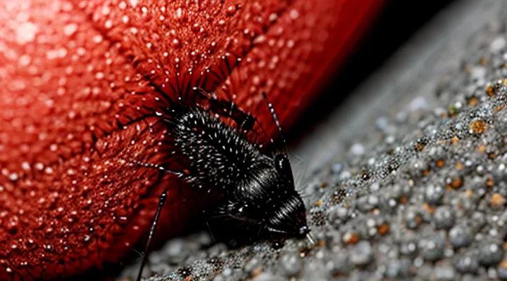
What Are Subcutaneous Mites?
Types of Mites Affecting Humans
Mites that invade human tissue vary in morphology, life cycle, and clinical impact. Understanding the specific species clarifies the mechanisms behind subcutaneous infestations.
The most frequently implicated organisms are:
- Sarcoptes scabiei – the causative agent of scabies; adult females burrow into the epidermis, laying eggs that develop within the skin layers. The resulting tunnels produce intense pruritus and secondary inflammation.
- Trombiculidae larvae (chiggers) – attach to the epidermis, inject digestive enzymes, and feed on liquefied host tissue. Their feeding sites become painful papules that may progress to deeper dermal involvement.
- Demodex folliculorum and Demodex brevis – inhabit hair follicles and sebaceous glands; overgrowth can lead to folliculitis, rosacea‑like eruptions, and occasional subepidermal cyst formation.
- Pyemotes tritici – a predatory mite that, when opportunistically transferred to humans, penetrates the skin and causes erythematous, pruritic lesions often mistaken for bites.
- Dermanyssus gallinae (bird mite) – feeds on avian hosts but can bite humans, embedding its mouthparts in the superficial dermis and producing localized swelling and itching.
- House dust mites (Dermatophagoides spp.) – primarily provoke allergic responses; they rarely penetrate the skin but may exacerbate existing dermatoses.
Each species employs a distinct method of tissue invasion: burrowing, enzymatic digestion, or superficial attachment. The resulting pathology ranges from superficial dermatitis to deeper, subcutaneous inflammation, depending on the mite’s anatomical adaptations and host immune response. Recognizing these differences guides accurate diagnosis and targeted treatment.
Life Cycle of Subcutaneous Mites
Subcutaneous mites progress through a defined series of developmental stages that enable them to invade and persist within the host’s skin. The cycle begins when adult females deposit eggs in the superficial dermis. Eggs hatch within 3‑5 days, releasing first‑stage larvae that lack functional mouthparts and remain confined to the epidermal layer. After a brief feeding period, larvae molt into nymphs, which develop functional chelicerae capable of penetrating deeper tissue. Nymphal stages, typically two in number, undergo successive molts every 5‑7 days, each molt increasing body size and reproductive capacity. Mature adults emerge after the final molt, possessing fully developed reproductive organs and the ability to produce large numbers of eggs, thereby perpetuating the infestation.
Key biological factors influencing the cycle include temperature, humidity, and host immune response. Optimal temperatures (20‑30 °C) accelerate development, while low humidity prolongs each stage. Host skin integrity and inflammatory reactions can limit mite penetration but often fail to eradicate established populations. Understanding each phase of the life cycle is essential for targeting interventions, such as topical acaricides applied during the larval and early nymphal stages, when the mites are most vulnerable.
Primary Causes of Infestation
Compromised Immune System
A weakened immune system markedly increases the risk of mite penetration into the deeper layers of the skin. When cellular immunity is compromised, the body’s ability to recognize and eliminate ectoparasites declines, allowing mites to survive and reproduce beneath the epidermis.
Common sources of immune suppression include:
- HIV infection and other immunodeficiency disorders
- Chemotherapy or radiation therapy
- Long‑term corticosteroid use
- Diabetes mellitus with poor glycemic control
- Severe malnutrition or protein‑energy deficiency
These conditions impair neutrophil function, reduce cytokine production, and alter the integrity of the skin barrier. The resulting environment favors mite attachment, burrowing, and colonisation, leading to persistent subcutaneous infestations that are difficult to treat.
Laboratory assessment often reveals reduced lymphocyte counts or functional deficits, confirming the link between immune compromise and mite proliferation. Restoring immune competence, when possible, diminishes mite load and supports conventional antiparasitic therapy.
Close Contact with Infested Individuals or Animals
Close physical interaction with people or animals harboring mites provides a direct pathway for larvae to penetrate the skin. Mites such as Sarcoptes scabiei and Trombiculidae species attach to the host’s surface, then migrate into the epidermis or dermis during prolonged skin‑to‑skin contact, grooming, or handling of infested fur. The transfer is most efficient when the host’s skin is compromised, moist, or covered by hair that retains the arthropods.
Transmission intensifies in environments where individuals share bedding, clothing, or equipment, because mites can survive off‑host for several hours. Contact with domestic pets, livestock, or wildlife that display active infestation increases the probability of acquiring subcutaneous stages, especially when protective barriers (gloves, clothing) are absent. Group living situations, such as dormitories, shelters, or farms, further amplify exposure due to repeated close proximity.
Typical scenarios that facilitate infestation include:
- Sleeping in the same bed with an infected person or animal.
- Carrying or caring for pets with visible mite activity.
- Participating in sports or activities that involve close bodily contact.
- Sharing towels, uniforms, or protective gear without proper sanitation.
Preventive measures focus on minimizing direct contact with known infested hosts, implementing routine examination of animals, and applying acaricidal treatments when infestation is confirmed. Early detection and isolation of affected individuals interrupt the transmission chain and reduce the risk of subcutaneous mite invasion.
Environmental Factors
Subcutaneous mite infestations develop when conditions favor mite survival and penetration of the skin. Environmental parameters determine the abundance of mites and the likelihood of human exposure.
- High relative humidity supports mite reproduction and prolongs larval activity.
- Warm temperatures (20‑30 °C) accelerate development cycles, increasing population density.
- Dense vegetation provides shelter and breeding sites, especially in moist understory layers.
- Soil rich in organic matter retains moisture and serves as a reservoir for mite stages.
- Presence of reservoir hosts (rodents, small mammals) amplifies mite numbers and facilitates transmission to humans.
- Seasonal rainfall patterns create temporary habitats that boost mite proliferation.
- Human activities that disturb soil or vegetation (agricultural work, construction) increase contact with infested substrates.
- Climate change shifts temperature and precipitation regimes, expanding suitable habitats into previously cooler regions.
These factors collectively create environments where mites thrive, enhancing the risk of subcutaneous infestation.
Predisposing Factors
Age and Vulnerability
Age is a primary determinant of susceptibility to subcutaneous mite infestations. Infants and young children possess thin dermal layers, limited immune competence, and underdeveloped skin barrier functions, all of which facilitate mite penetration and colonisation. Elderly individuals experience epidermal thinning, reduced collagen synthesis, and diminished cellular immunity, creating conditions favorable for mite invasion.
Key age‑related vulnerabilities include:
- Dermal thickness: Reduced in both early childhood and senescence, allowing easier mite migration beneath the skin.
- Immune response: Immaturity in children and immunosenescence in seniors diminish the capacity to recognise and eliminate mite antigens.
- Comorbidities: Chronic diseases common in older adults (e.g., diabetes, vascular insufficiency) impair wound healing and increase tissue susceptibility.
- Behavioral factors: Infants often remain in close contact with contaminated bedding; elderly patients may have limited mobility, leading to prolonged exposure to infested environments.
Understanding these age‑specific risk factors enables targeted prevention, early diagnosis, and appropriate therapeutic interventions for subcutaneous mite infestations.
Pre-existing Skin Conditions
Pre‑existing dermatological disorders create an environment that favors the colonization and penetration of subcutaneous mites. Disrupted epidermal barriers allow mites to bypass superficial defenses, while altered skin microflora reduces competition and facilitates mite survival.
Common conditions that increase susceptibility include:
- Atopic dermatitis – chronic inflammation and itching compromise skin integrity, providing entry points for mites.
- Psoriasis – hyperkeratosis and scaling create niches where mites can hide and migrate deeper.
- Seborrheic dermatitis – excess sebum alters the lipid composition of the stratum corneum, enhancing mite adhesion.
- Chronic ulcers or wounds – exposed dermal tissue offers direct access for mite burrowing.
- Lichen planus – interface dermatitis weakens the dermo‑epidermal junction, permitting mite infiltration.
These disorders often involve immune dysregulation, which impairs the host’s ability to recognize and eliminate ectoparasites. Additionally, topical therapies such as corticosteroids or immunosuppressants can further diminish local immune responses, inadvertently encouraging mite proliferation beneath the skin surface.
Poor Hygiene Practices
Poor personal and environmental hygiene creates conditions that favor the penetration of mites into the dermal layer. Inadequate washing of skin, infrequent changing of clothing, and failure to launder bedding allow mite populations to accumulate and seek a host. When skin surfaces remain moist and unclean, mites encounter reduced barriers and increased opportunities for attachment and migration beneath the stratum corneum.
Key hygiene deficiencies that increase the risk of subdermal mite colonisation include:
- Irregular bathing or showering, especially after exposure to contaminated environments.
- Use of worn or soiled garments without regular laundering at temperatures sufficient to kill arthropods.
- Accumulation of dust, debris, and organic matter in living spaces, providing habitats for mite development.
- Neglect of personal items such as towels, socks, and shoes, which retain moisture and serve as reservoirs.
- Inadequate cleaning of surfaces that come into direct contact with skin, such as bedsheets, mattress covers, and upholstery.
These practices diminish the mechanical removal of mites and the disruption of their life cycle, allowing larvae to infiltrate the epidermis and advance to the subcutaneous tissue. Consistent hygiene protocols—daily cleansing, routine laundering at ≥60 °C, and regular environmental sanitation—interrupt this process and substantially lower infestation incidence.
Mechanisms of Infestation
Mite Penetration of the Skin Barrier
Mite penetration of the skin barrier occurs when microscopic arthropods overcome the outermost protective layers and reach the viable epidermis or dermis. Successful invasion requires a combination of structural, biochemical, and environmental conditions that weaken or bypass the stratum corneum.
The stratum corneum consists of tightly packed keratinocytes embedded in a lipid matrix. Mites exploit this barrier through:
- Mechanical forces generated by specialized mouthparts that scrape or pierce corneocytes.
- Production of proteolytic enzymes (e.g., keratinases, collagenases) that degrade keratin and extracellular matrix proteins.
- Secretion of lipolytic compounds that disrupt intercellular lipids, increasing permeability.
- Chemotactic attraction to host-derived cues such as heat, carbon dioxide, and skin secretions, guiding mites toward vulnerable sites.
Host factors that facilitate penetration include:
- Compromised barrier integrity due to cuts, abrasions, dermatological diseases, or chronic inflammation.
- Reduced lipid content from excessive washing, harsh detergents, or topical steroids.
- Immunosuppression, which diminishes the skin’s innate defense mechanisms and allows mites to establish deeper colonization.
Environmental contributors enhance the likelihood of subcutaneous mite infestations:
- High humidity and temperature, which promote mite activity and survival on the skin surface.
- Overcrowded living conditions, increasing exposure to infested individuals or contaminated bedding.
- Presence of animal reservoirs that harbor mite species capable of human transmission.
Collectively, these mechanisms explain how mites breach the skin barrier and cause infections beneath the superficial layers. Effective prevention requires maintaining barrier integrity, controlling environmental humidity, and minimizing exposure to known mite vectors.
Immunological Response to Mites
Subcutaneous mite infestations develop when mites breach the epidermal barrier and establish colonies within the dermis. The host’s immune system detects this intrusion through pattern‑recognition receptors on keratinocytes and resident dendritic cells. These cells release pro‑inflammatory cytokines (IL‑1β, TNF‑α) that recruit neutrophils and macrophages to the site of invasion.
The adaptive arm responds primarily via a Th2‑biased pathway. Antigen presentation by Langerhans cells triggers differentiation of CD4⁺ T cells into Th2 effectors, which secrete IL‑4, IL‑5 and IL‑13. These cytokines promote class‑switching of B cells to produce IgE specific for mite allergens. IgE binds to FcεRI receptors on mast cells and basophils; subsequent cross‑linking by mite antigens induces degranulation, releasing histamine, leukotrienes and proteases that exacerbate tissue edema and pruritus.
Eosinophils, attracted by IL‑5, infiltrate the dermal layer and release major basic protein and eosinophil cationic protein. These mediators damage mite cuticles and amplify inflammation, yet excessive eosinophilic activity can impair wound healing and facilitate further mite penetration.
Regulatory mechanisms attempt to limit damage. T regulatory cells produce IL‑10 and TGF‑β, dampening Th2 activity and curbing mast cell activation. However, in individuals with impaired regulatory function—due to genetic predisposition, immunosuppression, or chronic skin disease—the balance shifts toward uncontrolled Th2 responses, increasing susceptibility to deep mite colonization.
Key immunological components involved in the response to subdermal mites:
- Innate detection: keratinocyte Toll‑like receptors, dendritic cell cytokine release
- Cellular effectors: neutrophils, macrophages, eosinophils
- Adaptive drivers: Th2 cells, IgE production, mast cell degranulation
- Regulatory controls: T regulatory cells, anti‑inflammatory cytokines
When any of these elements are deficient or dysregulated, the host’s capacity to eliminate mites diminishes, allowing infestation to persist and spread within the dermal tissue.
Secondary Infections and Complications
Subcutaneous mite infestations frequently breach the epidermal barrier, creating portals for microbial invasion. Bacterial colonization of the compromised tissue commonly produces cellulitis, characterized by erythema, warmth, and edema that expand beyond the initial lesion. When bacterial load exceeds local defenses, abscess formation occurs, often requiring incision and drainage to prevent progression to deeper structures.
Secondary complications include:
- Lymphangitis, marked by tender, cord-like streaks extending toward regional lymph nodes.
- Necrotizing fasciitis, a rapidly advancing infection that destroys fascia and subcutaneous fat, demanding immediate surgical debridement.
- Systemic sepsis, presenting with fever, tachycardia, and hypotension, especially in immunocompromised hosts.
- Allergic dermatitis, triggered by mite antigens, leading to pruritic, eczematous eruptions that may persist after parasite eradication.
- Chronic scarring and hyperpigmentation, resulting from prolonged inflammation and tissue remodeling.
Delayed recognition of mite activity increases the likelihood of these outcomes. Prompt antimicrobial therapy, guided by culture results when available, mitigates bacterial spread. Adjunctive measures such as wound hygiene, topical antiseptics, and avoidance of secondary trauma reduce the risk of further infection. In severe cases, hospitalization for intravenous antibiotics and surgical intervention is warranted to preserve limb function and prevent systemic deterioration.
