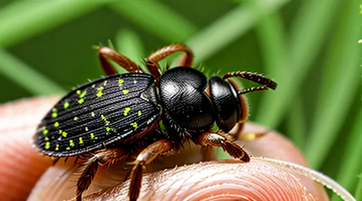
What Constitutes a «Tick Bite»?
Distinguishing Between «Bite» and «Attachment»
Ticks insert their mouthparts when they bite, creating a small puncture that may cause local irritation. The act of biting does not automatically lead to the prolonged feeding stage known as «Attachment». During attachment, the tick secures itself with cement-like substances, remains attached for hours to days, and expands its feeding tube to ingest blood.
Pathogen transmission from ticks generally requires the attachment phase. Most bacteria, viruses, and protozoa need time to migrate from the tick’s salivary glands into the host’s bloodstream. A brief bite without subsequent attachment rarely results in disease, although allergic reactions to tick saliva can occur immediately.
Key distinctions between «Bite» and «Attachment»:
- «Bite»: momentary puncture; may last seconds; limited saliva exposure.
- «Attachment»: sustained anchoring; lasts from several hours to days; enables pathogen transfer.
- Risk level: low for bite alone; elevated for attachment due to prolonged exposure.
- Clinical signs: mild skin irritation after bite; fever, rash, or joint pain may develop after attachment.
Potential Risks of Unattached Tick Encounters
Mechanical Irritation and Skin Reactions
Mechanical irritation occurs when a tick’s mouthparts press against the epidermis without penetrating deeper tissue. The pressure and movement of the mandibles can create a localized abrasion, often perceived as a mild prick or tingling sensation.
Typical cutaneous responses include:
- Erythema surrounding the contact point, usually 2‑5 mm in diameter.
- Small papular lesions that may develop within hours.
- Pruritus that intensifies during the first 24 hours.
- Transient edema limited to the immediate area.
The inflammatory reaction is mediated by histamine release from mast cells activated by the mechanical trauma. In most cases, the redness and swelling resolve spontaneously within 48‑72 hours. Persistent or expanding lesions, ulceration, or secondary bacterial infection warrant professional evaluation.
Patients should monitor the bite site for:
- Enlargement beyond the initial margin.
- Persistent pain or warmth indicative of cellulitis.
- Development of a necrotic center, which may suggest a different pathogen.
Prompt removal of the tick and thorough cleansing of the skin reduce the risk of complications. Even without attachment, the mechanical stimulus alone can provoke noticeable, though generally self‑limiting, dermatologic effects.
Transmitting Pathogens Without Full Attachment
Ticks that probe the skin without inserting their hypostome still pose a health risk. The act of probing introduces saliva, which may contain infectious agents, into the epidermis. Even brief contact can deposit pathogens on the wound surface, allowing entry through microabrasions created during the bite attempt.
Transmission without sustained feeding occurs through three primary mechanisms:
- Salivary injection during the exploratory phase of the bite.
- Regurgitation of infected gut contents when the tick attempts to feed.
- Mechanical transfer of pathogens from the tick’s external cuticle to the host’s skin.
Pathogens documented to be transmitted during the probing stage include:
- Borrelia burgdorferi complex (Lyme disease agents).
- Anaplasma phagocytophilum (causing anaplasmosis).
- Babesia microti (responsible for babesiosis).
- Rickettsia spp. (spotted fever group).
- Tick‑borne encephalitis virus (TBEV) in regions where the virus is endemic.
Risk assessment depends on several variables:
- Contact time: exposure lasting more than a few minutes increases the probability of pathogen transfer.
- Tick species: Ixodes ricinus, Dermacentor variabilis, and Amblyomma americanum are most frequently associated with early‑stage transmission.
- Life stage: nymphs, due to their small size, often remain undetected and may feed briefly before being removed.
- Pathogen prevalence in the local tick population.
Preventive measures focus on immediate removal and monitoring. Use fine‑tipped tweezers to grasp the tick as close to the skin as possible and extract without crushing the body. Clean the bite site with antiseptic. Observe the area for erythema, expanding rash, or flu‑like symptoms for up to four weeks. Seek medical evaluation if any systemic signs develop, as early antimicrobial therapy reduces disease severity.
The Role of Saliva in Disease Transmission
Tick saliva contains a complex mixture of proteins, enzymes, and bioactive molecules that are introduced into the host’s skin during probing. These substances suppress blood clotting, modulate immune responses, and create an environment conducive to pathogen survival.
Key components and their effects include:
- Anticoagulants that prevent platelet aggregation, allowing continuous blood flow.
- Anti‑inflammatory agents that reduce local swelling and pain, delaying host detection.
- Immunosuppressive factors that inhibit cytokine production and leukocyte activation.
- Molecules that bind and protect microorganisms, facilitating their entry into the bloodstream.
Even brief contact can deposit saliva onto the skin surface. Certain pathogens, such as Borrelia spp. and Rickettsia spp., are capable of transmission within minutes of saliva exposure. Others, requiring prolonged feeding, are less likely to be transmitted when the tick disengages quickly. Consequently, a non‑attached bite does not guarantee safety; the risk depends on the pathogen’s transmission dynamics and the volume of saliva transferred.
Risk assessment must consider saliva‑mediated transmission as a distinct pathway, separate from the mechanical injury of the bite. Protective measures include prompt removal of any attached arthropod, thorough skin cleansing after exposure, and monitoring for early symptoms of vector‑borne diseases.
Early-Stage Transmission: A Closer Look
Early‑stage transmission occurs when a tick attaches briefly and disengages before the typical feeding period. Most bacterial agents, such as «Borrelia burgdorferi», «Anaplasma phagocytophilum» and «Rickettsia spp.», require several hours of attachment to migrate from the mouthparts to the host’s bloodstream. Viral agents, for example the tick‑borne encephalitis virus, can be transferred more rapidly, yet still depend on a minimum of 15‑30 minutes of sustained feeding.
The probability of infection rises sharply after the first hour of attachment. Studies show that a bite removed within ten minutes rarely results in pathogen transfer. Laboratory experiments with laboratory‑reared nymphs confirm that transmission efficiency for Lyme‑disease spirochetes approaches zero before the 24‑hour mark, while the risk for certain viruses remains low but non‑negligible after fifteen minutes.
Key considerations for early‑stage exposure:
- Minimum attachment time for bacterial transmission: ≥ 4 hours (most pathogens).
- Minimum attachment time for viral transmission: ≥ 15 minutes (specific to tick‑borne encephalitis).
- Immediate removal reduces infection risk to near‑baseline levels.
- Tick species matters: Ixodes ricinus and Ixodes scapularis are primary vectors for bacterial agents; Dermacentor and Hyalomma species more often transmit viral agents.
Prompt removal, thorough skin inspection and monitoring for symptoms remain the primary preventive measures.
Assessing the Likelihood of Infection
A tick that merely touches the skin and does not remain attached presents a markedly reduced risk of pathogen transmission. Most bacteria, viruses, and protozoa carried by ticks require several hours of feeding to migrate from the tick’s salivary glands into the host’s bloodstream. Without sustained attachment, the mechanical transfer of infectious material is rare.
Key determinants of infection probability include:
- Tick species known to harbour specific pathogens (e.g., Ixodes scapularis for Borrelia burgdorferi).
- Local prevalence of the pathogen within the tick population.
- Duration of skin contact before the tick is removed.
- Host factors such as immune competence and skin integrity.
When a non‑attached bite occurs, immediate washing of the area with soap and water reduces any residual surface contamination. The skin should be inspected for erythema, a central punctum, or expanding rash. Symptoms such as fever, headache, or arthralgia emerging within weeks warrant clinical evaluation, especially in regions where tick‑borne diseases are endemic.
Prompt removal of the tick, even if it has not attached, coupled with vigilant symptom monitoring, provides an effective strategy to mitigate the already low likelihood of infection.
When to Seek Medical Attention
Identifying Concerning Symptoms After Tick Exposure
After a tick encounter, immediate removal does not guarantee the absence of health risks. Certain clinical manifestations may develop despite brief exposure, requiring prompt recognition.
Typical warning signs include:
- Expanding erythema at the bite site, especially a target‑shaped lesion larger than 5 cm.
- Sudden fever exceeding 38 °C, often accompanied by chills.
- Severe headache or neck stiffness without an alternative explanation.
- Muscular or joint pain that appears within days to weeks.
- Unexplained fatigue, dizziness, or nausea.
- Rash distinct from the bite site, such as maculopapular, petechial, or vesicular lesions.
- Neurological deficits, including facial palsy, tingling, or weakness.
Symptoms may emerge anywhere from 24 hours to several weeks after exposure, depending on the pathogen involved. Early signs frequently appear within the first week, while later manifestations, such as arthritis or neurological involvement, can develop after a month.
If any of the listed manifestations occur, medical assessment is mandatory. Diagnostic procedures typically involve serologic testing for Borrelia, Rickettsia, and other tick‑borne agents, supplemented by polymerase chain reaction assays when appropriate. Initiation of antimicrobial therapy should follow established clinical guidelines to reduce the likelihood of complications.
Prevention and Best Practices
Ticks can transmit pathogens even after brief contact, because saliva may be deposited before firm attachment. Reducing exposure and responding promptly lower the probability of infection.
Wearing long sleeves, tucking pants into socks, and applying EPA‑registered repellents containing DEET or picaridin create a physical and chemical barrier against crawling arthropods. Maintaining short grass and removing leaf litter around residential areas diminish habitat suitability for questing ticks. Regularly inspecting pets and treating them with veterinarian‑approved acaricides prevents wildlife from introducing ticks into the household.
If a tick is found on the skin before it secures a mouthpart, follow these steps:
- Use fine‑pointed tweezers to grasp the tick as close to the skin as possible.
- Pull upward with steady, even pressure; avoid twisting or crushing the body.
- Disinfect the bite site with an alcohol‑based solution or iodine.
- Record the date, location, and any visible species characteristics.
- Observe the area for erythema, swelling, or flu‑like symptoms for up to four weeks; seek medical evaluation if systemic signs develop.
Prompt removal and thorough site care, combined with environmental management, constitute the most effective strategy to prevent disease transmission from non‑attached tick encounters.
Proper Tick Removal Techniques
Proper tick removal reduces the risk of pathogen transmission, even when the arthropod has not yet attached firmly. Prompt, correct technique eliminates the possibility of saliva entering the wound and minimizes skin trauma.
- Use fine‑tipped, non‑toothed tweezers.
- Grasp the tick as close to the skin surface as possible.
- Apply steady, upward pressure without twisting or jerking.
- Release the tick once the mouthparts separate from the skin.
- Disinfect the bite area with an antiseptic solution.
- Wash hands thoroughly after the procedure.
After extraction, examine the tick for retained mouthparts; any fragments left in the skin may increase infection risk. Preserve the specimen in a sealed container if identification or laboratory testing is required. Observe the bite site for redness, swelling, or a bullseye rash, and monitor for fever or flu‑like symptoms over the next several weeks. Seek professional medical evaluation if any adverse signs develop.
Post-Exposure Monitoring Guidelines
A tick removed before attachment carries a low but measurable risk of pathogen transmission. Prompt post‑exposure monitoring reduces the chance of delayed diagnosis and severe outcomes.
After removal, the bite site should be cleansed with soap and water, and the incident recorded: date, geographic location, and, when possible, tick species. This information assists clinicians in assessing exposure risk.
Monitoring protocol:
- Inspect the bite area daily for up to 30 days. Look for expanding erythema, especially a target‑shaped rash, or any unusual skin changes.
- Record body temperature twice daily. Persistent fever above 38 °C warrants evaluation.
- Note systemic symptoms: headache, fatigue, muscle aches, joint pain, or neurological disturbances (e.g., tingling, weakness).
- For diseases with longer incubation, such as tick‑borne encephalitis, continue observation for up to 21 days; for Lyme disease, extend to 30 days.
Seek medical care immediately if any of the following appear:
- Expanding rash with central clearing (erythema migrans).
- Fever accompanied by severe headache, neck stiffness, or photophobia.
- Joint swelling or severe arthralgia.
- Neurological signs such as facial palsy, numbness, or confusion.
Healthcare providers may order serologic testing after the appropriate incubation period, typically 2–4 weeks for Lyme disease and 7–14 days for tick‑borne encephalitis. Maintaining a detailed exposure log simplifies diagnostic decisions and facilitates timely treatment.
