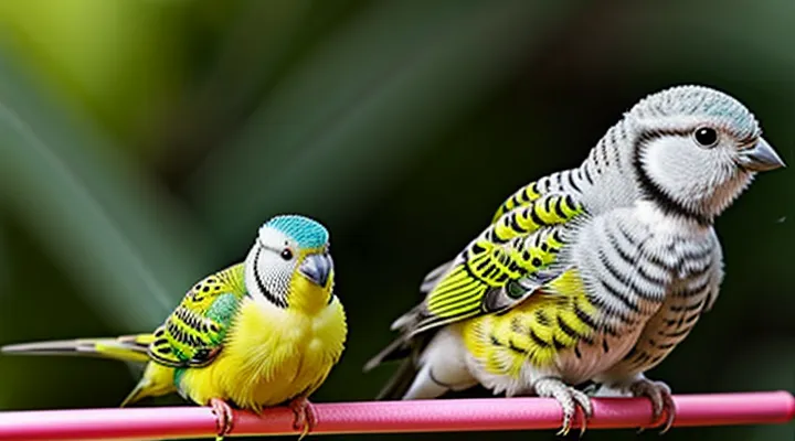
«Understanding Budgerigar Mites»
«Types of Mites Affecting Budgerigars»
«Knemidocoptes pila (Scaly Face Mite)»
Knemidocoptes pila, commonly known as the scaly‑face mite, infests the facial skin of budgerigars and may be confused with tick infestations because both produce visible lesions. The mite burrows into the stratum corneum, creating dry, crusted plaques around the beak, nostrils, and eyes. Diagnosis relies on visual inspection of characteristic scaly lesions and microscopic confirmation of the mite’s oval shape and short legs.
Effective treatment includes systemic acaricides and topical agents. Recommended protocols are:
- Oral ivermectin at 0.2 mg/kg once daily for three consecutive days; repeat after two weeks to eliminate residual stages.
- Topical selamectin applied to the feathered skin at the label dose, repeated after ten days.
- Manual removal of crusts with a soft brush before medication to improve drug penetration.
Supportive care involves maintaining optimal humidity (45–55 %) and providing a balanced diet enriched with vitamin A to promote skin regeneration. Environmental sanitation requires thorough cleaning of cages, perches, and feeding dishes with a diluted bleach solution (1 % sodium hypochlorite) and drying before reuse. Re‑infestation risk diminishes when the habitat is kept dry and free of organic debris.
Prevention focuses on regular health checks, prompt isolation of affected birds, and routine prophylactic use of a low‑dose acaricide during breeding season. Monitoring for recurrence includes weekly visual examinations for new crusts or itching behavior.
«Other Mites»
Budgerigars infested with ticks often harbor additional ectoparasites. Recognizing and managing these co‑infestations prevents secondary skin damage and supports overall health.
Common co‑existing mites include:
- «Scaly leg mite» (Knemidokoptes pilae) – causes crusty lesions on the tibiotarsal joint.
- «Fowl mite» (Ornithonyssus sylviarum) – feeds on blood, leading to anemia and feather loss.
- «Feather mite» (Family Psoroptidae) – resides within feather shafts, producing irritation and feather malformation.
- «Cheyletiella mite» (Cheyletiella spp.) – produces fine scales on the skin, often mistaken for dandruff.
Treatment protocols typically combine systemic acaricides with topical applications. Ivermectin administered orally at 0.2 mg/kg for three consecutive days eliminates both ticks and most mite species. For resistant infestations, topical permethrin (0.5 % solution) applied to the ventral surface and wing joints for five days enhances efficacy. Environmental decontamination—thorough cleaning of cages, perches, and nesting material—removes residual stages and reduces reinfestation risk. Regular health checks, including feather and skin inspection, ensure early detection of mite resurgence.
«Identifying Mite Infestation»
«Clinical Signs and Symptoms»
Tick infestation in budgerigars produces observable alterations that indicate parasitic attachment and blood loss. Early detection relies on recognizing these manifestations.
Typical manifestations include:
- Localized swelling or erythema at attachment sites, often on the head, neck, or ventral skin.
- Visible engorged ticks, ranging from 2 mm to 10 mm, embedded in feather follicles or skin folds.
- Excessive preening or feather plucking around the affected area.
- Anemia‑related pallor of the mucous membranes, particularly the comb and leg veins.
- Lethargy, reduced activity, and diminished appetite.
- Weight loss measurable over several days.
Severe presentations may involve:
- Hemorrhagic lesions surrounding the tick, indicating tissue necrosis.
- Secondary bacterial infection, evident by purulent discharge or foul odor.
- Acute drop in packed cell volume, potentially leading to cardiovascular compromise.
- Respiratory distress if systemic infection spreads.
Observation of these signs directs immediate intervention, including tick removal, supportive therapy, and antimicrobial administration when secondary infection is suspected. Regular health checks reduce the likelihood of advanced disease progression.
«Visual Examination»
The success of managing a tick infestation in a budgerigar relies on a thorough «Visual Examination». Direct observation allows early detection, assessment of parasite load, and determination of appropriate intervention.
During the examination, the bird is gently restrained on a soft surface. The examiner inspects the plumage, skin, and vent area, separating feathers to expose the underlying epidermis. Attention is given to the following regions:
- Crown and forehead, where ticks often attach near the beak.
- Wing joints and feather bases, common sites for engorged specimens.
- Tail and vent, areas prone to hidden infestations.
Ticks appear as small, oval, darkened bodies attached to the skin. Engorged females may swell to several millimeters, distorting feather placement. Early-stage larvae are less conspicuous, requiring close scrutiny of feather shafts and skin folds.
Visible signs of irritation, such as erythema, feather loss, or crusted lesions, indicate secondary inflammation. Documentation of the number, stage, and location of each tick guides the selection of removal techniques and topical treatments.
Prompt removal after a comprehensive visual assessment reduces the risk of anemia, secondary infection, and transmission of vector‑borne pathogens.
«Veterinary Diagnosis»
Ticks attached to a budgerigar produce localized erythema, swelling, and occasional feather loss. Birds may exhibit reduced appetite, lethargy, or anemia when infestation is heavy. Rapid identification of the ectoparasite is essential for effective management.
Physical examination begins with gentle restraint and thorough inspection of the plumage, especially the ventral and wing regions where ticks preferentially embed. Visual confirmation of the parasite’s morphology, including scutum shape and mouthpart orientation, distinguishes hard ticks («Ixodidae») from soft ticks («Argasidae»). Palpation of the attachment site assesses tissue reaction and determines whether the tick is engorged, a factor influencing disease transmission risk.
Laboratory diagnostics support clinical findings:
- Microscopic examination of a detached tick for species confirmation.
- Blood smear to detect hemoparasites such as «Babesia» or «Haemoproteus» that may be transmitted by ticks.
- Complete blood count to evaluate anemia, leukocytosis, or eosinophilia.
- Biochemical panel to monitor hepatic and renal function before therapeutic intervention.
Differential diagnosis includes mites, fungal infections, and traumatic skin lesions. Absence of characteristic tick morphology or negative blood smear directs attention toward alternative etiologies.
When diagnosis confirms tick infestation, removal of the parasite with fine forceps, followed by topical antiseptic application, reduces secondary infection. Systemic therapy may involve a single dose of an approved acaricide, such as ivermectin, adjusted for the bird’s weight. Post‑treatment monitoring includes repeat blood work to verify resolution of hematologic abnormalities and ensure no residual pathogen presence.
«Treatment Protocols»
«Topical Medications»
«Ivermectin Application»
Ivermectin is a macrocyclic lactone widely employed to eliminate ectoparasites, including ticks, in small parrots. Its efficacy derives from binding to glutamate‑gated chloride channels, causing paralysis and death of the parasite.
Recommended dosage for budgerigars ranges from 0.2 mg kg⁻¹ to 0.4 mg kg⁻¹, administered orally or subcutaneously. Single‑dose treatment is sufficient for most tick species; repeat dosing after 7 days addresses any newly hatched larvae.
Administration procedure
- Weigh the bird accurately; calculate the exact dose.
- Dissolve the appropriate amount of «Ivermectin» in a small volume of water for oral use, or draw the calculated dose into a sterile syringe for subcutaneous injection.
- Deliver the solution gently to avoid stress; ensure complete ingestion or proper injection technique.
- Record the date, dose, and route for each individual.
Precautions include avoiding use in birds younger than three weeks, in individuals with hepatic impairment, and in conjunction with other neurotoxic agents. Observe for signs of toxicity such as ataxia, tremors, or reduced appetite within 24 hours; discontinue treatment and seek veterinary assistance if symptoms appear.
Follow‑up consists of a physical examination 3–5 days post‑treatment to confirm tick removal, and a repeat assessment at two weeks to verify the absence of reinfestation. Environmental control measures—cleaning cages, treating perches, and eliminating rodent reservoirs—enhance long‑term success.
«Moxidectin Application»
Moxidectin is a macrocyclic lactone anthelmintic approved for ectoparasite control in psittacine birds, including budgerigars. Administration targets ticks that may infest the species, providing rapid immobilisation and death of the parasite.
Oral solution is the preferred formulation for small parrots. Recommended dosage ranges from 0.2 mg kg⁻¹ to 0.4 mg kg⁻¹ body weight, delivered as a single dose. The exact amount is calculated by weighing the bird and using a calibrated syringe to ensure accuracy. After dosing, the bird should be observed for at least 30 minutes for signs of vomiting or respiratory distress.
Moxidectin exhibits a high safety margin when used within the prescribed range. Species‑specific contraindications include birds with severe hepatic impairment or known hypersensitivity to macrocyclic lactones. Concurrent administration of other antiparasitic agents may increase neurotoxicity risk and should be avoided.
Efficacy assessment includes:
- Physical inspection of the plumage and skin for live ticks at 24 hours post‑treatment.
- Repeat examination at 7 days to confirm absence of reinfestation.
- Laboratory faecal analysis, if available, to verify parasite clearance.
Storage conditions require refrigeration at 2 °C–8 °C, protected from light. Shelf life after opening is limited to 30 days; any remaining product beyond this period must be discarded to prevent loss of potency.
«Moxidectin Application» thus provides a reliable, single‑dose protocol for eliminating tick infestations in budgerigars, provided dosage calculations are precise, contraindications are respected, and post‑treatment monitoring is performed.
«Systemic Medications»
Effective management of tick infestations in budgerigars frequently requires systemic medication to eliminate parasites residing within the host’s bloodstream. Systemic agents reach the tick after ingestion or absorption, ensuring rapid parasite death and reducing the risk of secondary infection.
«Systemic Medications» commonly employed include:
- Ivermectin: oral dose of 0.2 mg/kg body weight, repeated after 48 hours to cover emerging life stages.
- Selamectin: topical application at 0.2 mg/kg, administered once; systemic absorption provides coverage for up to two weeks.
- Doramectin: subcutaneous injection of 0.2 mg/kg, repeated after 72 hours for comprehensive control.
Key considerations:
- Weight accuracy: dosing errors can cause neurotoxicity; precise scales are mandatory.
- Drug interactions: avoid concurrent use of phenobarbital or other enzyme‑inducing agents that may lower plasma concentrations.
- Species sensitivity: budgerigars exhibit heightened susceptibility to macrocyclic lactones; monitor for ataxia, tremors, or depression for 24 hours post‑treatment.
- Withdrawal intervals: adhere to manufacturer guidelines when birds are destined for breeding or exhibition.
Monitoring after administration includes daily visual inspection of the skin and plumage for residual ticks, and evaluation of appetite and activity levels. Persistent infestation after two treatment cycles warrants veterinary reassessment and possible adjustment of the therapeutic regimen.
«Environmental Management»
«Cage Cleaning and Disinfection»
Effective tick control in budgerigars begins with rigorous cage hygiene. Regular removal of debris, droppings, and food remnants eliminates habitats where ticks can survive and reproduce.
- Remove all accessories, perches, and toys.
- Scrape surfaces to discard organic buildup.
- Wash items with warm water and mild detergent.
- Rinse thoroughly to avoid detergent residues.
After physical cleaning, apply a disinfectant proven safe for avian environments. Recommended agents include diluted chlorhexidine (0.05 % solution) or a quaternary ammonium compound following manufacturer’s concentration guidelines. Soak removable items for the prescribed contact time, then air‑dry completely before reassembly.
Disinfection of the cage interior should involve:
- Spraying the chosen solution onto all surfaces.
- Allowing the solution to remain for the minimum effective period (typically 10–15 minutes).
- Wiping with disposable paper towels to remove excess liquid.
- Ventilating the enclosure until fully dry.
Maintaining a clean and disinfected cage reduces the likelihood of reinfestation after tick removal. Routine cleaning every 2–3 days, combined with monthly deep disinfection, supports long‑term health of budgerigars and minimizes tick‑related complications.
«Preventative Measures»
The health of budgerigars depends on proactive steps that reduce exposure to ectoparasites. Regular cleaning of cages, perches, and feeding dishes removes detritus where ticks may hide. Bedding should be replaced weekly and washed at temperatures exceeding 60 °C to eliminate viable stages.
Routine inspection of each bird is essential. Visual checks of the vent, wings, and feather bases, performed at least twice weekly, reveal early attachment before reproduction. Any visible tick should be removed with fine forceps, taking care to extract the entire body.
Environmental control limits tick reservoirs. Treat surrounding vegetation with acaricidal sprays approved for avian use, following label instructions for concentration and re‑application intervals. Avoid the use of broad‑spectrum insecticides inside the aviary to prevent respiratory irritation.
Quarantine procedures protect established flocks. Newly acquired birds remain isolated for a minimum of 30 days, during which they undergo thorough examinations and, if necessary, prophylactic acaricide treatment. Transfer of equipment between quarantine and resident areas is prohibited.
Nutritional support strengthens the immune response. Diets rich in vitamins A, E, and essential fatty acids contribute to skin integrity, reducing the likelihood of successful tick attachment.
Scheduled veterinary assessments provide professional oversight. Veterinarians can administer long‑acting injectable or topical acaricides, monitor resistance patterns, and advise on updates to preventive protocols.
«Preventative Measures» therefore consist of sanitation, systematic observation, habitat treatment, quarantine, nutrition, and veterinary care, forming a comprehensive strategy against tick infestation in budgerigars.
«Post-Treatment Care»
«Monitoring for Recurrence»
Effective management of a tick infestation in a budgerigar requires systematic observation after the initial removal. Early detection of re‑appearance prevents secondary complications and reduces the need for repeated interventions.
«Monitoring for Recurrence» should incorporate the following elements:
- Visual inspection of the plumage and skin at least once daily, focusing on the vent, legs, and head where ticks commonly attach.
- Weighing the bird weekly; a sudden loss of weight may indicate hidden parasitic activity or associated illness.
- Examination of fecal samples every two weeks for tick ova or larvae, using a microscope to identify characteristic morphology.
- Environmental assessment of the cage and surrounding area weekly, including cleaning of perches, toys, and substrate, and application of an approved acaricide to the habitat if any signs of infestation are found.
- Documentation of findings in a logbook, noting dates, observations, and any treatment administered, to facilitate trend analysis.
Consistent application of these measures provides a reliable framework for identifying recurrence promptly and maintaining the health of the affected budgerigar.
«Nutritional Support»
Nutritional support accelerates recovery from ectoparasite infestation in budgerigars by supplying resources required for tissue repair, immune function, and stress mitigation.
High‑quality protein sources—such as boiled egg, cooked chicken, or commercial seed blends fortified with soy—provide essential amino acids for feather regeneration and wound healing.
Key micronutrients include:
- Vitamin A: promotes epithelial integrity; found in carrots, sweet potatoes, and fortified pellets.
- Vitamin D₃: enhances calcium absorption; supplied through exposure to natural sunlight or calibrated UVB lighting.
- Vitamin E: acts as an antioxidant; available in wheat germ oil or leafy greens.
- Calcium: supports skeletal strength during increased activity; delivered via cuttlebone or mineral blocks.
- Omega‑3 fatty acids: reduce inflammation; incorporated through flaxseed oil or fish oil droplets.
Adequate hydration prevents dehydration caused by blood loss and supports metabolic processes. Fresh water should be refreshed daily, and electrolyte solutions may be added during acute phases.
Feeding frequency should increase to three to four small meals per day, ensuring steady nutrient intake without overloading the digestive system.
Supplementation with probiotic blends can stabilize gut flora, reducing secondary infections.
Monitoring body weight and fecal consistency provides objective indicators of nutritional adequacy and overall health status.
«Stress Reduction»
Minimizing stress is essential when removing an ectoparasite from a budgerigar. Elevated cortisol levels impair immune response and increase the risk of injury during the procedure.
- Provide a quiet, dimly lit environment; eliminate sudden noises and movements.
- Use a soft, non‑slip surface to prevent the bird from slipping while being restrained.
- Pre‑warm the handling area to maintain normal body temperature and avoid hypothermia.
- Apply a calm, steady hand; avoid rapid or jerky motions that trigger panic.
Restraint should be gentle yet secure. A towel or lightweight cloth can be wrapped around the bird, leaving the head exposed. The caretaker’s fingers must support the wings and body without compressing the thorax. A fine‑pointed tweezers, sterilized with alcohol, grasps the tick close to the skin, avoiding tearing of the mouthparts. Immediate removal reduces the duration of discomfort and limits pathogen transmission.
After extraction, inspect the site for residual debris or bleeding. Administer a topical antiseptic approved for avian use, then monitor the bird for at least 24 hours. Offer a high‑quality seed mix and fresh water to encourage normal feeding behavior. Regular environmental enrichment, such as perches of varying diameters and safe chew toys, sustains low baseline stress levels and supports overall health.
