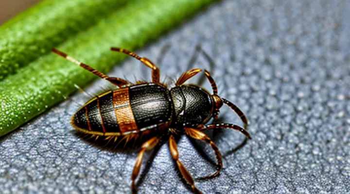
Key Characteristics of a Newly Attached Tick
Size and Shape Variations
A newly attached tick presents a compact, flattened body that conforms closely to the host’s skin. The overall length typically measures between 1 mm and 3 mm, depending on species and developmental stage. Adult females of larger species may reach up to 4 mm, while larvae rarely exceed 0.5 mm. The dorsal shield (scutum) remains visible as a distinct, hardened plate, often darker than the surrounding cuticle.
Shape changes occur rapidly after attachment. The ventral side expands to accommodate the feeding apparatus, producing a slight convex curvature. Mouthparts—chelicerae and hypostome—penetrate the epidermis and become visible as a small, protruding structure. The abdomen remains relatively undeveloped at this stage; significant swelling appears only after several hours of blood ingestion.
Key size and shape parameters observed in the initial attachment phase:
- Length: 0.5 mm – 4 mm, species‑dependent
- Width: 0.3 mm – 2 mm, proportionate to length
- Scutum visibility: continuous, unbroken plate
- Mouthpart orientation: ventrally directed, partially exposed
These measurements provide a reliable baseline for identifying a tick immediately after it secures itself on a host.
Color and Texture
A newly attached tick appears markedly different from its free‑living stage. The dorsal surface is generally a uniform, pale hue—ranging from light tan to a muted reddish‑brown—reflecting the blood it has begun to ingest. The ventral side often shows a slightly darker shade, sometimes with a faint pinkish tint caused by the expanding gut.
-
Color
-
Texture
These visual cues—uniform pale coloration on the back, a reddish abdomen, and a smooth, engorged texture—are reliable indicators that a tick has just secured its attachment.
Position on the Host
A newly attached tick presents a flattened dorsal shield that lies flush against the host’s skin. The mouthparts, or capitulum, are fully inserted, appearing as a small, dark point protruding from the body. The abdomen remains relatively unfilled, giving the insect a compact, oval shape. Legs extend outward from the ventral side, creating a stable tripod that supports the feeding position.
Typical locations on the host include:
- Scalp and neck region, where hair provides concealment.
- Axillary folds, offering warmth and humidity.
- Inguinal area, protected by clothing.
- Behind the ears, a common site for quick access to blood vessels.
In each site the tick aligns its capitulum upward, penetrating the epidermis at a shallow angle to maximize blood flow. The body’s ventral surface contacts the skin, leaving no visible gap. Leg placement varies to accommodate the curvature of the host’s surface, ensuring continuous attachment during movement.
Distinguishing a Tick from Other Pests
Common Misidentifications
A tick that has just begun feeding appears as a small, swollen, oval body attached firmly to the skin. The abdomen expands dramatically, often resembling a tiny, brownish or reddish balloon. The mouthparts, called the hypostome, are embedded in the skin and may be visible as a dark point at the rear of the body.
Misidentifying a newly attached tick is common. Errors arise because several other organisms or skin conditions share superficial similarities. Recognizing key differences prevents unnecessary alarm or inappropriate treatment.
- Spider or mite: Typically have distinct legs or segmented bodies; a feeding tick lacks visible legs and presents a smooth, rounded shape.
- Flea: Small, laterally flattened, and capable of jumping; a feeding tick is engorged, immobile, and firmly anchored.
- Scab or crust: Forms as a dry, flaky layer; a tick’s surface remains moist and retains a defined, rounded outline.
- Skin tag: Soft, flesh-colored protrusion with a stalk; a tick’s body is darker, more uniform, and the attachment point is a puncture rather than a stalk.
- Dermatofibroma or cyst: Firm, raised nodule without a central puncture; a tick shows a central dark point where the mouthparts penetrate.
Accurate identification relies on observing the engorged, smooth, oval form and the embedded mouthpart. When doubt persists, removal with fine-tipped tweezers and consultation with a medical professional are advisable.
Unique Anatomical Features
A newly attached tick appears flattened, with a dark, matte dorsal shield (scutum) that covers only the anterior portion of the body. The ventral side is concealed beneath a thin, translucent cuticle, making the overall silhouette compact and difficult to see against the host’s skin.
Key anatomical elements that distinguish an attached tick include:
- Capitulum – a forward‑projecting structure housing the mouthparts; its shape is species‑specific and remains visible as a small, raised protrusion.
- Hypostome – a barbed, spear‑like organ that penetrates the host’s skin, anchoring the parasite and facilitating blood intake.
- Palps – paired sensory appendages that flank the hypostome, aiding in locating blood vessels.
- Coxae and Legs – eight legs originate from the coxae; they are positioned close to the body, giving the tick a low profile while it feeds.
- Cement cone – a secretion that hardens around the mouthparts, securing the tick to the host’s epidermis.
During the early feeding phase, the abdomen (idiosoma) expands minimally, retaining a slender, elongated shape. The cuticle remains semi‑transparent, allowing the host’s skin tone to show through. As blood ingestion progresses, the abdomen swells dramatically, and the tick’s coloration shifts from dark brown to a lighter, more opaque hue, but these changes occur after the initial attachment period.
What to Do Upon Discovery
Safe Removal Techniques
A tick that has just embedded its mouthparts appears swollen at the head end, with a dark, rounded body and a visible attachment point where the hypostome penetrates the skin. Prompt, proper removal prevents pathogen transmission and minimizes tissue damage.
Safe removal steps:
- Grasp the tick as close to the skin as possible using fine‑point tweezers.
- Apply steady, downward pressure to pull the tick straight out without twisting.
- Avoid squeezing the abdomen; crushing can release infectious fluids.
- After extraction, cleanse the bite area with antiseptic solution.
- Preserve the tick in a sealed container if identification or testing is required.
- Monitor the site for signs of infection or rash over the next several weeks.
If the tick’s mouthparts remain embedded, do not dig them out with sharp instruments. Instead, apply a clean, damp cloth to the area and seek medical advice. Regularly inspect exposed skin after outdoor activities, especially in areas with dense vegetation, to detect newly attached ticks before they become engorged.
When to Seek Medical Attention
A tick that has just attached appears as a small, engorged arachnid firmly attached to the skin, often with its mouthparts embedded. The body may be swollen and darkened, and the surrounding skin can show a red halo or irritation.
Seek professional medical care if any of the following conditions occur:
- A rash develops that expands rapidly, forms a target or bull’s‑eye pattern, or is accompanied by fever.
- Flu‑like symptoms appear within weeks of the bite, such as headache, muscle aches, fatigue, or joint pain.
- The tick remains attached for more than 48 hours, especially if it is a known disease‑vector species.
- The bite site becomes increasingly painful, swollen, or shows signs of infection (pus, warmth, red streaks).
- You have a weakened immune system, are pregnant, or have a history of tick‑borne illnesses.
Prompt evaluation enables early testing and treatment, reducing the risk of complications from diseases transmitted by ticks.
