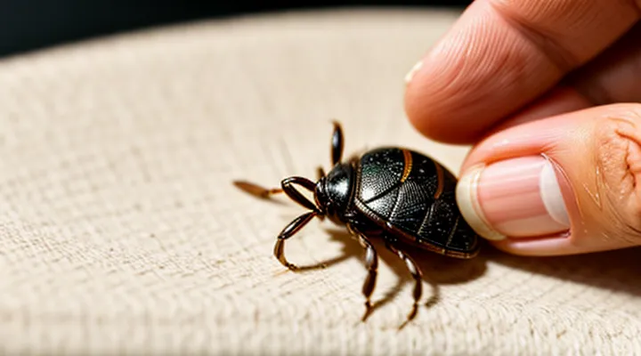
Initial Steps Before Tick Removal
Gathering Necessary Supplies
The Importance of «Fine-tipped Tweezers»
Removing a tick promptly reduces the risk of pathogen transmission. The tool used determines whether the parasite is extracted intact or whether mouthparts remain embedded, which can cause infection. Fine‑tipped tweezers meet the requirements for safe removal.
These tweezers have a narrow, pointed tip that fits around the tick’s head without crushing the body. The metal construction allows a firm, controlled grip. The design minimizes contact with surrounding skin, decreasing the chance of bruising.
Advantages of fine‑tipped tweezers include:
- Precise alignment with the tick’s mouthparts, enabling a clean pull.
- Ability to apply steady, linear force, preventing the head from breaking off.
- Simple sterilization with alcohol or boiling water.
- Compact size for use on any body area, including scalp and interdigital spaces.
Effective use follows a short sequence: grasp the tick as close to the skin as possible, align the tweezers parallel to the surface, pull upward with steady pressure, and avoid twisting. After extraction, disinfect the bite site and the tweezers. Dispose of the tick in a sealed container for possible testing.
Choosing fine‑tipped tweezers ensures the entire parasite is removed in one motion, lowers the likelihood of secondary infection, and complies with medical recommendations for tick removal.
Instruments to Strictly «Avoid»
When removing a tick from a person, certain tools must be excluded because they increase the risk of incomplete extraction, pathogen transmission, or tissue damage.
- Fine‑point tweezers with serrated or rubber‑coated tips – gripping surfaces can crush the tick’s body, forcing internal fluids into the wound.
- Bare fingers or ungloved hands – lack of precision leads to slippage and potential loss of the tick’s mouthparts.
- Pincers, pliers, or nail clippers – excessive force and broad jaws compress the tick, often leaving the hypostome embedded.
- Heat sources such as matches, cigarettes, or hot needles – cause the tick to release saliva while still attached, raising infection probability.
- Chemicals (oil, petroleum jelly, alcohol) applied directly to the tick – induce regurgitation of pathogens and do not aid in removal.
- Squeezing the abdomen or applying pressure to the tick’s body – forces infected fluids into the host’s skin.
Only instruments designed for delicate, firm grasp without crushing, such as straight, non‑serrated fine‑point tweezers, should be employed. Avoiding the listed items ensures complete removal, minimizes tissue trauma, and reduces the chance of disease transmission.
Preparing the Removal Environment
Prepare a clean, well‑lit area where the tick removal will take place. Choose a surface that can be disinfected easily, such as a hard table covered with a disposable barrier. Ensure adequate lighting, preferably a lamp with adjustable brightness, to visualize the attachment point clearly.
Gather the necessary instruments before beginning. Required items include:
- Fine‑point tweezers or forceps made of stainless steel
- Antiseptic wipes or solution (e.g., 70 % isopropyl alcohol)
- Disposable gloves
- A small container with a lid for the extracted tick
- A bandage or sterile gauze for post‑removal care
Sanitize the work surface and the instruments with the antiseptic solution. Wear gloves to protect both the patient and the handler from potential pathogens. Position the patient comfortably, exposing the affected skin while maintaining privacy. Keep the container for the tick within reach to avoid cross‑contamination after removal.
The Essential Removal Technique
Positioning the Removal Tool
Grasping as Close to the Skin as Possible
Grasp the tick as close to the skin as possible. Holding the mouthparts near the entry point prevents the parasite’s head from breaking off, which could leave infected tissue behind.
- Use fine‑point tweezers or a specialized tick‑removal tool.
- Position the tips around the tick’s head, not the body.
- Apply steady, gentle pressure to lift the tick straight upward.
- Avoid twisting, jerking, or squeezing the abdomen.
After extraction, clean the bite area with antiseptic, wash hands thoroughly, and monitor the site for signs of infection such as redness, swelling, or a rash. If any symptoms develop, seek medical advice promptly.
Why «Twisting or Jerking» is Dangerous
Removing a tick by twisting or jerking can leave the mouthparts embedded in the skin. Retained parts create a portal for bacteria, increase inflammation, and may trigger local infection.
The following risks arise from improper extraction:
- Incomplete removal – head and hypostome remain, providing a conduit for pathogen entry.
- Elevated disease transmission – prolonged attachment of mouthparts raises the chance of transmitting bacteria, viruses, or protozoa.
- Tissue trauma – sudden force tears skin, causing bleeding, pain, and possible scarring.
- Delayed healing – damaged tissue requires more time to close, exposing the wound to environmental contaminants.
A controlled, steady pull with fine‑point tweezers, keeping the tick’s body aligned with the skin, minimizes these hazards and ensures complete removal.
Applying Consistent Upward Pressure
Applying consistent upward pressure is a critical element of safe tick extraction. The technique minimizes the risk of the tick’s mouthparts remaining embedded in the skin, which can lead to infection or prolonged irritation.
To implement the method, follow these precise actions:
- Grip the tick as close to the skin as possible using fine‑point tweezers or a specialized tick removal tool.
- Align the instrument with the tick’s body, avoiding squeezing its abdomen.
- Apply steady, upward force while maintaining the grip; do not rock, twist, or jerk the tick.
- Continue the pressure until the tick releases its attachment and separates cleanly.
- After removal, clean the bite area with antiseptic and inspect the tick for any retained parts.
Consistent pressure ensures that the anchoring structures—hypostome and cement—are overcome without breaking the tick’s body. Interrupting the force or using irregular motions increases the chance of mouthpart fragmentation. The method works for all common tick species on humans, provided the tool is appropriately sized and the operator maintains a firm, uninterrupted pull.
Strategies When Parts Are Left Behind
When a tick is detached, fragments of its mouthparts may remain embedded in the skin. Retained parts can cause local inflammation, infection, or prolonged exposure to tick‑borne pathogens. Prompt, precise action reduces these risks.
- Inspect the bite site closely after removal. Look for any visible protrusion or irregularity.
- If a fragment is visible, grasp it with fine‑point tweezers, positioning the tips as close to the skin as possible.
- Apply steady, gentle pressure to pull the fragment out in line with the skin surface; avoid twisting or jerking motions that could enlarge the wound.
- Disinfect the area with an antiseptic solution such as chlorhexidine or iodine.
- Cover the site with a clean dressing and monitor for redness, swelling, or discharge over the next 48 hours.
If the fragment cannot be seen or extracted easily, do not dig or use blunt objects. Instead:
- Clean the area thoroughly.
- Apply a topical antibiotic ointment.
- Seek medical evaluation, especially if the bite is in a sensitive region (e.g., scalp, eyelid) or the individual has a compromised immune system.
Medical professionals may employ a sterile needle or scalpel to excise the residual tissue, followed by suturing if necessary. After removal, document the incident, note any symptoms, and consider testing for tick‑borne diseases according to local health guidelines.
Handling the Tick and the Bite Site
Disposing of the Removed Parasite
Methods for «Killing the Tick»
Removing a tick safely begins with ensuring the parasite is dead before extraction. Killing the tick eliminates the risk of saliva or gut contents contaminating the wound and reduces the chance of pathogen transmission.
- Mechanical crushing: Place a fine‑pointed pair of tweezers over the tick’s head, apply steady pressure to crush the body, then lift the dead insect away. This method requires immediate disposal in a sealed container.
- Freezing: Apply a commercially available cryogenic spray or a small ice pack directly to the tick for 30–60 seconds. The rapid temperature drop immobilizes and kills the arthropod without compromising its outer shell.
- Chemical immobilization: Use a topical solution containing 70 % isopropyl alcohol or a veterinary‑approved acaricide. Saturate the tick for at least 10 seconds; the chemical penetrates the cuticle, causing rapid death.
- Thermal destruction: Heat a metal instrument (e.g., a scalpel tip) with a lighter until red‑hot, then touch the tick’s body briefly. The heat denatures proteins, killing the parasite instantly.
After the tick is confirmed dead, grasp the mouthparts with fine‑pointed tweezers as close to the skin as possible, pull upward with steady, even force, and avoid twisting. Clean the bite area with antiseptic and monitor for signs of infection. Dispose of the dead tick in a sealed bag or flush it down the toilet.
When to Save the Specimen for Testing
Removing a tick from a human host requires prompt, careful extraction and, in certain circumstances, preservation of the arthropod for laboratory analysis. Retaining the specimen is justified only when the risk of pathogen transmission is significant or when epidemiological data are needed. Save the tick for testing if any of the following conditions apply:
- The bite occurred in an area endemic for tick‑borne diseases such as Lyme disease, Rocky Mountain spotted fever, or ehrlichiosis.
- The patient exhibits symptoms consistent with a tick‑borne infection (fever, rash, arthralgia, or neurologic signs) within weeks of the attachment.
- The tick was attached for longer than 24 hours, increasing the probability of pathogen acquisition.
- The species cannot be identified confidently during removal, and species‑specific risk assessment is required.
- Public health authorities request specimens for surveillance or outbreak investigation.
If none of these criteria are met, discarding the tick after removal is acceptable. When preservation is indicated, place the tick in a sealed container with a moist cotton ball, label with date, location, and host details, and refrigerate (4 °C) or freeze (‑20 °C) until submission to a qualified laboratory. Proper handling prevents degradation of potential pathogens and ensures reliable diagnostic results.
Sanitizing the Affected Area
After a tick is removed, the bite site must be cleaned promptly to reduce the risk of bacterial infection and pathogen transmission. Begin by washing the area with mild soap and running water for at least 20 seconds, then pat dry with a disposable towel.
- Apply a broad‑spectrum antiseptic (e.g., povidone‑iodine, chlorhexidine, or alcohol‑based solution) directly to the wound.
- Allow the antiseptic to remain in contact for the duration recommended on the product label (usually 30 seconds to 1 minute) before covering.
- If a sterile dressing is needed, place it over the treated area to protect against external contaminants.
Observe the site for signs of redness, swelling, or discharge over the next 24–48 hours. Should any inflammatory response develop, seek medical evaluation and consider a topical antibiotic or prescription treatment as directed by a healthcare professional.
Recognizing Complications and Red Flags
Localized Reactions and Signs of Secondary Infection
After extracting a tick, examine the bite area at least once daily for the first week. Look for redness, swelling, or a small crater that persists beyond 24 hours.
Common benign responses include a faint pink ring, mild itching, or a transient rash that fades within a few days. These signs usually indicate normal irritation from the bite.
Signs that a secondary infection may be developing are:
- Increasing redness extending beyond the immediate perimeter of the wound
- Warmth or throbbing sensation at the site
- Purulent discharge or visible pus
- Swelling that enlarges rather than recedes
- Fever, chills, or malaise accompanying the local reaction
If any of these symptoms appear, clean the area with antiseptic, apply a sterile dressing, and seek medical evaluation promptly. Early antimicrobial treatment can prevent complications such as cellulitis or Lyme‑related manifestations.
Indicators of Potential Tick-Borne Illness
Monitoring for the Characteristic «Bulls-eye Rash»
After a tick is detached, the most reliable indicator that the bite may have transmitted an infection is the development of a target‑shaped skin lesion. This rash, commonly called a bullseye rash, appears in a predictable window and signals the need for prompt evaluation.
The lesion typically emerges within 3 – 30 days after removal. Early appearance (around day 5) suggests rapid pathogen transmission, while later onset may indicate slower progression. Absence of the rash does not guarantee safety; some infections manifest without cutaneous signs.
Key visual features include:
- Central clearing surrounded by a red ring, often 5 – 30 mm in diameter.
- Expansion over several days, sometimes forming concentric rings.
- Mild itching or tenderness, but usually no severe pain.
Monitoring protocol:
- Inspect the bite site daily for at least four weeks.
- Photograph any skin changes to track size and pattern.
- Record the date of tick removal and note any systemic symptoms (fever, fatigue, joint pain).
- Contact a healthcare professional immediately if the rash appears, expands rapidly, or is accompanied by flu‑like signs.
Early detection of the bullseye rash enables timely antimicrobial therapy, reducing the risk of complications. Consistent self‑examination combined with clear documentation provides the most effective surveillance after tick extraction.
General Systemic Symptoms Requiring Intervention
Ticks attached to the skin can transmit pathogens that trigger systemic responses. When a bite leads to signs that extend beyond the local site, immediate medical evaluation is warranted. The following manifestations indicate that simple removal is insufficient and professional intervention is required.
- Fever exceeding 38 °C (100.4 °F)
- Severe headache or neck stiffness
- Generalized muscle or joint pain, especially if persistent or worsening
- Nausea, vomiting, or diarrhea not attributable to other causes
- Rash that expands rapidly, appears as a “bull’s‑eye,” or presents as multiple lesions
- Swelling of lymph nodes distant from the bite site
- Sudden onset of confusion, dizziness, or fainting
- Signs of anaphylaxis: difficulty breathing, swelling of the face or throat, rapid heartbeat, or drop in blood pressure
These systemic indicators suggest possible infection with agents such as Borrelia burgdorferi, Anaplasma phagocytophilum, or other tick‑borne pathogens. Prompt antimicrobial therapy, supportive care, or emergency treatment may be necessary to prevent complications. If any of the listed symptoms develop after a tick is extracted, seek medical attention without delay.
