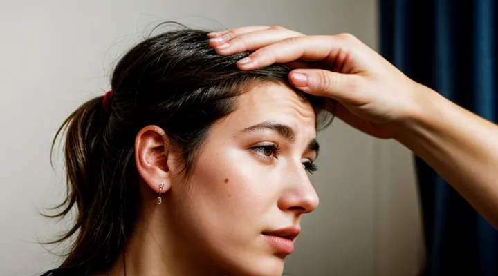
Understanding Sarcoptes Scabiei
«The Scabies Mite Life Cycle»
The scabies mite, Sarcoptes scabiei, completes its development on human skin. An adult female burrows into the stratum corneum, creates a tunnel, and deposits 2–3 eggs daily for up to 30 days. The life cycle proceeds as follows:
- Egg: 2–4 days; hatches into a larva within the tunnel.
- Larva: six-legged; remains in the tunnel for 3–4 days, then molts.
- Nymph: eight-legged; undergoes two successive molts over 3–4 days each, maturing into an adult.
- Adult: male and female; females return to the surface to create new burrows and continue egg production.
All stages occur on the host’s epidermis; the mite cannot survive off the body for more than 48 hours. The scalp presents a less favorable environment because hair density, sebaceous secretions, and temperature differ from typical infestation sites such as the wrists, elbows, and intertriginous areas. Nevertheless, if the mite reaches the scalp, it follows the same developmental pattern, although infestation density tends to be lower. Consequently, the mite’s life cycle does not restrict it to any specific body region, but ecological factors make sustained colonization of the head uncommon.
«Preferred Habitats on the Human Body»
Scabies mites (Sarcoptes scabiei var. hominis) require warm, moist skin surfaces where they can burrow and reproduce. Their preferred locations on an adult host include:
- Interdigital spaces of the hands
- Wrists and forearms
- Axillary folds
- Waistline and belt area
- Genital region and buttocks
- Under the breasts in females
These sites share high skin temperature, thin stratum corneum, and limited hair density, facilitating mite penetration and egg laying.
The scalp presents several deterrents: dense hair reduces direct skin contact, the scalp’s temperature is slightly lower than core body temperature, and sebum production creates a less favorable environment. Consequently, infestations on the head are uncommon in immunocompetent individuals. In cases of severe immunosuppression, extensive crusted scabies, or in infants whose hair is sparse, mites may be found on the scalp and ears.
Thus, while the head is not a typical habitat, it can become colonized under specific pathological conditions that alter the protective barriers normally preventing mite survival.
Scabies in Atypical Locations
«Scalp Scabies: A Rare Presentation»
Scabies mites (Sarcoptes scabiei var. hominis) typically colonize the skin’s stratum corneum, preferring warm, moist intertriginous zones. The scalp is an uncommon site because hair shafts impede mite movement and the sebaceous environment differs from preferred habitats. Nevertheless, infestations confined to the scalp have been documented, especially in infants, immunocompromised patients, and individuals with extensive dermatological disease.
Clinical presentation of scalp scabies includes:
- Intense pruritus, often worsening at night
- Fine, grayish‑white papules or crusted lesions over the hairline, occipital region, and behind the ears
- Visible burrows parallel to hair growth, occasionally revealed by dermoscopy as a “jet‑liner” sign
- Secondary bacterial infection in severe cases, leading to impetigo or cellulitis
Diagnosis relies on:
- Direct microscopic examination of skin scrapings showing mites, eggs, or fecal pellets
- Dermoscopic identification of characteristic mite morphology
- Exclusion of other pruritic scalp disorders (e.g., pediculosis, seborrheic dermatitis) through clinical correlation
Effective management consists of topical scabicidal agents such as 5 % permethrin cream applied to the scalp and surrounding hairline, left for 8–12 hours before washing. In refractory or crusted forms, oral ivermectin (200 µg/kg) administered in two doses 1–2 weeks apart enhances eradication. Adjunctive measures include washing bedding at ≥60 °C, treating close contacts, and applying antiseptics to prevent bacterial superinfection.
While the scalp does not provide an optimal environment for Sarcoptes scabiei, rare cases confirm that mites can survive and reproduce on the scalp under specific host conditions. Prompt recognition and targeted therapy prevent progression to extensive crusted scabies and limit transmission.
«Factors Contributing to Atypical Scabies»
Scabies mites normally inhabit the skin’s intertriginous zones, but several conditions allow them to colonize the scalp and other atypical regions.
Immunosuppression, whether caused by HIV infection, organ transplantation, or systemic corticosteroids, reduces the host’s ability to limit mite proliferation. The resulting high mite burden can overwhelm the usual distribution limits, leading to infestation of the hair‑bearing scalp.
Advanced age, particularly in institutionalized elderly patients, often coincides with reduced grooming capacity and compromised skin integrity. These factors facilitate mite migration to the head.
Crusted (Norwegian) scabies presents with millions of mites, far exceeding the load seen in classic scabies. The massive population overwhelms the normal confinement to flexural areas, causing widespread involvement that includes the scalp.
Dermatologic conditions that disrupt the skin barrier—psoriasis, eczema, seborrheic dermatitis—create microenvironments favorable for mite survival on the scalp surface.
Poor hygiene and crowded living conditions increase exposure to heavily infested individuals, raising the probability of atypical colonization.
The following factors most commonly contribute to scabies appearing on the head:
- Immunocompromised status (HIV, transplant, immunosuppressive therapy)
- Elderly patients with limited self‑care
- Crusted scabies with extremely high mite numbers
- Coexisting skin disorders that compromise the barrier
- Overcrowding and inadequate sanitation
Understanding these contributors clarifies why scabies mites can persist in the head region despite their usual preference for other body sites.
Differential Diagnosis for Scalp Conditions
«Common Scalp Ailments»
Scalp disorders encompass a range of conditions that affect hair follicles, sebaceous glands, and the surrounding skin. Among them, the presence of the scabies mite on the scalp is uncommon but not impossible. The mite normally colonizes warm, moist areas such as the wrists, elbows, and groin. Survival on the scalp requires a suitable environment—adequate humidity, minimal hair grooming, and, often, compromised host immunity. Infants, individuals with severe eczema, or patients receiving immunosuppressive therapy may present with scalp infestation.
Typical scalp problems include:
- Dandruff (dry scalp flaking)
- Seborrheic dermatitis (inflamed, oily scaling)
- Psoriasis (thick, silvery plaques)
- Folliculitis (inflamed hair follicles)
- Pediculosis (head‑lice infestation)
- Scabies (mite‑induced burrowing lesions)
When scabies involves the scalp, lesions appear as tiny, intensely itchy papules, frequently accompanied by linear burrows at the base of hair shafts. Diagnosis relies on microscopic identification of mites, eggs, or fecal pellets from skin scrapings. Effective treatment mirrors that for body scabies: topical permethrin 5 % applied to the entire scalp and left for the recommended duration, or oral ivermectin when topical therapy is impractical. Adjunctive measures—regular washing of bedding, clothing, and personal items—prevent reinfestation.
In summary, while the scalp is not the primary habitat for the scabies mite, specific host factors can enable colonization, adding scabies to the list of common scalp ailments that require targeted diagnosis and therapy.
«Distinguishing Scabies from Other Conditions»
Scabies infestations most often affect the wrists, interdigital spaces, and trunk, but occasional involvement of the scalp and hairline occurs, especially in infants and immunocompromised individuals. Recognizing scabies when it appears on the head requires careful separation from other dermatologic conditions that produce similar lesions.
Typical scabies signs include:
- Intense nocturnal pruritus triggered by pressure or heat.
- Linear or serpentine burrows visible under a dermatoscope or magnifying lens.
- Small, erythematous papules often grouped around hair follicles on the scalp.
- Presence of mites, eggs, or fecal pellets on skin scrapings examined microscopically.
Key differentiators from common mimickers:
- Pediculosis capitis (head lice) – causes itching and nits attached to hair shafts; lacks burrows and microscopic mites in skin layers.
- Seborrheic dermatitis – presents with greasy scaling and erythema; pruritus is milder, and no burrows are observed.
- Atopic dermatitis – shows chronic eczematous patches, often with a history of asthma or allergic rhinitis; burrows are absent.
- Tinea capitis (fungal infection) – produces alopecia, broken hairs, and kerion formation; confirmed by KOH preparation, not by mite detection.
- Contact dermatitis – limited to areas of allergen exposure, with vesicles or edema rather than burrows.
Diagnostic confirmation relies on:
- Skin scraping from active lesions, placed on a glass slide with mineral oil, and examined at 100–400× magnification for Sarcoptes scabiei.
- Dermoscopic visualization of the "jet‑liner" sign, indicating the mite’s anterior portion within a burrow.
- Response to a standard scabicidal regimen (e.g., permethrin 5% cream) applied to the entire scalp and neck; rapid symptom resolution supports the diagnosis.
Accurate differentiation prevents unnecessary antifungal or antibacterial treatments and ensures timely implementation of scabicidal therapy, which is essential for controlling infestation and limiting spread to other body regions.
Diagnosis and Treatment of Scalp Scabies
«Diagnostic Methods for Scabies»
Scabies diagnosis relies on direct observation of the mite, its eggs, or fecal material. Clinical inspection identifies characteristic linear burrows and intense pruritus, especially at typical sites such as wrists, interdigital spaces, and, in rare cases, the scalp of infants or immunocompromised adults. Confirmatory tests include:
- Skin scraping examined under light microscopy to reveal live mites, ova, or feces.
- Dermoscopy (entodermoscopy) that magnifies burrows and displays the “delta wing” sign of the mite’s anterior body.
- Burrow ink test, where a drop of ink highlights the tunnel for easier visualization.
- Polymerase chain reaction (PCR) targeting mite DNA for high‑sensitivity detection in ambiguous presentations.
- Confocal laser scanning microscopy, providing in‑vivo imaging of mite structures without biopsy.
Laboratory culture is not performed because the mite cannot survive outside the host. When head involvement is suspected, careful examination of the hairline and scalp skin, combined with microscopy of scalp scrapings, confirms or excludes infestation. Accurate diagnosis guides timely treatment and prevents spread.
«Topical and Oral Treatments»
Scabies infestations of the scalp require prompt pharmacologic intervention because the mite can survive in the dense hair environment. Effective management combines topical acaricides with systemic agents to eradicate both surface and concealed parasites.
Topical options:
- Permethrin 5 % cream applied to the entire scalp, left for 8–14 hours before washing; repeat after one week.
- Benzyl benzoate 25 % lotion, spread over hair and scalp, left for 12 hours; a second application after 48 hours enhances cure rates.
- Crotamiton 10 % lotion, applied nightly for three consecutive nights; suitable for patients with permethrin intolerance.
Oral agents:
- Ivermectin 200 µg/kg as a single dose, repeated after 7 days; dosage may be increased for severe or refractory cases.
- Albendazole 400 mg daily for three days, used when ivermectin is contraindicated or ineffective.
Adjunct measures:
- Wash bedding, clothing, and towels at 60 °C or seal in plastic bags for at least 72 hours.
- Avoid scalp scratching to prevent secondary infection; consider topical corticosteroids if inflammation is pronounced.
Combining a properly applied topical preparation with a single dose of oral ivermectin yields the highest eradication rates for head scabies, minimizing the risk of persistent infestation.
Prevention and Management
«Personal Hygiene Practices»
Personal hygiene directly affects the likelihood of scabies mites colonizing the scalp and hair. Regular washing with medicated or antibacterial shampoo removes debris and reduces mite density. Thorough drying of hair and scalp eliminates the moist environment that supports mite survival.
Key practices include:
- Daily shampooing, preferably with products containing permethrin or benzyl benzoate for known infestations.
- Frequent combing of hair with a fine-toothed lice comb to dislodge mites and eggs.
- Cleaning of hair accessories, pillowcases, and hats at temperatures above 60 °C or using a disinfectant spray.
- Avoiding shared headgear and close scalp contact with infected individuals.
Consistent application of these measures limits mite transmission and prevents the establishment of a viable population on the head.
«Environmental Decontamination»
Scabies mites typically colonize warm, protected skin areas such as the wrists, elbows, and genital region. The scalp provides a less favorable environment because hair density, sebum production, and frequent washing reduce mite survival. Nevertheless, cases of head involvement have been documented, especially in infants and immunocompromised individuals where the mite may temporarily occupy hair follicles.
Effective environmental decontamination limits reinfestation and supports treatment success. Key actions include:
- Washing bedding, clothing, and towels in hot water (≥ 60 °C) for at least 10 minutes, followed by high‑heat drying.
- Sealing non‑washable items in plastic bags for a minimum of 72 hours, the duration required for mite mortality without a host.
- Vacuuming carpets, upholstered furniture, and mattresses thoroughly; discarding vacuum bags or cleaning canisters immediately.
- Applying a 0.1 % benzyl benzoate or permethrin solution to surfaces that cannot be laundered, adhering to manufacturer safety guidelines.
Prompt removal of contaminated items, combined with appropriate topical or oral acaricidal therapy, eliminates the risk of head colonization and prevents further spread within the household.
